

PD-1 deficiency augments bone marrow failure in a minor-histocompatibility antigen mismatch lymphocyte infusion model
- PMID: 29524567
- PMCID: PMC5962409
- DOI: 10.1016/j.exphem.2018.03.001
PD-1 deficiency augments bone marrow failure in a minor-histocompatibility antigen mismatch lymphocyte infusion model
Abstract
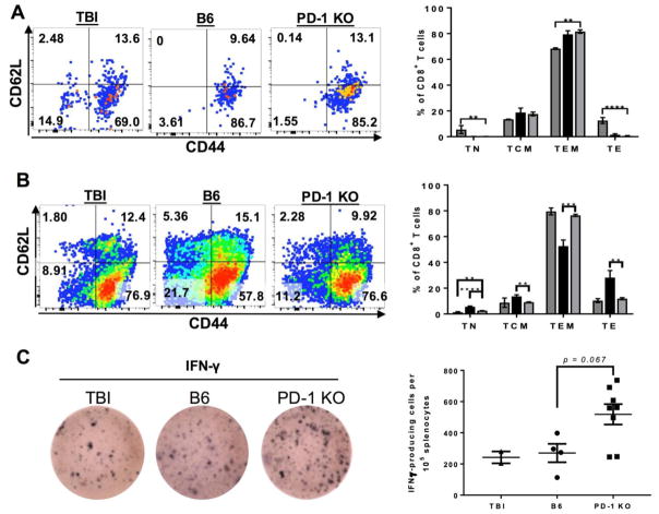
Although PD-1 blockade has revolutionized cancer immunotherapy, immune-related adverse events (irAEs) present life-threatening complications. Recent reports of aplastic anemia (AA) as irAEs implicate PD-1/PD-L1 as important in preventing immune-mediated destruction of the hematopoietic niche. Infusion of PD-1-deficient (PD-1 knockout [KO]) lymph node (LN) cells into minor-antigen mismatched mice resulted in early mortality, as well as more severe bone marrow (BM) hypoplasia, anemia, and BM microarchitecture disruption in PD-1 KO LN-infused mice relative to mice that received B6 LN cell infusion. Mice that received PD-1 KO LN cells had more CD8+ T-cell infiltration of the BM and greater expansion of H60-specific CD8+ T cells than did their B6 LN-infused counterparts. In the spleen, CD8+ T cells were skewed to an effector memory phenotype, suggesting accelerated differentiation of PD-1 KO T cells. Our data suggest that PD-1 dysregulation has a role in murine BM failure and vigilance in irAE monitoring may be desirable to treat early AA and related cytopenias.
Published by Elsevier Inc.
Conflict of interest statement
The authors have no financial conflicts of interest.
Figures


Similar articles
-
Minor antigen h60-mediated aplastic anemia is ameliorated by immunosuppression and the infusion of regulatory T cells.J Immunol. 2007 Apr 1;178(7):4159-68. doi: 10.4049/jimmunol.178.7.4159. J Immunol. 2007. PMID: 17371972
-
A mouse model of lymphocyte infusion-induced bone marrow failure.Exp Hematol. 2004 Dec;32(12):1163-72. doi: 10.1016/j.exphem.2004.08.006. Exp Hematol. 2004. PMID: 15588941
-
[The characterization analysis of pathogenic T cells in immune-mediated aplastic anemia mouse model].Zhonghua Xue Ye Xue Za Zhi. 2022 Jul 14;43(7):587-593. doi: 10.3760/cma.j.issn.0253-2727.2022.07.010. Zhonghua Xue Ye Xue Za Zhi. 2022. PMID: 36709137 Free PMC article. Chinese.
-
Graft-versus-host disease impairs vaccine responses through decreased CD4+ and CD8+ T cell proliferation and increased perforin-mediated CD8+ T cell apoptosis.J Immunol. 2013 Feb 1;190(3):1351-9. doi: 10.4049/jimmunol.1200391. Epub 2012 Dec 28. J Immunol. 2013. PMID: 23275602 Free PMC article.
-
Real-time T-cell profiling identifies H60 as a major minor histocompatibility antigen in murine graft-versus-host disease.Blood. 2002 Dec 15;100(13):4259-65. doi: 10.1182/blood-2002-05-1299. Epub 2002 Aug 22. Blood. 2002. PMID: 12393464
Cited by
-
Spectrum of Immune Checkpoint Inhibitor Anemias: Results From a Single Center, Early-Phase Clinical Trials Case Series Experience.J Hematol. 2022 Jun;11(3):113-120. doi: 10.14740/jh1006. Epub 2022 Jun 2. J Hematol. 2022. PMID: 35837373 Free PMC article.
-
Hematologic Complications of Immune Checkpoint Inhibitors.Oncologist. 2019 May;24(5):584-588. doi: 10.1634/theoncologist.2018-0574. Epub 2019 Feb 28. Oncologist. 2019. PMID: 30819785 Free PMC article.
-
The Efficacy and Safety of Immune Checkpoint Inhibitors in Patients With Cancer and Preexisting Autoimmune Disease.Front Oncol. 2021 Feb 22;11:625872. doi: 10.3389/fonc.2021.625872. eCollection 2021. Front Oncol. 2021. PMID: 33692958 Free PMC article. Review.
References
-
- Brahmer JR, Drake CG, Wollner I, Powderly JD, Picus J, Sharfman WH, Stankevich E, et al. Phase I Study of Single-Agent Anti–Programmed Death-1 (MDX-1106) in Refractory Solid Tumors: Safety, Clinical Activity, Pharmacodynamics, and Immunologic Correlates. Journal of Clinical Oncology. 2010;28:3167–3175. - PMC - PubMed
-
- Friedman CF, Proverbs-Singh TA, Postow MA. Treatment of the Immune-Related Adverse Effects of Immune Checkpoint Inhibitors: A Review. JAMA Oncol. 2016;2:1346–1353. - PubMed
-
- Makarious D, Horwood K, Coward JIG. Myasthenia gravis: An emerging toxicity of immune checkpoint inhibitors. European Journal of Cancer. 2017;82:128–136. - PubMed
Publication types
MeSH terms
Substances
Grants and funding
LinkOut - more resources
Full Text Sources
Other Literature Sources
Medical
Research Materials
