. 2018 Apr 5;102(4):517-527.
doi: 10.1016/j.ajhg.2018.02.008. Epub 2018 Mar 8.

Identification and Rescue of Splice Defects Caused by Two Neighboring Deep-Intronic ABCA4 Mutations Underlying Stargardt Disease

Affiliations

Identification and Rescue of Splice Defects Caused by Two Neighboring Deep-Intronic ABCA4 Mutations Underlying Stargardt Disease

Silvia Albert et al. Am J Hum Genet. .

Abstract

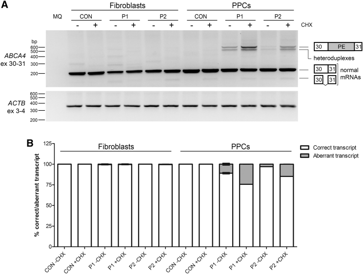
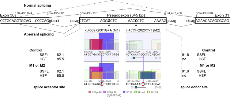
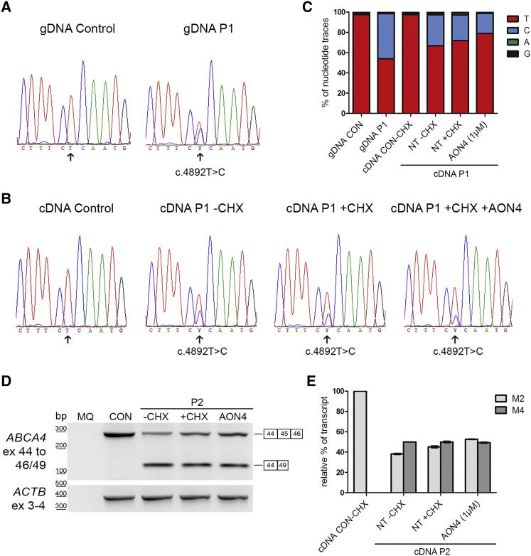
Sequence analysis of the coding regions and splice site sequences in inherited retinal diseases is not able to uncover ∼40% of the causal variants. Whole-genome sequencing can identify most of the non-coding variants, but their interpretation is still very challenging, in particular when the relevant gene is expressed in a tissue-specific manner. Deep-intronic variants in ABCA4 have been associated with autosomal-recessive Stargardt disease (STGD1), but the exact pathogenic mechanism is unknown. By generating photoreceptor precursor cells (PPCs) from fibroblasts obtained from individuals with STGD1, we demonstrated that two neighboring deep-intronic ABCA4 variants (c.4539+2001G>A and c.4539+2028C>T) result in a retina-specific 345-nt pseudoexon insertion (predicted protein change: p.Arg1514Leufs36), likely due to the creation of exonic enhancers. Administration of antisense oligonucleotides (AONs) targeting the 345-nt pseudoexon can significantly rescue the splicing defect observed in PPCs of two individuals with these mutations. Intriguingly, an AON that is complementary to c.4539+2001G>A rescued the splicing defect only in PPCs derived from an individual with STGD1 with this but not the other mutation, demonstrating the high specificity of AONs. In addition, a single AON molecule rescued splicing defects associated with different neighboring mutations, thereby providing new strategies for the treatment of persons with STGD1. As many genes associated with human genetic conditions are expressed in specific tissues and pre-mRNA splicing may also rely on organ-specific factors, our approach to investigate and treat splicing variants using differentiated cells derived from individuals with STGD1 can be applied to any tissue of interest.

Keywords: ABCA4; Stargardt disease; antisense oligonucleotide; deep-intronic mutation; exonic splicing enhancer; induced pluripotent stem cells; nonsense-mediated decay; photoreceptor precursor cells; pseudoexon; splicing modulation.


Figures

Figure 1
Figure 1
Identification of the Splice Defect Caused by Variants M1 and M2 (A) Reverse transcription (RT)-PCR on mRNA extracted from control, P1 (carrying variants M1: c.4539+2001G>A and M3: c.4892T>C), and P2 (carrying variants M2: c.4539+2028C>T and M4: c.6148−698_6670delinsTGTGCACCTCCCTAG)-derived fibroblasts and photoreceptor precursor cells (PPCs) using primers in exons 30 and 31 of ABCA4. For both P1- and P2-derived PPCs, some aberrantly spliced bands were detected, especially after cycloheximide (CHX) treatment (+). Actin (ACTB) RT-PCR was used as a control. Heteroduplexes contain transcripts with the pseudoexon together with the correct spliced transcript (exons 30 and 31) and the truncated splice variant of exon 30 that lacks the 3′ 73 nt of exon 30 (r.4467_4539del [p.Cys1490Glufs12]). (B) Semi-quantification of the ratio of correctly and aberrantly spliced ABCA4 transcript for each cell line with and without CHX. Error bars indicate standard deviation.
Figure 2
Figure 2
In Silico Characterization of the Effect Caused by Two Deep-Intronic Variants Schematic representation of the boundaries of the 345-bp pseudoexon, with the location of the variants M1 (c.4539+2001G>A) and M2 (c.4539+2028C>T), the genomic positions of the splice sites, the splicing events detected, and the splice site predictions for both acceptor and donor sites. The dashed line represents the splicing from a cryptic splice donor site in exon 30 at position g.94,495,074 (GRCh37/hg19) to the normal splice acceptor site of exon 31 (r.4467_4539del [p.Cys1490Glufs12]). The predicted values of the splice acceptor and donor sites in the control and mutant situations did not show any difference. In the middle panels, the effects of the variants enhancing or creating new ESE motifs are depicted. Abbreviations: SSFL, SpliceSiteFinder-like; HSF, Human Splicing Finder; nd, not detected.
Figure 3
Figure 3
Effect of Antisense Oligonucleotide Delivery at mRNA in Photoreceptor Precursor Cells (A) Schematic representation of the pseudoexon, indicating the location of the variants, the SC35 motifs with the highest scores, and the positions of the antisense oligonucleotides (AONs). (B) RNA analysis on AON-treated cells. RT-PCR from exon 30 to exon 31 of ABCA4 mRNA in control, P1 (M1: c.4539+2001G>A and M3: c.4892T>C), and P2 (M2: c.4539+2028C>T and M4: c.6148−698_6670delinsTGTGCACCTCCCTAG)-derived photoreceptor precursor cells (PPCs) upon AON delivery. Actin (ACTB) mRNA amplification was used to normalize samples. Abbreviations: NT-, non-treated and in the absence of cycloheximide (CHX); NT+, non-treated in the presence of CHX; A1, AON1; A2, AON2; A3, AON3; A4, AON4; S, SON; and MQ, PCR negative control. Heteroduplexes contain transcripts with the pseudoexon together with the correct spliced transcript (exons 30 and 31) and the truncated splice variant of exon 30 that lacks the 3′ 73 nt of exon 30 (r.4467_4539del [p.Cys1490Glufs12]). (C) Semi-quantification of the ratio of correctly versus aberrantly spliced transcripts in all P1 and P2 samples. (D) Percentage of correction of each AON compared to the NT+ based on the ratio observed in (C). Statistical differences in the efficacy of the AONs for M1 and M2 are indicated with an asterisk (p < 0.05 using Mann-Whitney test). All histograms illustrate the average ± standard deviation.
Figure 4
Figure 4
AON Rescue in Photoreceptor Precursor Cells Based on the Analysis of the mRNA Alleles Associated with Variants M1/M3 and M2/M4 (A–C) Assessment of the mRNA product carrying variant M3, c.4892T>C (p.Leu1631Pro) which is in trans with M1. Chromatograms (A) of the reverse sequence of genomic DNA (gDNA) in a control and individual P1 carrying M1 and M3. Chromatograms (B) of the sequence of the cDNA of the photoreceptor precursor cells (PPC) from the control cell line and from P1 PPCs in the absence (−) or presence (+) of cycloheximide (CHX) and 1 μM of AON4 (+AON4). The arrow indicates the position of the double peak where the M3 variant is located. (C) Graphical representation of the traces of each nucleotide in the sequence using ContigExpress software. (D and E) Semi-quantification of mRNA products associated with variants M2 and M4 in P2. (D) P2 PPCs carry M2 and c.6148–698_6670delinsTGTGCACCTCCCTAG (p.Val2050_Gln2243del) in trans. RT-PCR of the control (CON) and P2 PPCs without (−) or with (+) CHX and AON4 at 1 μM using the same forward primer in exon 44 and a reverse primer in exon 46 (that is not present in M4 genomic DNA nor in the mRNA product) and a reverse primer in exon 49. The upper band represents the correct spliced transcript from exon 44 to 46 corresponding to the M2 allele, while the lower band is the resulting transcript caused by the multi-exon deletion. An RT-PCR product of ACTB was used as loading control. (E) Graphical representation of the semi-quantification of the bands observed in (D). The mRNA product observed in the control (exons 44 to 46) was set at 100%, while the mRNA product from the M4 allele (exons 44 to 49) was set at 50% in the P2-PPCs –CHX condition. The mRNA product from allele M4 is not sensitive to NMD suppression. The correctly spliced transcripts from allele M2 increase upon NMD suppression (+CHX versus −CHX) of cultured PPCs and increase even more upon AON4 treatment. Error bars represent standard deviation for each condition.

References

    1. Fishman G.A., Stone E.M., Grover S., Derlacki D.J., Haines H.L., Hockey R.R. Variation of clinical expression in patients with Stargardt dystrophy and sequence variations in the ABCR gene. Arch. Ophthalmol. 1999;117:504–510. - PubMed
    1. Stargardt K. Über familiäre, progressive Degeneration in der Maculagegend des Auges. Graefes Arch. Clin. Exp. Ophthalmol. 1909;71:534–550.
    1. Allikmets R., Singh N., Sun H., Shroyer N.F., Hutchinson A., Chidambaram A., Gerrard B., Baird L., Stauffer D., Peiffer A. A photoreceptor cell-specific ATP-binding transporter gene (ABCR) is mutated in recessive Stargardt macular dystrophy. Nat. Genet. 1997;15:236–246. - PubMed
    1. Zernant J., Xie Y.A., Ayuso C., Riveiro-Alvarez R., Lopez-Martinez M.A., Simonelli F., Testa F., Gorin M.B., Strom S.P., Bertelsen M. Analysis of the ABCA4 genomic locus in Stargardt disease. Hum. Mol. Genet. 2014;23:6797–6806. - PMC - PubMed
    1. Molday R.S., Beharry S., Ahn J., Zhong M. Binding of N-retinylidene-PE to ABCA4 and a model for its transport across membranes. Adv. Exp. Med. Biol. 2006;572:465–470. - PubMed

Publication types