Review
. 2018 Apr;39(4):328-340.
doi: 10.1016/j.it.2018.02.003. Epub 2018 Mar 8.

Innate Antiviral Immunity in the Skin

Affiliations
Review

Innate Antiviral Immunity in the Skin

Chelsea Handfield et al. Trends Immunol. 2018 Apr.

Abstract

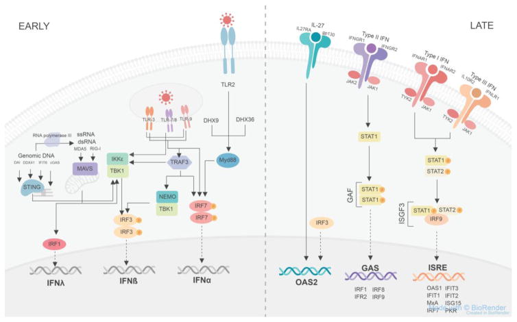
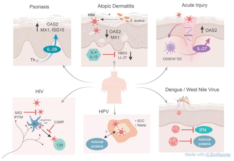
Barrier sites such as the skin play a critical role in immune defense. They must maintain homeostasis with commensals and rapidly detect and limit pathogen invasion. This is accomplished in part through the production of endogenous antimicrobial peptides and proteins, which can be either constitutive or inducible. Here, we focus particularly on the control of innate antiviral proteins and present the basic aspects of their regulation in the skin by interferons (IFNs), IFN-independent immunity, and environmental factors. We also discuss the activity and (dys-)function of antiviral proteins in the context of skin-tropic viruses and highlight the relevance of the innate antiviral pathway as a potential therapeutic avenue for vulnerable patient populations and skin diseases with high risk for virus infections.


Figures

Figure 1, Key Figure
Figure 1, Key Figure. Innate antiviral immunity signaling pathways
In the early phase, viral nucleic acids are recognized by toll-like receptors (TLR) on the plasma and endosomal membranes receptors or within the cytoplasm by melanoma differentiation-associated protein 5 (MDA5), retinoic-acid-inducible gene I (RIG-I), DNA-dependent activator of IFN-regulatory factors (DAI), DDX41, interferon (IFN)-induced protein with tetratricopeptide repeats 6 (IFIT6), cyclic GMP-AMP synthase (cGAS), DexH-box helicase 9 (DHX9), DHX36 or TNFα receptor-associated factor 3 (TRAF3). Signaling from TLR2, DHX9, and DHX36 goes through myeloid differentiation primary response gene 88 (Myd88) which stimulates phosphorylated IFN-regulatory factor 7 (IRF7) to homodimerize and enter the nucleus to increase the transcription of IFNα. Signaling from the endosomal TLRs can also stimulate phosphorylated IRF7 homodimerization, while an alternative signaling pathway is through the TANK binding kinase-1 (TBK1) and IκB kinase ε (IKKε) complex, which stimulates phosphorylated IRF3 homodimerization to induce the transcription of IFNβ. TLR3 can also signal through TRAF3, which induces the phosphorylation of IRF3 via the TBK1 and NF-κB essential modulator (NEMO) complex. The TBK1 and IKKε complex is also formed in response to genomic DNA binding to DAI, DDX41, IFIT6 and cGAS via STING, which is associated with the endoplasmic reticulum. During the replication of genomic DNA, RNA polymerase III forms single stranded RNA replicates that can be detected by MDA5 or RIG-I. MDA5 and RIG-I also sense viral dsRNA and ssRNA and then bind to mitochondrial antiviral signaling protein (MAVS), associated with the mitochondrial membrane, to stimulate the formation of the TBK1 and IKKe complex or MAVS binding stimulates IRF1 to translocate to the nucleus and induce the transcription of IFNλ. In the late phase, the cytokine interleukin-27 (IL-27) binds to its transmembrane plasma receptor composed of interleukin 27 receptor A (IL27RA) and glycoprotein 130 (gp130) stimulating the transcription of oligoadenylate synthetase 2 (OAS2) in an IFN-independent manner. An alternative signal to induce OAS2 independent of IFNs occurs via IRF3. Type II IFNs bind to their receptor consisting of IFNγ receptor 1 (IFNGR1) and IFNGR2; each subunit is associated with janus kinase 2 (JAK2) on the cytoplasmic domain. This activates signal transducers and activators of transcription 1 (STAT1) to become phosphorylated and homodimerize forming IFNγ activation factor (GAF). GAF then translocates to the nucleus and binds to GAF site within the promoter region of target genes to induce the transcription of IRF1, IRF2, IRF8, and IRF9. Type I IFNs bind to their receptor composed of IFNα receptor 1 (IFNAR1) associated with cytoplasmic tyrosine kinase 2 (TYK2) and IFNAR2 associated with cytoplasmic JAK1. Type III IFNs bind to their receptor composed of interleukin 10 receptor 2 (IL10R2) associated with cytoplasmic TYK2 and IFNλ receptor 1 (IFNLR1) associated with cytoplasmic JAK1. Both type I and III IFNs binding to their respective receptors leads to the formation of the phosphorylated STAT1 and phosphorylated STAT2 heterodimer complex which then conjugates with IRF9 to form the IFN-stimulated gene factor 3 (ISGF3) complex. ISGF3 then translocates to the nucleus and binds to the IFN-stimulated response element (ISRE) site within the promoter region of target genes to induce the transcription of OAS1, IFIT1, myxovirus-resistance A (MxA), IRF7, IFIT3, IFIT2, interferon-stimulated protein of 15kDa (ISG15), and protein kinase R (PKR).
Figure 2
Figure 2. Models of antiviral dysregulation within skin diseases
Antiviral immunity is dysregulated among a broad spectrum of skin conditions (clockwise): Psoriasis is a chronic inflammatory skin condition characterized by high Th17 numbers. These Th17 cells produce IL-29 which subsequently induces antiviral proteins including OAS2, MX1 and ISG15 [5]. However, innate immune regulators of antiviral proteins in psoriatic skin has not been defined. Skin affected by atopic dermatitis, also known as atopic eczema, shows increased expression of Th2-type cytokines interleukin-4 (IL-4) and IL-13 which inhibit antimicrobial proteins human beta defensin 3 (HBD3) and cathelicidin (LL-37), which have broad-spectrum antimicrobial activity including antibacterial and antiviral function [54, 113]. AD skin also shows decreased expression of professional antiviral proteins 2′5′-oligoadenylate synthase 2 (OAS2) and myxovirus-resistance 1 protein (MX1). Patients with AD have a defective skin barrier and are frequently colonized with Staphylococcus aureus (S. aureus) which can enhance Herpes simplex virus (HSV) infections via toxin production and enhances viral cell entry and replication [63]. In response to acute mechanical skin injury, double-stranded RNA serves as a danger associated molecular pattern (DAMP) stimulating CD301b+ dendritic cells (DC) to release IL-27, which induces the production of OAS2 in keratinocytes [7]. The Aedes mosquito transmits many emerging viruses such as Dengue and West Nile virus into human skin allowing these viruses to penetrate permissive epithelial cells including keratinocytes [–116]. Many viruses produce proteins that are able to counter the actions of interferon (IFN) and antiviral proteins [6, 110, 117]. Human papillomavirus (HPV) causes cutaneous warts and increases the risk of developing squamous cell carcinoma (SCC) typically in immunocompromised patients. Whether this virus is countered by type I or II IFNs or through IFN-independent pathways has yet to be determined. Also, the control of potentially oncogenic viruses like HPV and Merkel Cell polyomavirus (not shown here) by antiviral proteins is not well known. While there is no risk of transmission if the skin is not broken, contact between HIV-infected fluids with broken skin, wounds, or open sores on mucous membranes can lead to HIV transmission. HIV can initially infects the skin, specifically Langerhans cells within the epidermis as well as dermal dendritic cells, which then present HIV antigen to T cells [–121]. Antiviral proteins including MX2 and interferon-induced transmembrane proteins (IFITM) can counter HIV and may be able to prevent the transmission of the virus. Whether the neuropeptide calcitonin-gene related peptide (CGRP) released from neurons promotes proteasomal degradation of HIV within Langerhans cells in the skin, similar to what has been shown in mucosal Langerhans cells [122]is currently unknown and hypothetical.

Similar articles

Cited by

References

    1. Chan YK, Gack MU. Viral evasion of intracellular DNA and RNA sensing. Nat Rev Microbiol. 2016;14(6):360–73. - PMC - PubMed
    1. Iwasaki A. Exploiting Mucosal Immunity for Antiviral Vaccines. Annu Rev Immunol. 2016;34:575–608. - PubMed
    1. Urb M, Sheppard DC. The role of mast cells in the defence against pathogens. PLoS Pathog. 2012;8(4):e1002619. - PMC - PubMed
    1. Paludan SR, et al. Recognition of herpesviruses by the innate immune system. Nat Rev Immunol. 2011;11(2):143–54. - PMC - PubMed
    1. Wolk K, et al. IL-29 is produced by T(H)17 cells and mediates the cutaneous antiviral competence in psoriasis. Sci Transl Med. 2013;5(204):204ra129. - PubMed

Publication types