. 2018 Sep;138(9):1962-1972.
doi: 10.1016/j.jid.2018.03.001. Epub 2018 Mar 9.

Lack of Type 2 Innate Lymphoid Cells Promotes a Type I-Driven Enhanced Immune Response in Contact Hypersensitivity

Affiliations

Lack of Type 2 Innate Lymphoid Cells Promotes a Type I-Driven Enhanced Immune Response in Contact Hypersensitivity

David A Rafei-Shamsabadi et al. J Invest Dermatol. 2018 Sep.

Abstract

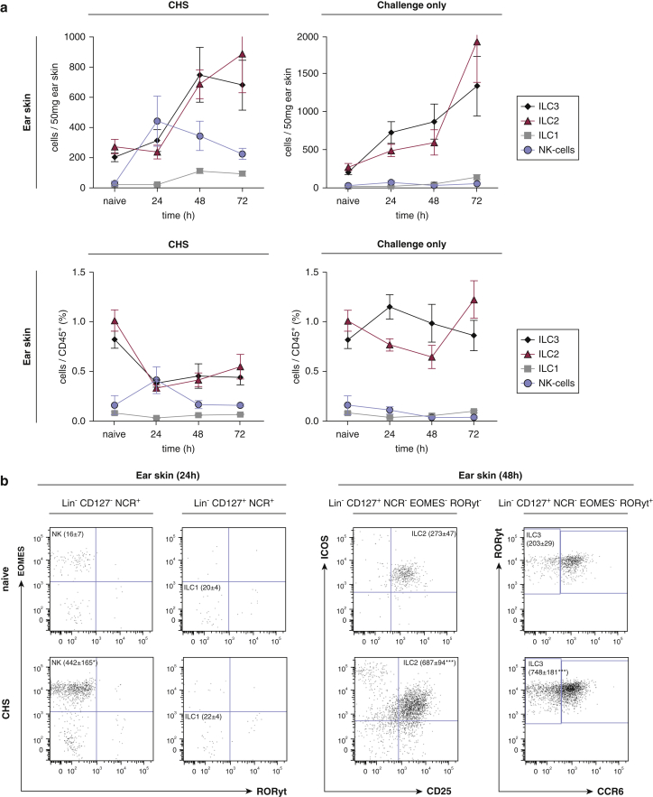
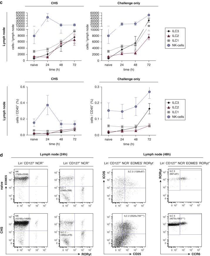
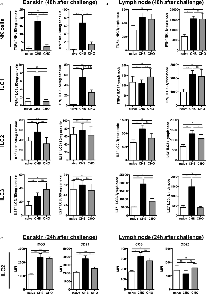
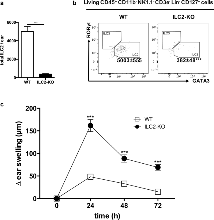
Allergic contact dermatitis and its animal model, contact hypersensitivity, are T-cell-mediated inflammatory skin diseases that require activation of the innate immune system. Here we investigate the role of innate lymphoid cells (ILCs) during the elicitation phase of 2,4,6-trinitrochlorobenzene-induced contact hypersensitivity using EomesGfp/+ x Rorc(γt)-CreTg x Rosa26RYfp/+ reporter mice. Ear swelling responses, cutaneous ILC numbers, and cytokine production were determined at different time points. Functional analyses were performed in a CD90.1/.2 congenic adoptive transfer model that allowed selective antibody-mediated depletion of ILCs before hapten challenge, and in Rorasg/floxIl7rCre/+ mice, which lack ILC2. Hapten challenge induced early increases of natural killer cells in skin and ear draining lymph nodes corresponding to the peak ear swelling response. In contrast, ILC1, 2, and 3 showed a delayed increase in numbers corresponding to the contact hypersensitivity resolution phase. Hapten challenge induced increased marker cytokines in all ILC subtypes and an activated phenotype in ILC2. Depletion of all ILC resulted in a significantly enhanced ear swelling response. Similarly, ILC2-deficient mice (Rorasg/floxIl7rCre/+) displayed increased ear swelling responses on hapten challenge, suggesting that ILC2 act as negative regulators in the type 1-dominated immune response of contact hypersensitivity.


Figures

Figure 1
Figure 1
On hapten challenge NK cells increase in ear skin before ILC2 and ILC3. (a, c) Kinetics of absolute cell numbers (upper panel) and relative cell numbers (lower panel) for all ILC subsets in the (a) ears and (c) lymph nodes of mice sensitized and challenged with TNCB (CHS) (left graphs) and mice only challenged with TNCB (challenge only) (right graphs). (b, d) Representative concatenate dot plots visualizing changes in numbers of NK cells and ILC1 at 24 hours and ILC2 and ILC3 at 48 hours, respectively, in CHS mice compared with naïve mice. Values are shown as absolute cell numbers per 50 mg ear skin and percentages of living CD45+ leukocytes, respectively. Data are shown as mean ± standard error of the mean, pooled data from three independent experiments with at least n ≥ 5 mice per group. Concatenate dot plots show data of pooled samples of naive and CHS groups of mice, respectively (n ≥ 5 mice per group). *P < 0.05, **P < 0.01, ***P < 0.001, and ****P < 0.0001. EOMESGfp RORγt-fm mice were used for these experiments. CHS, contact hypersensitivity; EOMES, eomesodermin; NCR, natural cytotoxicity triggering receptor, that is, NK1.1 and NKp46; NK, natural killer; ILC, innate lymphoid cell; TNCB, 2,4,6-trinitrochlorobenzene.
Figure 1
Figure 1
On hapten challenge NK cells increase in ear skin before ILC2 and ILC3. (a, c) Kinetics of absolute cell numbers (upper panel) and relative cell numbers (lower panel) for all ILC subsets in the (a) ears and (c) lymph nodes of mice sensitized and challenged with TNCB (CHS) (left graphs) and mice only challenged with TNCB (challenge only) (right graphs). (b, d) Representative concatenate dot plots visualizing changes in numbers of NK cells and ILC1 at 24 hours and ILC2 and ILC3 at 48 hours, respectively, in CHS mice compared with naïve mice. Values are shown as absolute cell numbers per 50 mg ear skin and percentages of living CD45+ leukocytes, respectively. Data are shown as mean ± standard error of the mean, pooled data from three independent experiments with at least n ≥ 5 mice per group. Concatenate dot plots show data of pooled samples of naive and CHS groups of mice, respectively (n ≥ 5 mice per group). *P < 0.05, **P < 0.01, ***P < 0.001, and ****P < 0.0001. EOMESGfp RORγt-fm mice were used for these experiments. CHS, contact hypersensitivity; EOMES, eomesodermin; NCR, natural cytotoxicity triggering receptor, that is, NK1.1 and NKp46; NK, natural killer; ILC, innate lymphoid cell; TNCB, 2,4,6-trinitrochlorobenzene.
Figure 2
Figure 2
Cytokine expression by ILC in ear skin and ear draining lymph nodes during the elicitation phase of CHS. Cytokine production of all ILC subsets in the (a) ear skin and (b) ear draining lymph nodes at 48 hours after antigen challenge in CHS and challenge-only mice compared with naïve mice. (c) ICOS and CD25 expression of ILC2 isolated from ear skin (c, left graphs) and ear draining lymph nodes (c, right graphs) at 24 hours after allergen challenge in CHS and challenge-only mice. Values are shown as absolute cell numbers per 50 mg ear skin and per total ear draining lymph node, respectively. Data are shown as mean ± standard error of the mean, pooled data of three independent experiments with n ≥ 5 mice per group. *P < 0.05, **P < 0.01, ***P < 0.001, and ****P < 0.0001. EOMESGfp RORγt-fm mice were used for these experiments. CHO, challenge only; CHS, contact hypersensitivity; EOMES, eomesodermin; ICOS, inducible T-cell costimulator; ILC, innate lymphoid cell; MFI, mean fluorescence intensity; NCR, natural cytotoxicity triggering receptor; NK, natural killer; ns, not significant; TNF, tumor necrosis factor.
Figure 3
Figure 3
Depletion of all ILC subsets leads to an enhanced ear swelling response. (a) Verification of ILC depletion in ear draining lymph nodes (a, upper panel) and ear skin (a, lower panel) after administering anti-CD90.2 (200 μg) every other day (4 days total) to CD90.2 Rag1–/– recipient mice that were reconstituted with FACS-sorted CD3, CD8+, and CD4+ T cells from CD90.1+ donor mice. Values in (a) are displayed as absolute cell numbers per 50 mg ear skin and percent age of cells of total leukocytes in the ear draining lymph nodes, respectively. (b) Representative dot plots showing depletion of CD90.2+ leukocytes in ear draining lymph nodes (b, upper panel) and ear skin (b, lower panel). (c) Ear swelling response after the last allergen challenge in ILC-depleted versus isotype-treated mice shown as delta from baseline ear thickness. Data in (a) and (c) are shown as mean ± standard error of the mean, pooled data of three independent experiments with n ≥ 5 mice per group. *P < 0.05, **P < 0.01, ***P < 0.001, and ****P < 0.0001. ILC, innate lymphoid cell.
Figure 4
Figure 4
Lack of ILC2 leads to increased CHS responses. (a) Number of ILC2 in the skin of ILC2-KO mice compared with wild-type mice. (b) Representative dot blots of ILC2 in the ear skin of naïve and ILC2-KO mice. (c) Ear swelling response of ILC2-KO compared with wild-type mice up to 72 hours after allergen challenge displayed as delta from baseline ear thickness. Values in (a) are displayed as absolute cell numbers per 50 mg ear skin. Data in (b) and (c) are shown as mean ± standard deviation, pooled data of two independent experiments with at least n ≥ 5 mice per group. *P < 0.05, **P < 0.01, ***P < 0.001, and ****P < 0.0001. ILC2 were first gated on live, CD45+ CD11b NK1.1 CD3e, and then lineage (CD19, B220, CD5, Gr-1, FreR1a, Ter119, F4/80) and CD127+. CHS, contact hypersensitivity; ILC, innate lymphoid cell; KO, knockout; NK, natural killer; WT, wild type.

References

    1. Arnold S.J., Sugnaseelan J., Groszer M., Srinivas S., Robertson E.J. Generation and analysis of a mouse line harboring GFP in the Eomes/Tbr2 locus. Genesis. 2009;47:775–781. - PMC - PubMed
    1. Artis D., Spits H. The biology of innate lymphoid cells. Nature. 2015;517:293–301. - PubMed
    1. Bando J.K., Liang H.-E., Locksley R.M. Identification and distribution of developing innate lymphoid cells in the fetal mouse intestine. Nat Immunol. 2015;16:153–160. - PMC - PubMed
    1. Batista M.D., Ho E.L., Kuebler P.J., Milush J.M., Lanier L.L., Kallas E.G. Skewed distribution of natural killer cells in psoriasis skin lesions. Exp Dermatol. 2013;22:64–66. - PMC - PubMed
    1. Bi J., Cui L., Yu G., Yang X., Chen Y., Wan X. NK cells alleviate lung inflammation by negatively regulating group 2 innate lymphoid cells. J Immunol. 2017;198:3336–3344. - PubMed

Publication types

LinkOut - more resources