Relationship between morphological development and sex hormone receptor expression of mammary glands with age in male rats
- PMID: 29526866
- PMCID: PMC6083026
- DOI: 10.1538/expanim.17-0134
Relationship between morphological development and sex hormone receptor expression of mammary glands with age in male rats
Abstract
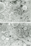
The aim of this study is to investigate the changes with age on morphology and sex hormone receptor expression in the mammary glands of male Sprague-Dawley rats, focusing on male-specific cells, "oxyphilic cells", observed after sexual maturity. The mammary glands of male rats at 14, 21, 35, 50, 75 and 100 days old were examined by gross observation, microscopic observation using whole mount specimens, histological and immunohistochemical sections. Grossly, mammary glands showed brown color at 50-100 days old. In whole mount specimens, terminal end buds (TEBs) were observed at 14-50 days old and the number of TEBs was highest at 35 days old. Histologically, the male mammary glands contained small epithelial cells with scanty cytoplasm at 14-35 days old while ductal and lobular epithelial cells were changed into oxyphilic cells with abundant cytoplasm at 50-100 days old. Immunohistochemicaly, androgen receptor (AR), estrogen receptor (ER) and progesterone receptor (PgR) expressions were found in both mammary glands found at a young age and oxyphilic cells. In oxyphilic cells, AR expression was dominant compared to ER and PgR expressions and increased with age. From these results, the development at 50-100 days old might be strongly related to AR. Ultrastructural observation of oxyphilic cells confirmed a number of lipid droplets, deformed and/or enlarged mitochondria, lysosomes and peroxisomes in their cytoplasm.
Keywords: development; hormone receptor; male rat; mammary gland; oxyphilic cell.
Figures


Similar articles
-
Pattern of distribution of cells positive for estrogen receptor alpha and progesterone receptor in relation to proliferating cells in the mammary gland.Breast Cancer Res Treat. 1999 Feb;53(3):217-27. doi: 10.1023/a:1006186719322. Breast Cancer Res Treat. 1999. PMID: 10369068
-
Effect of prenatal and prepubertal genistein exposure on N-methyl-N-nitrosourea-induced mammary tumorigenesis in female Sprague-Dawley rats.In Vivo. 2003 Jul-Aug;17(4):349-57. In Vivo. 2003. PMID: 12929590
-
Exploring the spatial dimension of estrogen and progesterone signaling: detection of nuclear labeling in lobular epithelial cells in normal mammary glands adjacent to breast cancer.Diagn Pathol. 2014;9 Suppl 1(Suppl 1):S11. doi: 10.1186/1746-1596-9-S1-S11. Epub 2014 Dec 19. Diagn Pathol. 2014. PMID: 25565114 Free PMC article.
-
[Dual effects of androgens on mammary gland].Bull Cancer. 2008 May;95(5):495-502. doi: 10.1684/bdc.2008.0631. Bull Cancer. 2008. PMID: 18541513 Review. French.
-
ER and PR signaling nodes during mammary gland development.Breast Cancer Res. 2012 Jul 19;14(4):210. doi: 10.1186/bcr3166. Breast Cancer Res. 2012. PMID: 22809143 Free PMC article. Review.
Cited by
-
New Advances in Targeted Therapy of HER2-Negative Breast Cancer.Front Oncol. 2022 Mar 4;12:828438. doi: 10.3389/fonc.2022.828438. eCollection 2022. Front Oncol. 2022. PMID: 35311116 Free PMC article. Review.
-
A mammary fibroadenoma with terminal end buds-like structures in a 7-week-old male SD rat.J Toxicol Pathol. 2023 Apr;36(2):131-138. doi: 10.1293/tox.2022-0071. Epub 2022 Dec 12. J Toxicol Pathol. 2023. PMID: 37101956 Free PMC article.
References
-
- Bocchinfuso W.P., Hively W.P., Couse J.F., Varmus H.E., Korach K.S.1999. A mouse mammary tumor virus-Wnt-1 transgene induces mammary gland hyperplasia and tumorigenesis in mice lacking estrogen receptor-alpha. Cancer Res. 59: 1869–1876. - PubMed
MeSH terms
Substances
LinkOut - more resources
Full Text Sources
Other Literature Sources
Research Materials

