

The mechanisms underlying the effects of AMH on Müllerian duct regression in male mice
- PMID: 29526868
- PMCID: PMC5938179
- DOI: 10.1292/jvms.18-0023
The mechanisms underlying the effects of AMH on Müllerian duct regression in male mice
Abstract
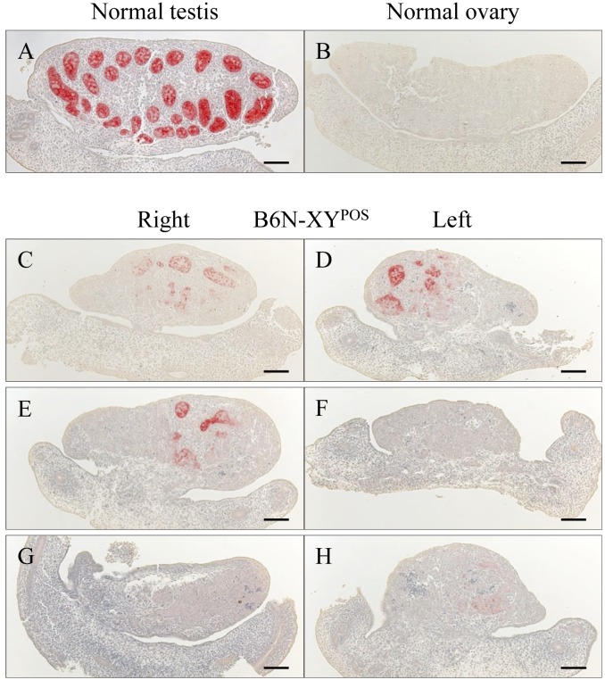
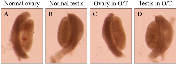
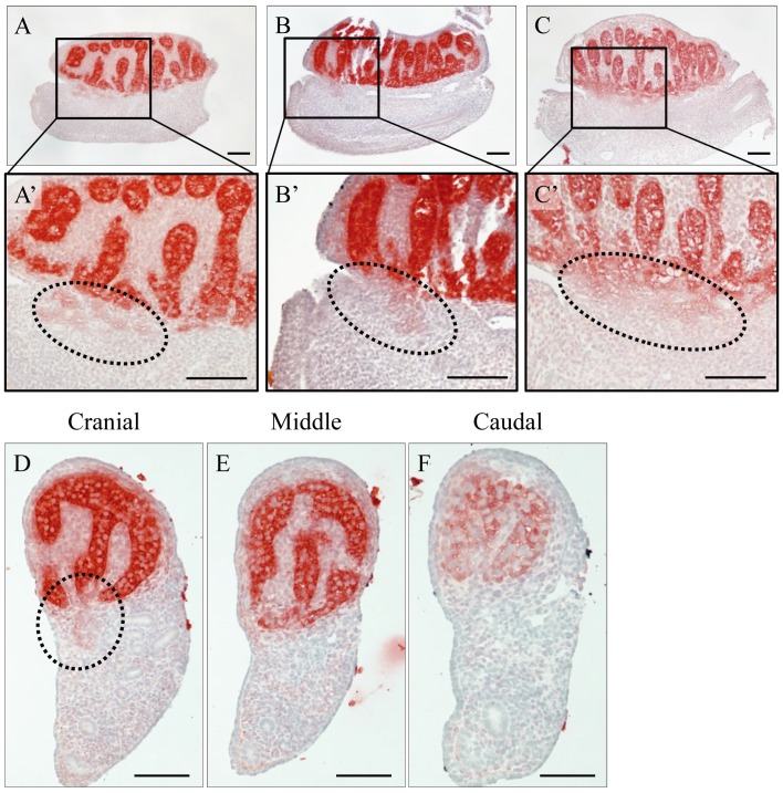
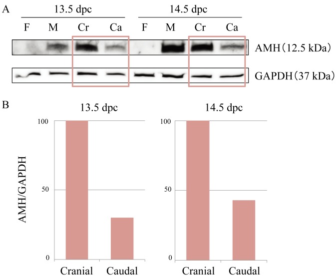
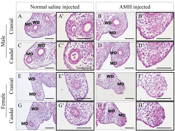
Anti-Müllerian hormone (AMH) produced in the developing testis induces the regression of the Müllerian duct, which develops into the oviducts, uterus and upper vagina. In our true hermaphrodite mouse with an ovary on one side and a testis on the other (O/T), the oviduct and uterus are present only on the ovary side, and nothing derived from the Müllerian duct is present on the testis side. Here, we investigate the mechanism underlying the unilateral Müllerian duct regression and the mode of AMH signaling, by performing immunohistology, Western blotting, and organ culture analyses. The histological analysis revealed that during the start of the Müllerian duct regression, the duct in the O/T mice was clearly regressed on the AMH-positive testis side compared to the AMH-negative ovary side. The immunohistochemistry showed a diffuse immunoreaction of AMH in the interstitium surrounding the testis cord and boundary region between the testis and mesonephros, especially in the cranial portion. Western blotting revealed that the amount of AMH in the cranial half of the mesonephros was larger than that in the caudal half. AMH injected into the gonads in organ culture induced the regression of the Müllerian duct via the interstitium of the organ. These results suggest that AMH acts on the Müllerian duct in male mice by exuding into the interstitium surrounding the testis cord and infiltrating through the cranial region from the testis to the mesonephros.
Keywords: AMH (anti-Müllerian hormone); Müllerian duct regression; secretion manner; sexual differentiation; true hermaphrodite.
Figures


Similar articles
-
β-Catenin is essential for Müllerian duct regression during male sexual differentiation.Development. 2011 May;138(10):1967-75. doi: 10.1242/dev.056143. Epub 2011 Apr 13. Development. 2011. PMID: 21490063 Free PMC article.
-
Direct diffusion of anti-Müllerian hormone from both the cranial and caudal regions of the testis during early gonadal development in mice.Dev Dyn. 2024 Mar;253(3):296-311. doi: 10.1002/dvdy.662. Epub 2023 Oct 3. Dev Dyn. 2024. PMID: 37787412
-
Anti-Müllerian Hormone Is Required for Chicken Embryonic Urogenital System Growth but Not Sexual Differentiation.Biol Reprod. 2015 Dec;93(6):138. doi: 10.1095/biolreprod.115.131664. Epub 2015 Oct 28. Biol Reprod. 2015. PMID: 26510867
-
The in vivo roles of müllerian-inhibiting substance.Curr Top Dev Biol. 1994;29:171-87. doi: 10.1016/s0070-2153(08)60550-5. Curr Top Dev Biol. 1994. PMID: 7828438 Review.
-
AMH and AMHR2 Involvement in Congenital Disorders of Sex Development.Sex Dev. 2022;16(2-3):138-146. doi: 10.1159/000518273. Epub 2021 Aug 31. Sex Dev. 2022. PMID: 34515230 Review.
Cited by
-
Establishment of an organ culture system to maintain the structure of mouse Müllerian ducts during development.J Vet Med Sci. 2024 Mar 16;86(3):300-307. doi: 10.1292/jvms.23-0492. Epub 2024 Jan 24. J Vet Med Sci. 2024. PMID: 38267037 Free PMC article.
-
Mini review: Asymmetric Müllerian duct development in the chicken embryo.Front Cell Dev Biol. 2024 Feb 5;12:1347711. doi: 10.3389/fcell.2024.1347711. eCollection 2024. Front Cell Dev Biol. 2024. PMID: 38380340 Free PMC article. Review.
-
Effect of AMH on primordial follicle populations in mouse ovaries and human pre-pubertal ovarian xenografts during doxorubicin treatment.Front Cell Dev Biol. 2024 Aug 27;12:1449156. doi: 10.3389/fcell.2024.1449156. eCollection 2024. Front Cell Dev Biol. 2024. PMID: 39258229 Free PMC article.
-
Expression patterns of sex steroid receptors in developing mesonephros of the male mouse: three-dimensional analysis.Cell Tissue Res. 2023 Sep;393(3):577-593. doi: 10.1007/s00441-023-03796-0. Epub 2023 Jun 19. Cell Tissue Res. 2023. PMID: 37335379
-
Genetic differences between C57BL/6 substrains affect the process of testis differentiation in YPOS mice.J Vet Med Sci. 2019 Apr 27;81(4):608-611. doi: 10.1292/jvms.18-0621. Epub 2019 Mar 4. J Vet Med Sci. 2019. PMID: 30828038 Free PMC article.
References
-
- Allard S., Adin P., Gouédard L., di Clemente N., Josso N., Orgebin-Crist M. C., Picard J. Y., Xavier F.2000. Molecular mechanisms of hormone-mediated Müllerian duct regression: involvement of β-catenin. Development 127: 3349–3360. - PubMed
-
- Baarends W. M., van Helmond M. J. L., Post M., van der Schoot P. J. C. M., Hoogerbrugge J. W., de Winter J. P., Uilenbroek J. T. J., Karels B., Wilming L. G., Meijers J. H. C., Themmen A. P. N., Grootegoed J. A.1994. A novel member of the transmembrane serine/threonine kinase receptor family is specifically expressed in the gonads and in mesenchymal cells adjacent to the müllerian duct. Development 120: 189–197. - PubMed
MeSH terms
Substances
LinkOut - more resources
Full Text Sources
Other Literature Sources
Molecular Biology Databases