

Aceclofenac-Induced Erythema Annulare Centrifugum
- PMID: 29527030
- PMCID: PMC5838759
- DOI: 10.4103/ijd.IJD_728_16
Aceclofenac-Induced Erythema Annulare Centrifugum
Abstract
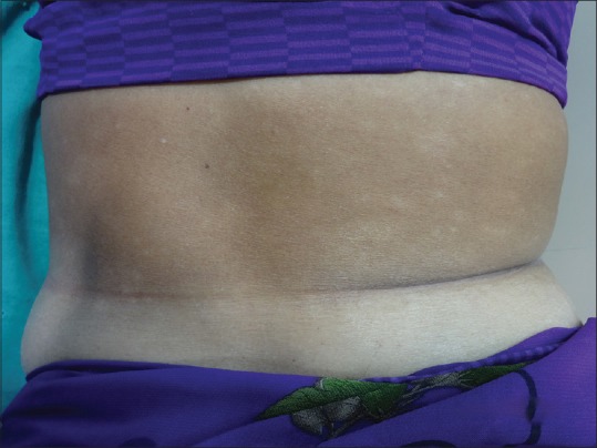
Erythema annulare centrifugum (EAC) is characterised by slowly enlarging annular erythematous lesions and is thought to represent a clinical reaction pattern to infections, medications, and rarely, underlying malignancy. Causative drugs include chloroquine, cimetidine, gold sodium thiomalate, amitriptyline, finasteride, etizolam etc. We present a case of 40-year-old woman who presented to us with a 10 days history of nonpruritic, peripherally growing annular erythematous eruption. She had a history of recent onset of joint pain, for which she was taking aceclofenac 90 mg once a day for 5 days prior to the onset of the rash. This was confirmed on biopsy as EAC. The rash promptly subsided after stopping the drug. We report this case as there was no previous report of aceclofenac induced EAC.
Keywords: Aceclofenac; drug induced; erythema annulare centrifugum.
Conflict of interest statement
There are no conflicts of interest. What is new? Acelofenac induced EAC has not been reported previously.Strong suscipion can help identify and withdraw the culprit drug.Withdrawal of the drug aids in resolution of lesions.
Figures


References
-
- Ziemer M, Eisendle K, Zelger B. New concepts on erythema annulare centrifugum: A clinical reaction pattern that does not represent a specific clinicopathological entity. Br J Dermatol. 2009;160:119–26. - PubMed
-
- Ashurst PJ. Erythema annulare centrifugum due to hydroxychloroquine sulfate and chloroquine sulfate. Arch Dermatol. 1967;95:37–9. - PubMed
-
- Kuroda K, Yabunami H, Hisanaga Y. Etizolam-induced superficial erythema annulare centrifugum. Clin Exp Dermatol. 2002;27:34–6. - PubMed
-
- Al Hammadi A, Asai Y, Patt ML, Sasseville D. Erythema annulare centrifugum secondary to treatment with finasteride. J Drugs Dermatol. 2007;6:460–3. - PubMed
-
- Naranjo CA, Busto U, Sellers EM, Sandor P, Ruiz I, Roberts EA, et al. A method for estimating the probability of adverse drug reactions. Clin Pharmacol Ther. 1981;30:239–45. - PubMed
LinkOut - more resources
Full Text Sources
Other Literature Sources