

Animal Toxins as Therapeutic Tools to Treat Neurodegenerative Diseases
- PMID: 29527170
- PMCID: PMC5829052
- DOI: 10.3389/fphar.2018.00145
Animal Toxins as Therapeutic Tools to Treat Neurodegenerative Diseases
Abstract
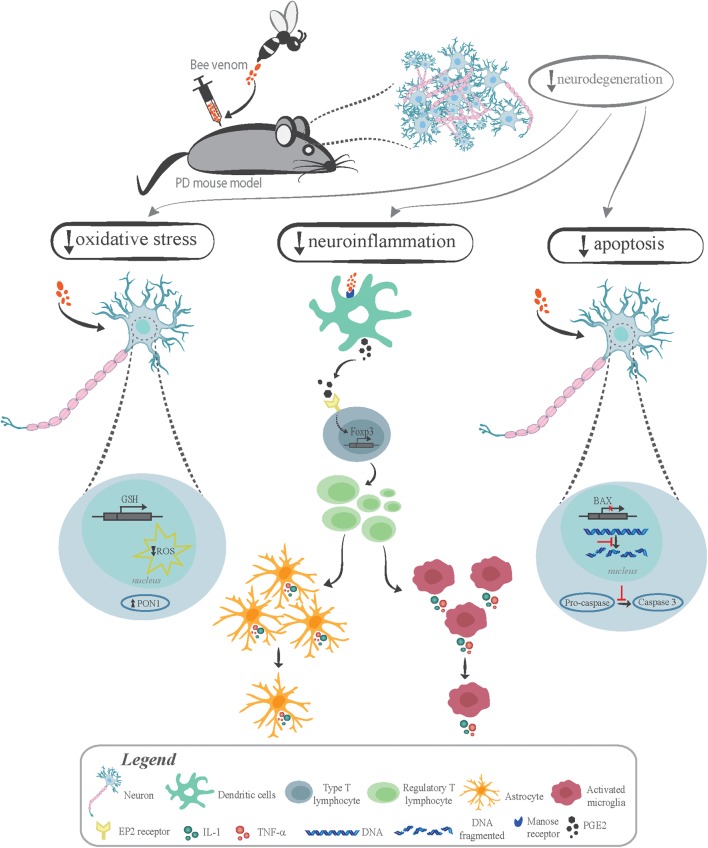
Neurodegenerative diseases affect millions of individuals worldwide. So far, no disease-modifying drug is available to treat patients, making the search for effective drugs an urgent need. Neurodegeneration is triggered by the activation of several cellular processes, including oxidative stress, mitochondrial impairment, neuroinflammation, aging, aggregate formation, glutamatergic excitotoxicity, and apoptosis. Therefore, many research groups aim to identify drugs that may inhibit one or more of these events leading to neuronal cell death. Venoms are fruitful natural sources of new molecules, which have been relentlessly enhanced by evolution through natural selection. Several studies indicate that venom components can exhibit selectivity and affinity for a wide variety of targets in mammalian systems. For instance, an expressive number of natural peptides identified in venoms from animals, such as snakes, scorpions, bees, and spiders, were shown to lessen inflammation, regulate glutamate release, modify neurotransmitter levels, block ion channel activation, decrease the number of protein aggregates, and increase the levels of neuroprotective factors. Thus, these venom components hold potential as therapeutic tools to slow or even halt neurodegeneration. However, there are many technological issues to overcome, as venom peptides are hard to obtain and characterize and the amount obtained from natural sources is insufficient to perform all the necessary experiments and tests. Fortunately, technological improvements regarding heterologous protein expression, as well as peptide chemical synthesis will help to provide enough quantities and allow chemical and pharmacological enhancements of these natural occurring compounds. Thus, the main focus of this review is to highlight the most promising studies evaluating animal toxins as therapeutic tools to treat a wide variety of neurodegenerative conditions, including Alzheimer's disease, Parkinson's disease, brain ischemia, glaucoma, amyotrophic lateral sclerosis, and multiple sclerosis.
Keywords: animal venom; excitotoxicity; neurodegenerative disease; neuroinflammation; neuronal death; toxins.
Figures
Similar articles
-
Animal Venom Peptides as a Treasure Trove for New Therapeutics Against Neurodegenerative Disorders.Curr Med Chem. 2019;26(25):4749-4774. doi: 10.2174/0929867325666181031122438. Curr Med Chem. 2019. PMID: 30378475 Review.
-
Ion Channels-related Neuroprotection and Analgesia Mediated by Spider Venom Peptides.Curr Protein Pept Sci. 2023;24(5):365-379. doi: 10.2174/1389203724666230328133102. Curr Protein Pept Sci. 2023. PMID: 37018532 Review.
-
Pharmacological Alternatives for the Treatment of Neurodegenerative Disorders: Wasp and Bee Venoms and Their Components as New Neuroactive Tools.Toxins (Basel). 2015 Aug 18;7(8):3179-209. doi: 10.3390/toxins7083179. Toxins (Basel). 2015. PMID: 26295258 Free PMC article. Review.
-
Venoms as an adjunctive therapy for Parkinson's disease: where are we now and where are we going?Future Sci OA. 2020 Nov 30;7(2):FSO642. doi: 10.2144/fsoa-2020-0119. Future Sci OA. 2020. PMID: 33437512 Free PMC article. Review.
-
The deep-rooted origin of disulfide-rich spider venom toxins.Elife. 2023 Feb 9;12:e83761. doi: 10.7554/eLife.83761. Elife. 2023. PMID: 36757362 Free PMC article.
Cited by
-
Animal Venoms-Curse or Cure?Biomedicines. 2021 Apr 12;9(4):413. doi: 10.3390/biomedicines9040413. Biomedicines. 2021. PMID: 33921205 Free PMC article.
-
Bee venom attenuates neurodegeneration and motor impairment and modulates the response to L-dopa or rasagiline in a mice model of Parkinson's disease.Iran J Basic Med Sci. 2020 Dec;23(12):1628-1638. doi: 10.22038/ijbms.2020.46469.10731. Iran J Basic Med Sci. 2020. PMID: 33489038 Free PMC article.
-
Melittin Induces Local Order Changes in Artificial and Biological Membranes as Revealed by Spectral Analysis of Laurdan Fluorescence.Toxins (Basel). 2020 Nov 8;12(11):705. doi: 10.3390/toxins12110705. Toxins (Basel). 2020. PMID: 33171598 Free PMC article.
-
Cytotoxic Effect of Bee (A. mellifera) Venom on Cancer Cell Lines.J Pharmacopuncture. 2020 Dec 31;23(4):212-219. doi: 10.3831/KPI.2020.23.4.212. J Pharmacopuncture. 2020. PMID: 33408897 Free PMC article.
-
Scoliidines: Neuroprotective Peptides in Solitary Scoliid Wasp Venoms.Toxins (Basel). 2024 Oct 17;16(10):446. doi: 10.3390/toxins16100446. Toxins (Basel). 2024. PMID: 39453222 Free PMC article.
References
-
- Abhinav K., Stanton B., Johnston C., Hardstaff J., Orrell R. W., Howard R., et al. (2007). Amyotrophic lateral sclerosis in South-East England: a population-based study. The South-East England register for amyotrophic lateral sclerosis (SEALS Registry). Neuroepidemiology 29 44–48. 10.1159/000108917 - DOI - PubMed
-
- Adams R. A., Passino M., Sachs B. D., Nuriel T., Akassoglou K. (2004). Fibrin mechanisms and functions in nervous system pathology. Mol. Interv. 4 163–176. - PubMed
-
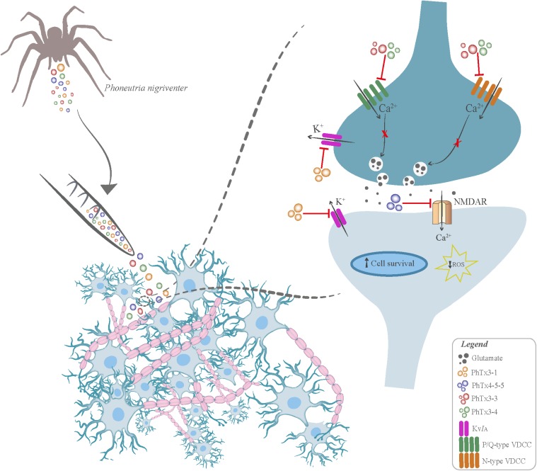
- Agostini R. M., do Nascimento Pinheiro A. C., Binda N. S., Romano Silva M. A., do Nascimento Cordeiro M., et al. (2011). Phoneutria spider toxins block ischemia-induced glutamate release and neuronal death of cell layers of the retina. Retina 31 1392–1399. 10.1097/IAE.0b013e318205b249 - DOI - PubMed
Publication types
LinkOut - more resources
Full Text Sources
Other Literature Sources