. 2018:12:84-90.
doi: 10.1038/s41566-017-0078-z. Epub 2018 Jan 15.

Wavefront shaping with disorder-engineered metasurfaces

Affiliations

Wavefront shaping with disorder-engineered metasurfaces

Mooseok Jang et al. Nat Photonics. 2018.

Abstract

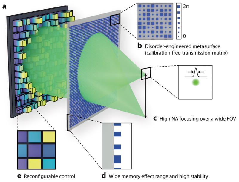
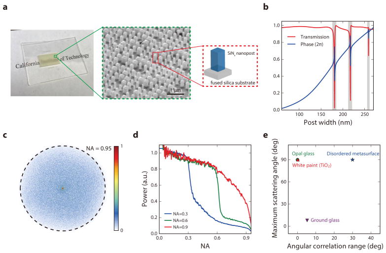
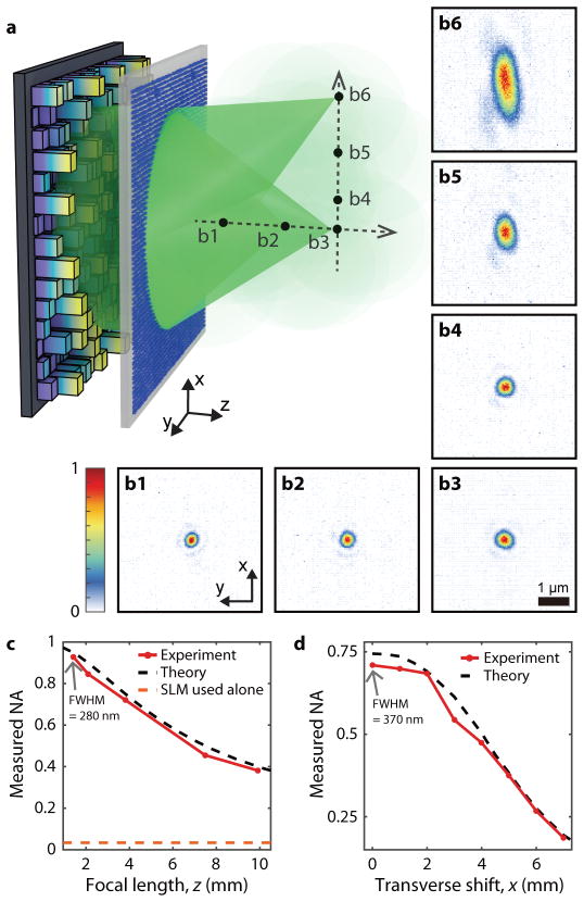
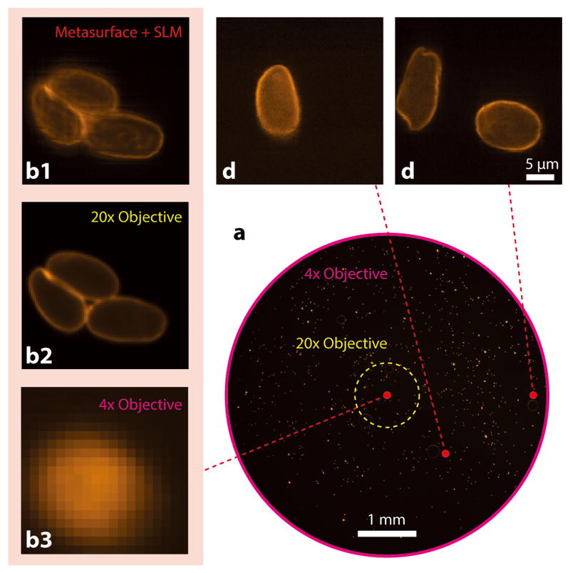
Recently, wavefront shaping with disordered media has demonstrated optical manipulation capabilities beyond those of conventional optics, including extended volume, aberration-free focusing and subwavelength focusing. However, translating these capabilities to useful applications has remained challenging as the input-output characteristics of the disordered media (P variables) need to be exhaustively determined via O(P) measurements. Here, we propose a paradigm shift where the disorder is specifically designed so its exact input-output characteristics are known a priori and can be used with only a few alignment steps. We implement this concept with a disorder-engineered metasurface, which exhibits additional unique features for wavefront shaping such as a large optical memory effect range in combination with a wide angular scattering range, excellent stability, and a tailorable angular scattering profile. Using this designed metasurface with wavefront shaping, we demonstrate high numerical aperture (NA > 0.5) focusing and fluorescence imaging with an estimated ~2.2×10⁸ addressable points in an ~8 mm field of view.


Conflict of interest statement

Competing financial interests The authors declare no competing financial interests.

Figures

Figure 1
Figure 1. Wavefront shaping assisted by a disorder-engineered metasurface
(a) The system setup consists of two planar components, an SLM and a disorder-engineered metasurface. (b) The disorder-engineered metasurface is implemented by fabricating the nanoposts with varying sizes, which correspond to different phase delays ϕ(x,y) on the metasurface. (c) The wide angular scattering range enables high NA focusing over a wide FOV. (d) The thin, planar nature of the disordered metasurface yields a large memory effect range and also makes the transmission matrix of the metasurface extraordinarily stable. (e) The SLM enables reconfigurable control of the expanded optical space available through the disordered metasurface.
Figure 2
Figure 2. Characterization of disorder-engineered metasurfaces
(a) Photograph and SEM image of a fabricated disorder-engineered metasurface. (b) Simulated transmission and phase of the SiNx nanoposts as a function of their width at a wavelength of 532 nm. These data are used as a look-up table for the metasurface design. (c) Measured 2D angular scattering profile of the disordered metasurface, normalized to the strongest scattered field component. (d) Measured 1D angular scattering profile of the disordered metasurfaces that were specifically designed to scatter the incident light to certain angular ranges (NA = 0.3, 0.6, 0.9). (e) Memory effect range and angular scattering range of the disordered metasurface compared with conventional random media such as white paint, opal glass, and ground glass diffusers.
Figure 3
Figure 3. Experimental demonstration of diffraction-limited focusing over an extended volume
(a) Schematic of optical focusing assisted by the disordered metasurface. The incident light is polarized along the x direction. (b16) Measured 2D intensity profiles for the foci reconstructed at the positions indicated in (a). b1–b3 are the foci along the optical axis at z = 1.4, 2.1, and 3.8, mm, respectively, corresponding to NAs of 0.95, 0.9, and 0.75. b3–b6 are the foci at x = 0, 1, 4, and 7 mm scanned on the fixed focal plane of z = 3.8 mm. Scale bar: 1 μm. (c) Measured NA (along x-axis) of the foci created along the optical axis (red solid line) compared with theoretical values (black dotted line). When the SLM is used alone, the maximum accessible NA is 0.033 (orange dotted line), based on the Nyquist-Shannon sampling theorem. (d) Measured NA (along x-axis) of the foci created along x axis at z = 3.8 mm (red solid line) compared with theoretical values (black dotted line). The number of resolvable focusing points within the 14-mm diameter FOV was estimated to be 4.3×10⁸.
Figure 4
Figure 4. Demonstration of disordered metasurface assisted microscope for high resolution wide-FOV fluorescence imaging of giardia lamblia cysts
(a) Low resolution bright field image captured by a conventional fluorescence microscope with a 4× objective lens (NA = 0.1). Scale bar: 1 mm. (b13) Fluorescence images captured at the center of the FOV. (b1) Scanned image obtained with a disordered metasurface lens. (b2) Ground truth fluorescence image captured with a 20× objective lens (NA = 0.5). (b3) Magnified low-resolution fluorescence image captured with the 4× objective. (c, d) Images obtained with the disorder metasurface-assisted microscope at (x, y) = (1, 1) and (2.5, 0) mm, respectively. This demonstrates that we can indeed use the system for high resolution and wide FOV imaging.

References

    1. Mosk AP, Lagendijk A, Lerosey G, Fink M. Controlling waves in space and time for imaging and focusing in complex media. Nat Photonics. 2012;6:283–292.
    1. Tyson RK. Principles of Adaptive Optics. CRC Press; 2010. pp. 177–196.
    1. Horstmeyer R, Ruan H, Yang C. Guidestar-assisted wavefront-shaping methods for focusing light into biological tissue. Nat Photonics. 2015;9:563–571. - PMC - PubMed
    1. Vellekoop IM, Lagendijk A, Mosk AP. Exploiting disorder for perfect focusing. Nat Photonics. 2010;4:320–322.
    1. Vellekoop IM, Aegerter CM. Scattered light fluorescence microscopy: imaging through turbid layers. Opt Lett. 2010;35:1245–1247. - PubMed