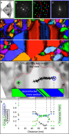
Towards 3D crystal orientation reconstruction using automated crystal orientation mapping transmission electron microscopy (ACOM-TEM)
- PMID: 29527435
- PMCID: PMC5827809
- DOI: 10.3762/bjnano.9.56
Towards 3D crystal orientation reconstruction using automated crystal orientation mapping transmission electron microscopy (ACOM-TEM)
Abstract
To relate the internal structure of a volume (crystallite and phase boundaries) to properties (electrical, magnetic, mechanical, thermal), a full 3D reconstruction in combination with in situ testing is desirable. In situ testing allows the crystallographic changes in a material to be followed by tracking and comparing the individual crystals and phases. Standard transmission electron microscopy (TEM) delivers a projection image through the 3D volume of an electron-transparent TEM sample lamella. Only with the help of a dedicated TEM tomography sample holder is an accurate 3D reconstruction of the TEM lamella currently possible. 2D crystal orientation mapping has become a standard method for crystal orientation and phase determination while 3D crystal orientation mapping have been reported only a few times. The combination of in situ testing with 3D crystal orientation mapping remains a challenge in terms of stability and accuracy. Here, we outline a method to 3D reconstruct the crystal orientation from a superimposed diffraction pattern of overlapping crystals without sample tilt. Avoiding the typically required tilt series for 3D reconstruction enables not only faster in situ tests but also opens the possibility for more stable and more accurate in situ mechanical testing. The approach laid out here should serve as an inspiration for further research and does not make a claim to be complete.
Keywords: 3D reconstruction; ACOM-TEM; STEM; in situ testing; quantitative crystallographic analysis.
Figures


References
-
- Legros M, Gianola D S, Hemker K J. Acta Mater. 2008;56:3380–3393. doi: 10.1016/j.actamat.2008.03.032. - DOI
-
- Jin M, Minor A M, Stach E A, Morris J W., Jr Acta Mater. 2004;52:5381–5387. doi: 10.1016/j.actamat.2004.07.044. - DOI
-
- Mompiou F, Legros M. Scr Mater. 2015;99:5–8. doi: 10.1016/j.scriptamat.2014.11.004. - DOI
LinkOut - more resources
Full Text Sources
Other Literature Sources
Molecular Biology Databases
