. 2017 Oct 28:17:835-846.
doi: 10.1016/j.nicl.2017.10.025. eCollection 2018.

Increased insula-putamen connectivity in X-linked dystonia-parkinsonism

Affiliations

Increased insula-putamen connectivity in X-linked dystonia-parkinsonism

Anne J Blood et al. Neuroimage Clin. .

Abstract

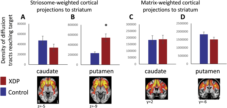
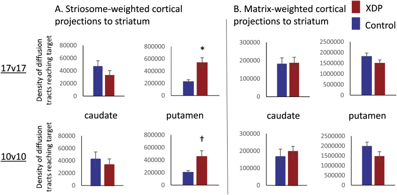
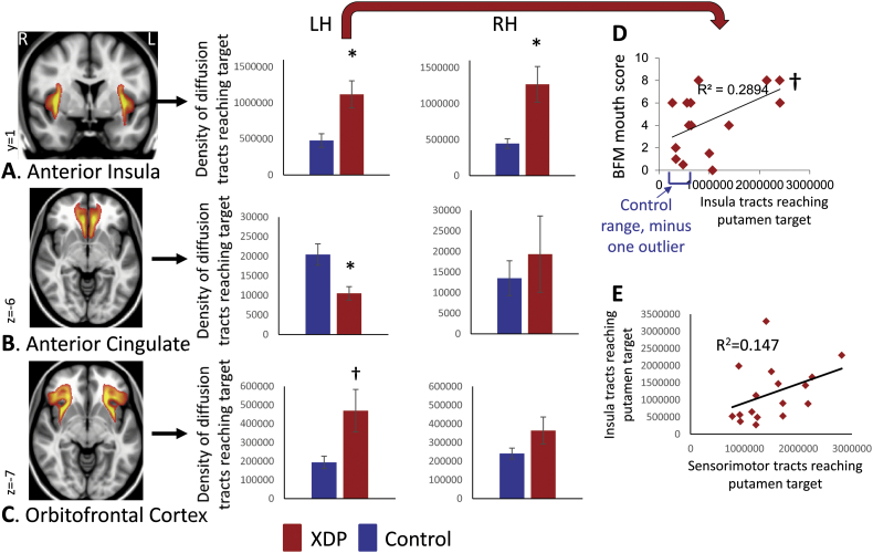
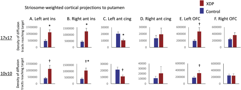
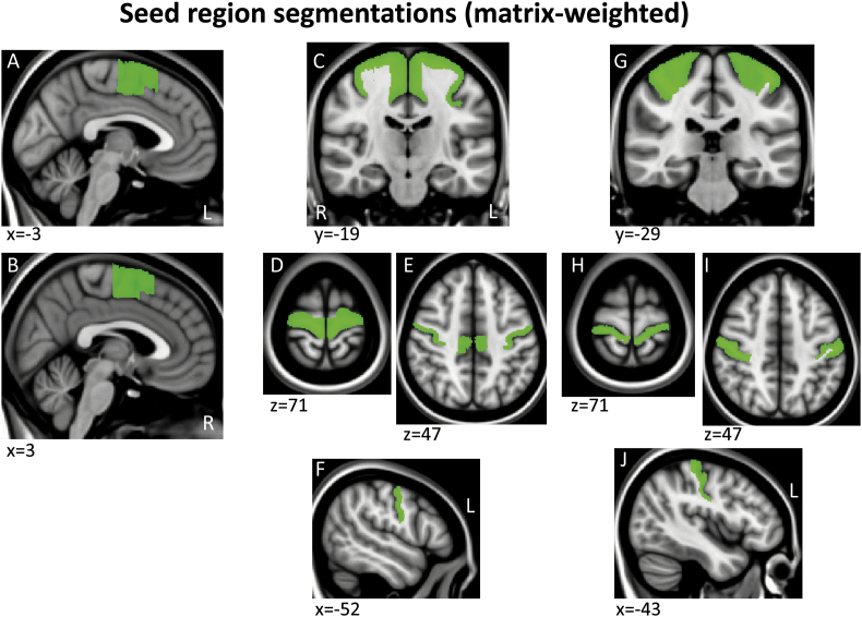
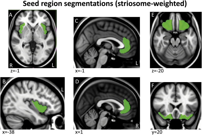
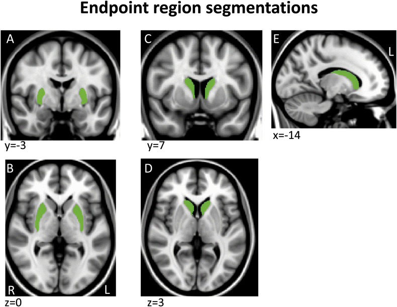
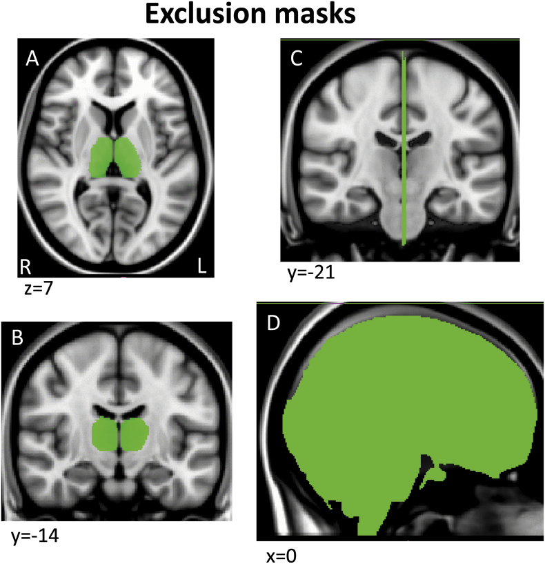
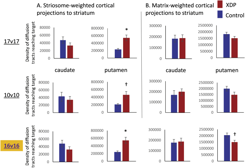
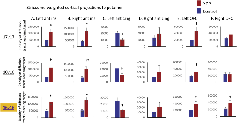
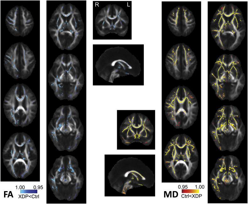
Preliminary evidence from postmortem studies of X-linked dystonia-parkinsonism (XDP) suggests tissue loss may occur first and/or most severely in the striatal striosome compartment, followed later by cell loss in the matrix compartment. However, little is known about how this relates to pathogenesis and pathophysiology. While MRI cannot visualize these striatal compartments directly in humans, differences in relative gradients of afferent cortical connectivity across compartments (weighted toward paralimbic versus sensorimotor cortex, respectively) can be used to infer potential selective loss in vivo. In the current study we evaluated relative connectivity of paralimbic versus sensorimotor cortex with the caudate and putamen in 17 individuals with XDP and 17 matched controls. Although caudate and putamen volumes were reduced in XDP, there were no significant reductions in either "matrix-weighted", or "striosome-weighted" connectivity. In fact, paralimbic connectivity with the putamen was elevated, rather than reduced, in XDP. This was driven most strongly by elevated putamen connectivity with the anterior insula. There was no relationship of these findings to disease duration or striatal volume, suggesting insula and/or paralimbic connectivity in XDP may develop abnormally and/or increase in the years before symptom onset.

Keywords: Matrix; Paralimbic; Sensorimotor; Striosomes; Tractography.


Figures

Fig. 1
Fig. 1
Comparison of XDP versus control tractography for a priori comparisons. Total termination tractography counts are shown for the striosome-weighted and matrix-weighted regions for the caudate (A,C) and putamen (B,D). XDP values are shown in red and control values in blue. Error bars indicate standard error of the mean. XDP: X-linked dystonia-parkinsonism. Images (from fslview) below graphs show representative sections through the left and right hemisphere averaged healthy control tractography for each of the four a priori tractography measures, superimposed on the MNI152_T1_1mm template. Note that these images are intended (and thresholded) to be qualitative only, and are included simply to illustrate the territory covered most prominently by each of the four measures; they do not convey information about magnitude or spatial extent. The MNI coordinate indicates the location of each image slice. Images are in radiologic format; L: left hemisphere and R: right hemisphere. *significant at p < 0.005; trend toward significance (within an order of magnitude of the significance p threshold).
Fig. 2
Fig. 2
Comparison of tractography measures for a priori comparisons in the 17 XDP versus 17 controls cohort, as compared with the 10 XDP versus 10 controls cohort. Total termination tractography counts from Fig. 1 (17 XDP versus 17 controls, top row) are compared with values for the smaller, more tightly matched cohort (bottom row). (A) Striosome-weighted region connectivity with the caudate (left) and putamen (right). (B) Matrix-weighted region connectivity with the caudate (left) and putamen (right). Error bars indicate standard error of the mean. *significant at p < 0.005; trend toward significance (within an order of magnitude of the significance p threshold). XDP: X-linked dystonia-parkinsonism.
Fig. 3
Fig. 3
Comparison of XDP versus control tractography for striosome-weighted connectivity with the putamen (posthoc comparisons). Total termination tractography counts are shown for (A) Anterior Insula, (B), Anterior/subcallosal Cingulate, and (C) Orbitofrontal Cortex connectivity with the left (LH) and right (RH) putamen. Images (from fslview) to the left of graphs show representative sections through averaged healthy control tractography maps used in posthoc tractography analyses, superimposed on the MNI152_T1_1mm template. Left and right hemisphere tractography are depicted together in the images, but were evaluated separately for statistics. Note that these images are intended (and thresholded) to be qualitative only, and are included simply to illustrate the territory covered most prominently by each of the measures; they do not convey information about magnitude or spatial extent. *significant at p < 0.005; trend toward significance (within an order of magnitude of the significance p threshold). (D) Relationship between left insula → putamen connectivity and mouth scores on the Burke Fahn Marsden (BFM) scale. (E) Absence of inverse relationship between sensorimotor → putamen and insula → putamen connectivity. XDP: X-linked dystonia-parkinsonism.
Fig. 4
Fig. 4
Comparison of tractography measures for posthoc comparisons in the 17 XDP versus 17 controls cohort, as compared with the 10 XDP versus 10 controls cohort. Total termination tractography counts from Fig. 1 (17 XDP versus 17 controls, top row) are compared with values for the smaller, more tightly matched cohort (bottom row). Individual striosome-weighted regions were individually assessed for connectivity with the ipsilateral putamen: (A) Left anterior insula, (B) Right anterior insula, (C) Left anterior/subcallosal cingulate, (D) Right anterior/subcallosal cingulate, (E) Left orbitofrontal cortex, (F) Right orbitofrontal cortex. Error bars indicate standard error of the mean. *significant at p < 0.005; trend toward significance (within an order of magnitude of the significance p threshold); * trend without ICV correction, but significant at p < 0.005 when ICV corrected. XDP: X-linked dystonia-parkinsonism. ant ins: anterior insula; ant cing: anterior/subcallosal cingulate; OFC: orbitofrontal cortex.
Supplementary Fig. 1
Example sections through the three matrix-weighted seed region segmentations shown on the MNI152_T1_1mm brain.
Supplementary Fig. 2
Example sections through the three striosome-weighted seed region segmentations shown on the MNI152_T1_1mm brain.
Supplementary Fig. 3
Example sections through the two endpoint region segmentations shown on the MNI152_T1_1mm brain.
Supplementary Fig. 4
Example sections through the two exclusion mask segmentations shown on the MNI152_T1_1mm brain.
Supplementary Fig. 5
Comparison of tractography measures for a priori comparisons in the 16 XDP versus 16 controls cohort, as compared with the 17 XDP versus 17 controls and 10 XDP versus 10 controls cohorts.
Supplementary Fig. 6
Comparison of tractography measures for posthoc comparisons in the 16 XDP versus 16 controls cohort, as compared with the 17 XDP versus 17 controls and 10 XDP versus 10 controls cohorts.
Supplementary Fig. 7
FA and MD map contrasts for the 17 XDP versus 17 healthy control cohort using TBSS and randomise with tfce correction for multiple comparisons across the FA-generated white matter skeleton.

Similar articles

Cited by

References

    1. Ackermann H., Riecker A. The contribution of the insula to motor aspects of speech production: a review and a hypothesis. Brain Lang. 2004;89:320–328. - PubMed
    1. Argyelan M., Carbon M., Niethammer M., Ulug A.M., Voss H.U., Bressman S.B., Dhawan V., Eidelberg D. Cerebellothalamocortical connectivity regulates penetrance in dystonia. J. Neurosci. 2009;29:9740–9747. - PMC - PubMed
    1. Arima T., Yanagi Y., Niddam D.M., Ohata N., Arendt-Nielsen L., Minagi S., Sessle B.J., Svensson P. Corticomotor plasticity induced by tongue-task training in humans: a longitudinal fMRI study. Exp. Brain Res. 2011;212:199–212. - PubMed
    1. Arndt S., Cohen G., Alliger R.J., Swayze V.W., 2nd, Andreasen N.C. Problems with ratio and proportion measures of imaged cerebral structures. Psychiatry Res. 1991;40:79–89. - PubMed
    1. Beaulieu C. The basis of anisotropic water diffusion in the nervous system — a technical review. NMR Biomed. 2002;15:435–455. - PubMed

Publication types

MeSH terms

Supplementary concepts