

Systematic comparison of different techniques to measure hippocampal subfield volumes in ADNI2
- PMID: 29527502
- PMCID: PMC5842756
- DOI: 10.1016/j.nicl.2017.12.036
Systematic comparison of different techniques to measure hippocampal subfield volumes in ADNI2
Abstract
Objective: Subfield-specific measurements provide superior information in the early stages of neurodegenerative diseases compared to global hippocampal measurements. The overall goal was to systematically compare the performance of five representative manual and automated T1 and T2 based subfield labeling techniques in a sub-set of the ADNI2 population.
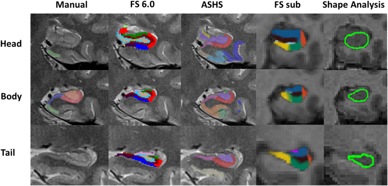
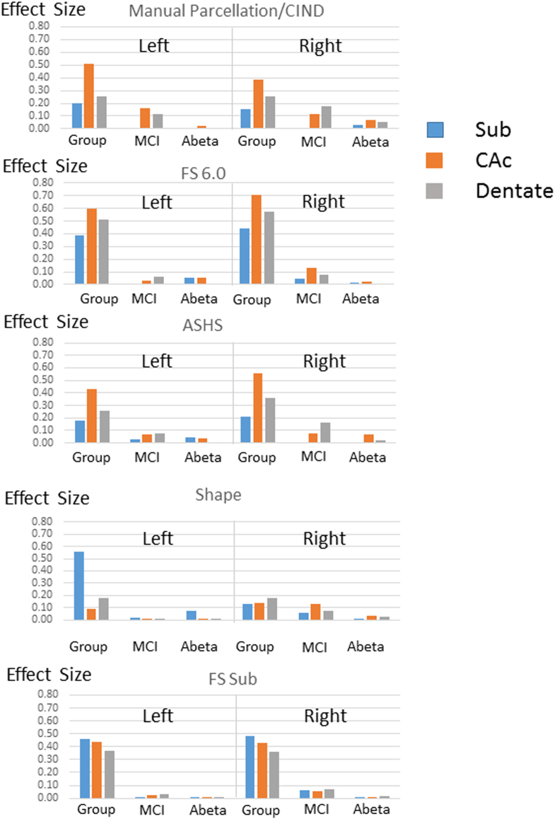
Methods: The high resolution T2 weighted hippocampal images (T2-HighRes) and the corresponding T1 images from 106 ADNI2 subjects (41 controls, 57 MCI, 8 AD) were processed as follows. A. T1-based: 1. Freesurfer + Large-Diffeomorphic-Metric-Mapping in combination with shape analysis. 2. FreeSurfer 5.1 subfields using in-vivo atlas. B. T2-HighRes: 1. Model-based subfield segmentation using ex-vivo atlas (FreeSurfer 6.0). 2. T2-based automated multi-atlas segmentation combined with similarity-weighted voting (ASHS). 3. Manual subfield parcellation. Multiple regression analyses were used to calculate effect sizes (ES) for group, amyloid positivity in controls, and associations with cognitive/memory performance for each approach.
Results: Subfield volumetry was better than whole hippocampal volumetry for the detection of the mild atrophy differences between controls and MCI (ES: 0.27 vs 0.11). T2-HighRes approaches outperformed T1 approaches for the detection of early stage atrophy (ES: 0.27 vs.0.10), amyloid positivity (ES: 0.11 vs 0.04), and cognitive associations (ES: 0.22 vs 0.19).
Conclusions: T2-HighRes subfield approaches outperformed whole hippocampus and T1 subfield approaches. None of the different T2-HghRes methods tested had a clear advantage over the other methods. Each has strengths and weaknesses that need to be taken into account when deciding which one to use to get the best results from subfield volumetry.
Figures
Similar articles
-
Multi-atlas segmentation of the whole hippocampus and subfields using multiple automatically generated templates.Neuroimage. 2014 Nov 1;101:494-512. doi: 10.1016/j.neuroimage.2014.04.054. Epub 2014 Apr 29. Neuroimage. 2014. PMID: 24784800
-
Automated subfield volumetric analysis of hippocampus in temporal lobe epilepsy using high-resolution T2-weighed MR imaging.Neuroimage Clin. 2016 Jun 13;12:57-64. doi: 10.1016/j.nicl.2016.06.008. eCollection 2016. Neuroimage Clin. 2016. PMID: 27489767 Free PMC article.
-
Hippocampal atrophy patterns in mild cognitive impairment and Alzheimer's disease.Hum Brain Mapp. 2010 Sep;31(9):1339-47. doi: 10.1002/hbm.20934. Hum Brain Mapp. 2010. PMID: 20839293 Free PMC article.
-
Structural imaging of hippocampal subfields in healthy aging and Alzheimer's disease.Neuroscience. 2015 Nov 19;309:29-50. doi: 10.1016/j.neuroscience.2015.08.033. Epub 2015 Aug 22. Neuroscience. 2015. PMID: 26306871 Review.
-
FreeSurfer-based segmentation of hippocampal subfields: A review of methods and applications, with a novel quality control procedure for ENIGMA studies and other collaborative efforts.Hum Brain Mapp. 2022 Jan;43(1):207-233. doi: 10.1002/hbm.25326. Epub 2020 Dec 27. Hum Brain Mapp. 2022. PMID: 33368865 Free PMC article. Review.
Cited by
-
In vivo hippocampal subfield volumes in bipolar disorder-A mega-analysis from The Enhancing Neuro Imaging Genetics through Meta-Analysis Bipolar Disorder Working Group.Hum Brain Mapp. 2022 Jan;43(1):385-398. doi: 10.1002/hbm.25249. Epub 2020 Oct 19. Hum Brain Mapp. 2022. PMID: 33073925 Free PMC article.
-
Test-retest reliability of hippocampal subfield volumes in a developmental sample: Implications for longitudinal developmental studies.J Neurosci Res. 2021 Oct;99(10):2327-2339. doi: 10.1002/jnr.24831. Epub 2021 Mar 22. J Neurosci Res. 2021. PMID: 33751637 Free PMC article.
-
Reduced hippocampal subfield volumes and memory function in school-aged children born preterm with very low birthweight (VLBW).Neuroimage Clin. 2019;23:101857. doi: 10.1016/j.nicl.2019.101857. Epub 2019 May 11. Neuroimage Clin. 2019. PMID: 31136968 Free PMC article.
-
Hippocampal subfield CA2+3 exhibits accelerated aging in Alcohol Use Disorder: A preliminary study.Neuroimage Clin. 2019;22:101764. doi: 10.1016/j.nicl.2019.101764. Epub 2019 Mar 14. Neuroimage Clin. 2019. PMID: 30904825 Free PMC article.
-
Atrophy patterns in hippocampal subregions and their relationship with cognitive function in fibromyalgia patients with mild cognitive impairment.Front Neurosci. 2024 May 23;18:1380121. doi: 10.3389/fnins.2024.1380121. eCollection 2024. Front Neurosci. 2024. PMID: 38846715 Free PMC article.
References
-
- Baldwin S.A., Gibson T., Callihan C.T., Sullivan P.G., Palmer E., Scheff S.W. Neuronal cell loss in the CA3 subfield of the hippocampus following cortical contusion utilizing optical discetor method for cell counting. J. Neurotrauma. 1997;14:385–398. - PubMed
-
- Bender A.R., Daugherty A.M., Raz N. Vascular risk moderates associations between hippocampal subfield volumes and memory. J. Cogn. Neurosci. 2013;25:1851–1862. - PubMed
-
- Bluemcke I., Beck H., Lie A.A., Wiestler O.D. Molecular neuropathology of human mesial temporal lobe epilepsy. Epilepsy Res. 1999;36:205–223. - PubMed
-
- Bluemcke I., Pauli E., Clusman H., Schramm J., Becker A., Elger C., Merschhemke M., Meenke H.J., Lehmann T., von Deimling A., Scheiwe C., Zentner J., Volk B., Romstoeck J., Stefan H., Hildebrandt M. A new clinico-pathological classification system for mesial temporal lobe sclerosis. Acta Neuropathol. 2007;113:235–244. - PMC - PubMed
Publication types
MeSH terms
Grants and funding
LinkOut - more resources
Full Text Sources
Other Literature Sources
Medical
