

doi: 10.1016/j.jacep.2017.08.009.
Detection of Fetal Arrhythmia Using Optically-Pumped Magnetometers
Affiliations
- PMID: 29527577
- PMCID: PMC5841248
- DOI: 10.1016/j.jacep.2017.08.009
Item in Clipboard
Detection of Fetal Arrhythmia Using Optically-Pumped Magnetometers
JACC Clin Electrophysiol.
2018 Feb.
No abstract available
Figures
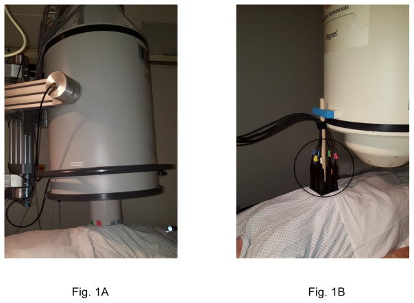
Photographs of (A) SQUID and (B) OPM fMCG systems. The OPM sensor array (circled) was supported by attaching it to a SQUID system.
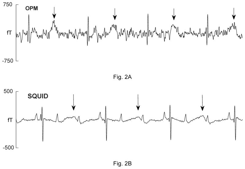
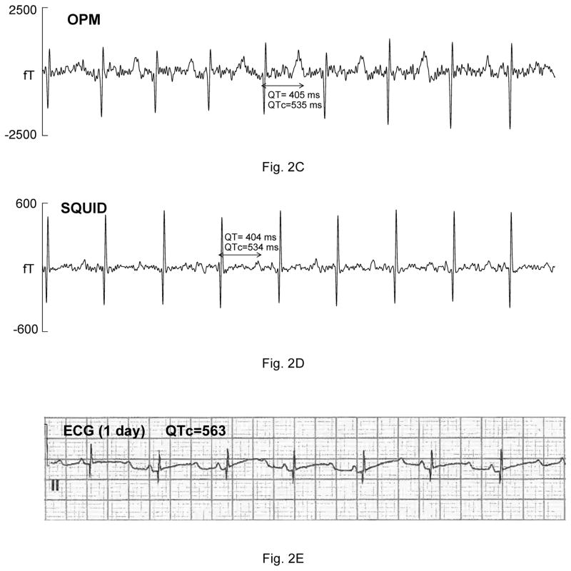
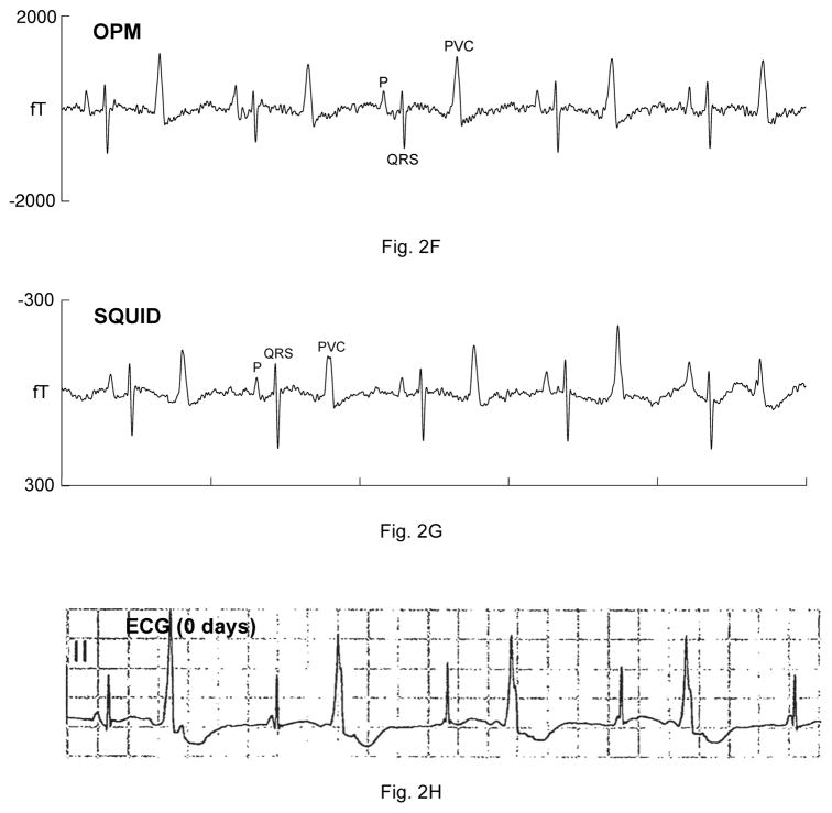
Comparison of OPM and SQUID recordings from four fetuses with sustained arrhythmia. Postnatal ECGs are shown for the last three. All of the tracings are 5 seconds long. A) and B) are from a fetus at 33-4/7 weeks’ with extreme QTc prolongation (QTc> 700 ms), resulting in 3:1 AV block. The T-waves are indicated by arrows. C) and D) are from a fetus at 29 weeks’ with QTc prolongation. F) and G) are from a fetus at 33-6/7 weeks’ with a complex, irregular rhythm. As shown, the predominant rhythm was ventricular bigeminy, in which a sinus beat alternates with a premature ventricular contraction. I) and J) are from a fetus at 30-1/7 weeks’ with a low atrial rhythm, characterized by a low heart rate, inverted P-wave, and short PR interval.
Comparison of OPM and SQUID recordings from four fetuses with sustained arrhythmia. Postnatal ECGs are shown for the last three. All of the tracings are 5 seconds long. A) and B) are from a fetus at 33-4/7 weeks’ with extreme QTc prolongation (QTc> 700 ms), resulting in 3:1 AV block. The T-waves are indicated by arrows. C) and D) are from a fetus at 29 weeks’ with QTc prolongation. F) and G) are from a fetus at 33-6/7 weeks’ with a complex, irregular rhythm. As shown, the predominant rhythm was ventricular bigeminy, in which a sinus beat alternates with a premature ventricular contraction. I) and J) are from a fetus at 30-1/7 weeks’ with a low atrial rhythm, characterized by a low heart rate, inverted P-wave, and short PR interval.
Comparison of OPM and SQUID recordings from four fetuses with sustained arrhythmia. Postnatal ECGs are shown for the last three. All of the tracings are 5 seconds long. A) and B) are from a fetus at 33-4/7 weeks’ with extreme QTc prolongation (QTc> 700 ms), resulting in 3:1 AV block. The T-waves are indicated by arrows. C) and D) are from a fetus at 29 weeks’ with QTc prolongation. F) and G) are from a fetus at 33-6/7 weeks’ with a complex, irregular rhythm. As shown, the predominant rhythm was ventricular bigeminy, in which a sinus beat alternates with a premature ventricular contraction. I) and J) are from a fetus at 30-1/7 weeks’ with a low atrial rhythm, characterized by a low heart rate, inverted P-wave, and short PR interval.
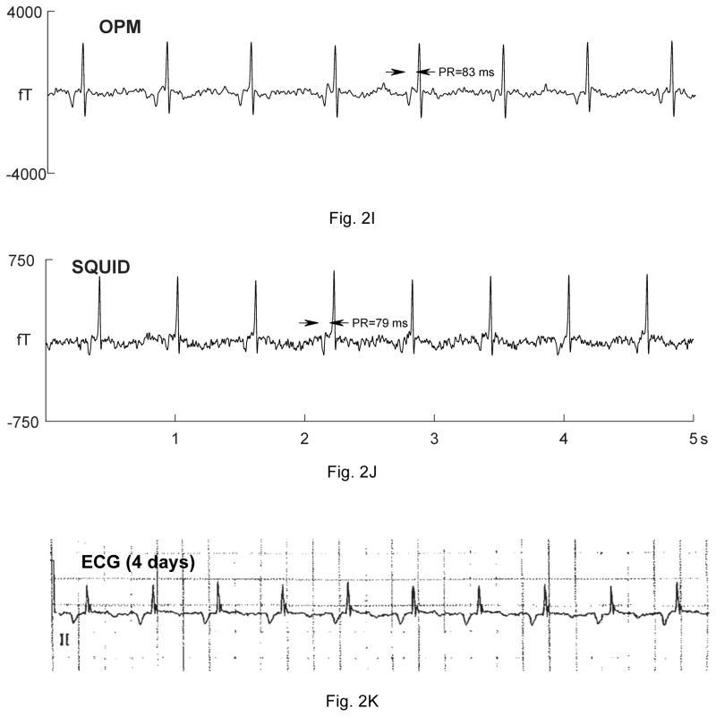
Comparison of OPM and SQUID recordings from four fetuses with sustained arrhythmia. Postnatal ECGs are shown for the last three. All of the tracings are 5 seconds long. A) and B) are from a fetus at 33-4/7 weeks’ with extreme QTc prolongation (QTc> 700 ms), resulting in 3:1 AV block. The T-waves are indicated by arrows. C) and D) are from a fetus at 29 weeks’ with QTc prolongation. F) and G) are from a fetus at 33-6/7 weeks’ with a complex, irregular rhythm. As shown, the predominant rhythm was ventricular bigeminy, in which a sinus beat alternates with a premature ventricular contraction. I) and J) are from a fetus at 30-1/7 weeks’ with a low atrial rhythm, characterized by a low heart rate, inverted P-wave, and short PR interval.
References
-
- Donofrio MT, Moon-Grady AJ, Hornberger LK, et al. Diagnosis and treatment of fetal cardiac disease: a scientific statement from the American Heart Association. Circulation. 2014;129:2183–242. - PubMed
-
- Fagaly RL. Neuromagnetic instrumentation. Adv Neurol. 1990;54:11–32. - PubMed
-
- Budker D, Romalis M. Optical magnetometry. Nat Phys. 2007;3:227–234.
Publication types
MeSH terms
Grants and funding
LinkOut - more resources
Full Text Sources
Other Literature Sources
Medical