. 2018 Sep;25(5):e12391.
doi: 10.1111/xen.12391. Epub 2018 Mar 12.

GalT-KO pig lungs are highly susceptible to acute vascular rejection in baboons, which may be mitigated by transgenic expression of hCD47 on porcine blood vessels

Affiliations

GalT-KO pig lungs are highly susceptible to acute vascular rejection in baboons, which may be mitigated by transgenic expression of hCD47 on porcine blood vessels

Hironosuke Watanabe et al. Xenotransplantation. 2018 Sep.

Abstract

Background: Despite recent progress in survival times of xenografts in non-human primates, there are no reports of survival beyond 5 days of histologically well-aerated porcine lung grafts in baboons. Here, we report our initial results of pig-to-baboon xeno-lung transplantation (XLTx).

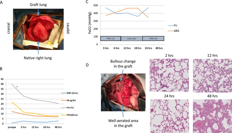
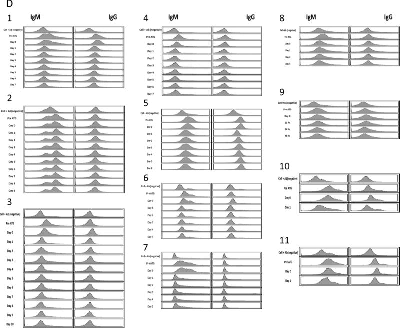
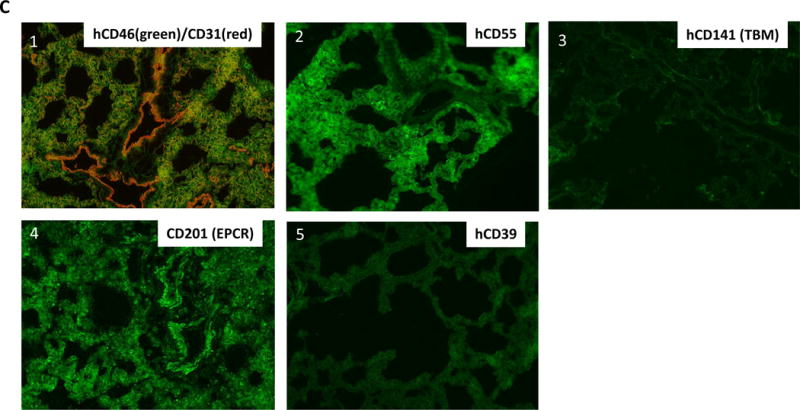
Methods: Eleven baboons received genetically modified porcine left lungs from either GalT-KO alone (n = 3), GalT-KO/humanCD47(hCD47)/hCD55 (n = 3), GalT-KO/hD47/hCD46 (n = 4), or GalT-KO/hCD39/hCD46/hCD55/TBM/EPCR (n = 1) swine. The first 2 XLTx procedures were performed under a non-survival protocol that allowed a 72-hour follow-up of the recipients with general anesthesia, while the remaining 9 underwent a survival protocol with the intention of weaning from ventilation.

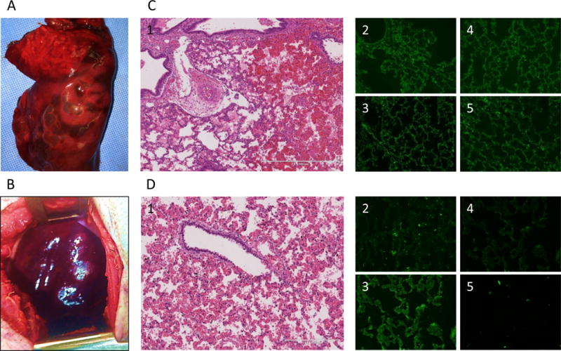
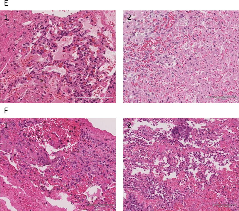
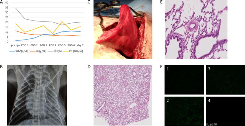
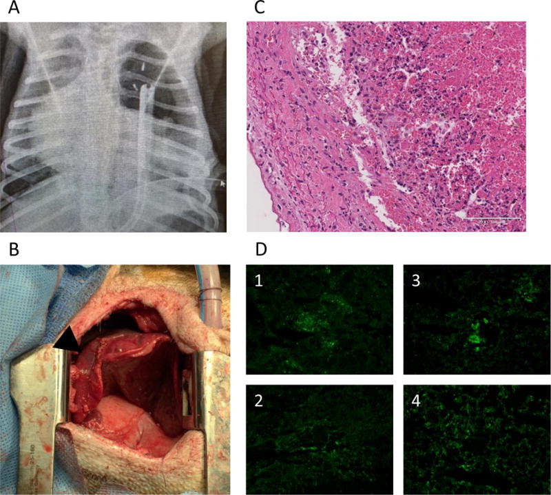
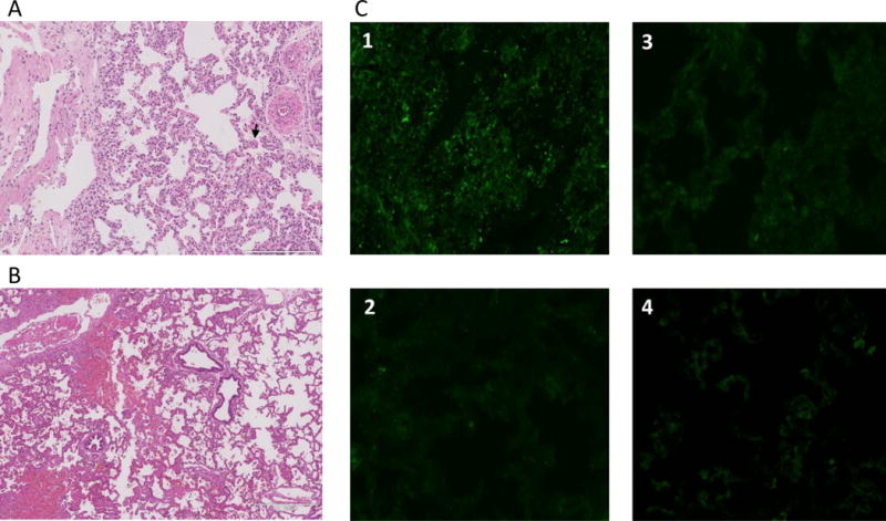
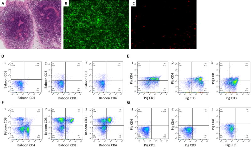
Results: Lung graft survivals in the 2 non-survival animals were 48 and >72 hours, while survivals in the other 9 were 25 and 28 hours, at 5, 5, 6, 7, >7, 9, and 10 days. One baboon with graft survival >7 days, whose entire lung graft remained well aerated, was euthanized on POD 7 due to malfunction of femoral catheters. hCD47 expression of donor lungs was detected in both alveoli and vessels only in the 3 grafts surviving >7, 9, and 10 days. All other grafts lacked hCD47 expression in endothelial cells and were completely rejected with diffuse hemorrhagic changes and antibody/complement deposition detected in association with early graft loss.

Conclusions: To our knowledge, this is the first evidence of histologically viable porcine lung grafts beyond 7 days in baboons. Our results indicate that GalT-KO pig lungs are highly susceptible to acute humoral rejection and that this may be mitigated by transgenic expression of hCD47.

Keywords: hCD47; lung transplantation; pig to baboon; xenotransplantation.


Figures

Figure 1
Figure 1
pDAF (CD55)-CD47 Expression Vector for Transgenic Pig Production. hCD47 and hCD55 expression cassettes are oriented in opposing directions. The hCD55 minigene is comprised of human genomic DNA including the native promoter/enhancer region, the 3′ UTR through the pA, exon 1 and intron 1. The cording sequence (CDS) sequence was substituted for genomic sequences covering the remaining exons. A small segment of plasmid backbone follows the CD55 minigene (not shown).
Figure 2
Figure 2
(A) Gross findings of native right lung and graft left lung of #13P64 immediately after reperfusion. Both lungs are well expanded and perfused. (B) CBC data during the experiment. All data (WBC, Hb, Hct and Platelets count) were stable till the termination, although one unit of pRBC was given at 2 hrs post-reperfusion (*). (C) PaO2 of systemic arterial blood gas and graft PV blood gas analysis. The PaO2 of both showed similar good value during the experiment. (D) Gross finding of the graft at the time of the termination and H&E findings of lung graft o at 2hr, 12hr, 24hr and 48 hr. Bullous change increased grossly. Thrombotic and hemorrhagic change increased with time.
Figure 3
Figure 3
(A) Systolic blood pressure and SpO2 of #13P85. Systolic blood pressure was > 90 mmHg and SpO2 was constantly > 95%. (B) CBC data during experiment. Although three pRBC transfusions (*) were required due to anemia, WBC and platelets were stable. (C) PaO2 of systemic arterial gas and graft PV gas. The PaO2 of both were of good value until the end of the experiment. (D) Gross finding of the graft lung at 72 hrs after and H&E findings of lung graft at 2, 24, 48 and 72 hrs after Tx. Although some superficial congestion due to atelectasis from compression by chest wall was found, the lung graft was well expanded and perfused grossly. Hemorrhagic change, congestion and microthrombi increased with time. However, there were still expanded alveoli and patent vessels 72 hrs after revascularization.
Figure 4
Figure 4
(A) necropsy finding of the lung graft of #14P27 and (B) 14P50. Severe congestion and hemorrhagic change were found. (C-1) HE finding at 25 hrs after Tx of #14P27. Hemorrhagic change and congestion increased. (C2-5) Immunofluorescence staining of IgM (C-2), IgG (C-3), C3 (C-4) and C5b (C-5) of the lung graft at 25 hrs after Tx. The positive deposition of IgM, IgG, C3 and C5b were detected. (D-1) H&E finding at 28 hrs after Tx of #14P50. Hemorrhagic changes and congestion were observed (D2-5) Immunofluorescence staining of IgM (D-2), IgG (D-3), C3 (D-4) and C5b (D-5) of the lung graft at 28 hrs after Tx. The positive deposits of IgM and IgG were detected. (E) The H&E finding of the lung graft of #12P53 (E-1) and 12P78 (E-2) at POD 5. Severe diffuse hemorrhagic changes were observed. (F) The H&E finding of the lung graft of #11P67 (F-1, POD 6) and 12P79 (F-2, POD 7), showing severe hemorrhagic changes.
Figure 4
Figure 4
(A) necropsy finding of the lung graft of #14P27 and (B) 14P50. Severe congestion and hemorrhagic change were found. (C-1) HE finding at 25 hrs after Tx of #14P27. Hemorrhagic change and congestion increased. (C2-5) Immunofluorescence staining of IgM (C-2), IgG (C-3), C3 (C-4) and C5b (C-5) of the lung graft at 25 hrs after Tx. The positive deposition of IgM, IgG, C3 and C5b were detected. (D-1) H&E finding at 28 hrs after Tx of #14P50. Hemorrhagic changes and congestion were observed (D2-5) Immunofluorescence staining of IgM (D-2), IgG (D-3), C3 (D-4) and C5b (D-5) of the lung graft at 28 hrs after Tx. The positive deposits of IgM and IgG were detected. (E) The H&E finding of the lung graft of #12P53 (E-1) and 12P78 (E-2) at POD 5. Severe diffuse hemorrhagic changes were observed. (F) The H&E finding of the lung graft of #11P67 (F-1, POD 6) and 12P79 (F-2, POD 7), showing severe hemorrhagic changes.
Figure 5
Figure 5
(A) CBC data during experiment of #14P4. (B) Chest X-ray finding and (C) gross finding of the graft through thoracotomy of #14P4 at POD 7. The lung graft was well expanded even with some inflammatory change. (D) H&E finding at POD 7 at low power and (E) at high power. The alveoli were well expanded and vessels were patent with minimal change. (F) Immunofluorescence staining of IgM (F-1), IgG (F-2), C3 (F-3) and C5b (F-4) of the lung graft. No deposition of these factors was seen.
Figure 6
Figure 6
(A) the chest X-ray finding and (B) gross appearance through thoracotomy of #13P17 at POD 7. The lower lobe expanded (allow), although the upper lobe was collapsed (arrow head). (C) H&E finding of the lung graft at POD 9. Severe hemorrhagic change and congestion in the graft lung were shown. (D) Immunofluorescence staining (IgM, IgG, C3 and C5b) of the lung graft at POD 9. The positive deposition of IgM, IgG, C3 and C5b were detected.
Figure 7
Figure 7
(A) HE finding of the lung graft of #13P44 at POD7. Well-aerated alveolar spaces with focal mincrothrombi were observed. (B) HE finding of the lung graft of #13P44 at POD10 revealed that aerated area remained approximately 50% of lower lobe while hemorrhagic changes developed. (C) Immunofluorescence staining of IgM (C-1) IgG (C-2), C3 (C-3) and C5b (C-4) of the lung graft of 13P44 at POD 10. Deposition of anti-pig IgM was detected but not IgG, C3 or C5b. (D) Anti Non-Gal antibody levels of baboon recipients assessed by FCM from pre XLTx until the end of the experiment: (1) #14P4, (2)#13P17, (3) #13P44, (4)#12P79, (5)#11P67, (6)#12P53, (7)#12P78, (8)#13P85 (9)#13P64 (10)#14P50 and (11)#14P27 by FCM. All recipients had preformed anti-pig non-Gal NAb. However, no elicited anti-pig non-Gal Ab was observed in sera from any of recipients.
Figure 7
Figure 7
(A) HE finding of the lung graft of #13P44 at POD7. Well-aerated alveolar spaces with focal mincrothrombi were observed. (B) HE finding of the lung graft of #13P44 at POD10 revealed that aerated area remained approximately 50% of lower lobe while hemorrhagic changes developed. (C) Immunofluorescence staining of IgM (C-1) IgG (C-2), C3 (C-3) and C5b (C-4) of the lung graft of 13P44 at POD 10. Deposition of anti-pig IgM was detected but not IgG, C3 or C5b. (D) Anti Non-Gal antibody levels of baboon recipients assessed by FCM from pre XLTx until the end of the experiment: (1) #14P4, (2)#13P17, (3) #13P44, (4)#12P79, (5)#11P67, (6)#12P53, (7)#12P78, (8)#13P85 (9)#13P64 (10)#14P50 and (11)#14P27 by FCM. All recipients had preformed anti-pig non-Gal NAb. However, no elicited anti-pig non-Gal Ab was observed in sera from any of recipients.
Figure 8
Figure 8
(A-1) Double-staining of hCD47 (green) and CD31 (red) of the lung graft of #14P4. hCD47 expression was positive both on alveolar epithelium (Alv) and vascular endothelium (EC) as well as median layers of vessels. Arrows indicate the vessels. (A-2) hCD47 expression of the lung graft of #13P44. Double staining of hCD47 and CD31 showed hCD47 expression was positive both on Alv and EC. (A-3) Double staining of hCD47/CD31 of the lung graft of 13P17. hCD47 expression was positive both on Alv and in vessels. (A-4) hCD47 expression of the lung graft of #12P53. Double staining of hCD47 and CD31 showed hCD47 expression was positive on Alv, but not on EC. Arrows indicate the lack of expression of hCD47 on EC. (B-1) Double staining of hCD55 (green) and CD31 (red) of the lung graft of #14P4. The expression was positive both on Alv and in vessels. Arrow indicates the vessel. (B-2) Double staining of hCD55 (green) and CD31 (red) of the lung graft of 13P44. hCD55 expression was undetectable. (B-3) Double-staining of hCD46 and CD31 of the lung graft of #13P17. hCD46 expression was positive both on Alv and in vessels. (B-4) Double staining of hCD46 and CD31 of the lung graft of #12P53. hCD46 expression was positive on both Alv and EC. (C): Immunofluorescence staining of the lung graft of #14P50. (C-1) Double staining of hCD46 and CD31 of the lung graft of #14P50. hCD46 expression was both positive on Alv and in vessels. (C-2) hCD55 was positive only on Alv, but not in vessels. (C-3) hCD141(TBM) expression of the lung graft of #14P50 showing no expression in the lung. (C-4) CD201 (EPCR) was positive both on Alv and EC. (C-5) hCD39 expression of the lung graft of #14P50 was undetectable.
Figure 8
Figure 8
(A-1) Double-staining of hCD47 (green) and CD31 (red) of the lung graft of #14P4. hCD47 expression was positive both on alveolar epithelium (Alv) and vascular endothelium (EC) as well as median layers of vessels. Arrows indicate the vessels. (A-2) hCD47 expression of the lung graft of #13P44. Double staining of hCD47 and CD31 showed hCD47 expression was positive both on Alv and EC. (A-3) Double staining of hCD47/CD31 of the lung graft of 13P17. hCD47 expression was positive both on Alv and in vessels. (A-4) hCD47 expression of the lung graft of #12P53. Double staining of hCD47 and CD31 showed hCD47 expression was positive on Alv, but not on EC. Arrows indicate the lack of expression of hCD47 on EC. (B-1) Double staining of hCD55 (green) and CD31 (red) of the lung graft of #14P4. The expression was positive both on Alv and in vessels. Arrow indicates the vessel. (B-2) Double staining of hCD55 (green) and CD31 (red) of the lung graft of 13P44. hCD55 expression was undetectable. (B-3) Double-staining of hCD46 and CD31 of the lung graft of #13P17. hCD46 expression was positive both on Alv and in vessels. (B-4) Double staining of hCD46 and CD31 of the lung graft of #12P53. hCD46 expression was positive on both Alv and EC. (C): Immunofluorescence staining of the lung graft of #14P50. (C-1) Double staining of hCD46 and CD31 of the lung graft of #14P50. hCD46 expression was both positive on Alv and in vessels. (C-2) hCD55 was positive only on Alv, but not in vessels. (C-3) hCD141(TBM) expression of the lung graft of #14P50 showing no expression in the lung. (C-4) CD201 (EPCR) was positive both on Alv and EC. (C-5) hCD39 expression of the lung graft of #14P50 was undetectable.
Figure 9
Figure 9
H&E findings of the VTL graft (A) and IF staining finding of the VTL graft; (B): anti pig Ab and (C): anti human Ab. (A) Cellularity was rich in medulla while poor in cortex. (B) Pig CD4 positive cells (green color) were present in the medulla. (C) Baboon CD3 weak positive cells (red color) were observed in the cortex. FCM analysis of the VTL (D and E) and peripheral LN of the #12P79 (F and G). (D1-3) FCM analysis of the VTL graft by using anti-human Abs showed CD3 low positive CD4CD8 double negative cells derived from baboon were present in the VTL. (E1-2) FCM analysis of the VTL by using anti-pig Abs showed CD1 positive/CD3 high positive CD4 positive cells were present in the VTL, indicating mature pig thymocytes existed in the VTL and the VTL was viable. (F and G) FCM analysis of the peripheral LN of #12P79. Cells in baboon LN were CD4 single positive or CD8 single positive baboon cells, different from the finding of the VTL (F). A small percent (<0.5%) of pig chimerisn was observed in the baboon LN. (G)

Similar articles

Cited by

References

    1. Klassen DK, Edwards LB, Stewart DE, Glazier AK, Orlowski JP, Berg CL. The OPTN Deceased Donor Potential Study: Implications for Policy and Practice. American journal of transplantation : official journal of the American Society of Transplantation and the American Society of Transplant Surgeons. 2016;16(6):1707–1714. doi: 10.1111/ajt.13731. - DOI - PubMed
    1. Yusen RD, Edwards LB, Kucheryavaya AY, Benden C, Dipchand AI, Goldfarb SB, et al. The Registry of the International Society for Heart and Lung Transplantation: Thirty-second Official Adult Lung and Heart-Lung Transplantation Report–2015; Focus Theme: Early Graft Failure. The Journal of heart and lung transplantation : the official publication of the International Society for Heart Transplantation. 2015;34(10):1264–1277. doi: 10.1016/j.healun.2015.08.014. - DOI - PubMed
    1. Keeshan BC, Rossano JW, Beck N, Hammond R, Kreindler J, Spray TL, et al. Lung transplant waitlist mortality: height as a predictor of poor outcomes. Pediatr Transplant. 2015;19(3):294–300. doi: 10.1111/petr.12390. - DOI - PubMed
    1. Sachs DH. The pig as a potential xenograft donor. Veterinary immunology and immunopathology. 1994;43(1–3):185–191. - PubMed
    1. Sato M, Kagoshima A, Saitoh I, Inada E, Miyoshi K, Ohtsuka M, et al. Generation of alpha-1,3-Galactosyltransferase-Deficient Porcine Embryonic Fibroblasts by CRISPR/Cas9-Mediated Knock-in of a Small Mutated Sequence and a Targeted Toxin-Based Selection System. Reprod Domest Anim. 2015;50(5):872–880. doi: 10.1111/rda.12565. - DOI - PubMed

Publication types

LinkOut - more resources