. 2018 Mar 12;14(3):e1007244.
doi: 10.1371/journal.pgen.1007244. eCollection 2018 Mar.

Müller glia-derived PRSS56 is required to sustain ocular axial growth and prevent refractive error

Affiliations

Müller glia-derived PRSS56 is required to sustain ocular axial growth and prevent refractive error

Seyyedhassan Paylakhi et al. PLoS Genet. .

Abstract

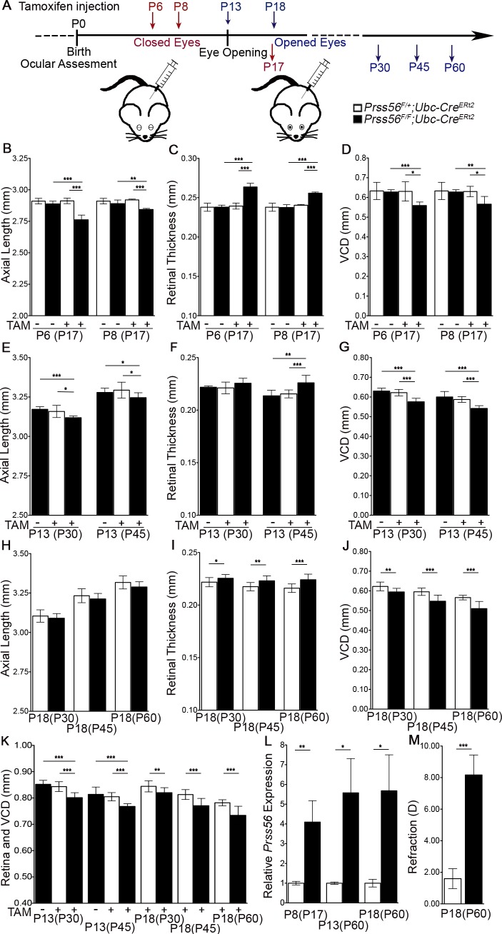
A mismatch between optical power and ocular axial length results in refractive errors. Uncorrected refractive errors constitute the most common cause of vision loss and second leading cause of blindness worldwide. Although the retina is known to play a critical role in regulating ocular growth and refractive development, the precise factors and mechanisms involved are poorly defined. We have previously identified a role for the secreted serine protease PRSS56 in ocular size determination and PRSS56 variants have been implicated in the etiology of both hyperopia and myopia, highlighting its importance in refractive development. Here, we use a combination of genetic mouse models to demonstrate that Prss56 mutations leading to reduced ocular size and hyperopia act via a loss of function mechanism. Using a conditional gene targeting strategy, we show that PRSS56 derived from Müller glia contributes to ocular growth, implicating a new retinal cell type in ocular size determination. Importantly, we demonstrate that persistent activity of PRSS56 is required during distinct developmental stages spanning the pre- and post-eye opening periods to ensure optimal ocular growth. Thus, our mouse data provide evidence for the existence of a molecule contributing to both the prenatal and postnatal stages of human ocular growth. Finally, we demonstrate that genetic inactivation of Prss56 rescues axial elongation in a mouse model of myopia caused by a null mutation in Egr1. Overall, our findings identify PRSS56 as a potential therapeutic target for modulating ocular growth aimed at preventing or slowing down myopia, which is reaching epidemic proportions.


Conflict of interest statement

The authors have declared that no competing interests exist

Figures

Fig 1
Fig 1. Prss56-/- eyes exhibit reduced ocular axial length and hyperopia.
(A) Representative images of slit-lamp examination by broad-beam illumination to assess ocular structures including the iris, pupil, and lens at 1 and 3 months of age. Prss56-/- eyes did not exhibit any obvious structural abnormalities and were indistinguishable from Prss56+/- eyes. (B) Representative OCT images demonstrating a reduction in ocular size in Prss56-/- compared to Prss56+/- mice (shown are P30 eyes). The red, blue and yellow lines indicate ocular axial length (AL), vitreous chamber depth (VCD) and anterior chamber depth (ACD), respectively. (C-D) Prss56-/- eyes exhibit a modest but highly significant reduction in axial length (C) and equatorial diameter (D) at P15 and P25. (E) A significant reduction in VCD and increase in ACD was detected in Prss56-/- compared to Prss56+/- eyes (shown are data from P30 eyes). (F) Consistent with reduced ocular size, Prss56-/- mice display a hyperopic refraction compared to Prss56+/- littermates (shown are data from 2-months old mice). (G) Retinal thickness was significantly increased in Prss56-/- compared to Prss56+/- eyes. Values are presented as mean ± SD, *** p<0.001, t-test. C and D: N > 10 per group; E and F: N ≥ 6 per group; and G: N≥ 4. (H-I) Representative B-scan images of eyes from an unaffected individual (H) and an individual with a PRSS56 mutation (I). VCD is substantially reduced in the eye of the individual carrying a mutant PRSS56 allele compared to a normal emmetropic eye.
Fig 2
Fig 2. Lineage tracing of Prss56 expressing cells during ocular development.
(A) Prss56Cre/+ mice were crossed to R26tdTomato/+ reporter mice that express tdTomato following CRE-mediated excision of a stop codon to label Prss56 expressing cells and their derivatives. (B-F) Representative images showing lineage tracing of Prss56 expressing cells (red) in Prss56Cre/+; R26tdTomato/+ eyes throughout ocular development. (B) tdTomato expression is first detected in the retina at embryonic day (E) 16.5 in retinal progenitor cells (RPCs). (C, D) The number of tdTomato positive RPCs increases with age, shown are (C) E18.5 and (D) P2 retinas. (E) By P7, when retinal laminar organization is visible, tdTomato expression was predominantly observed in cells exhibiting characteristic features of Müller glia, with cell bodies located in the inner nuclear layer and apicobasal processes extending across the retina. tdTomato expression was also detected in the inner segment of rod photoreceptors. (F) tdTomato expression continues to be detected in Müller cells and rod photoreceptors following complete maturation of retinal cell types at P13. Interestingly, tdTomato-labeled cells were enriched in the peripheral region and relatively sparser in the central region of the retina. (G) Ki67 immunolabeling of P0 Prss56Cre/+; R26tdTomato/+ eyes demonstrate Ki67 expression in tdTomato positive retinal cells. E, embryonic day; GCL, ganglionic cell layer; INL, inner nuclear layer; ONBL, outer neuroblastic layer; ONL, outer nuclear layer; P, postnatal day. Scale bars: 100μm (B-F) and 50μm (G).
Fig 3
Fig 3. Earliest Prss56 expression occurs in late retinal progenitor cells.
(A, B) Representative images of Prss56Cre/+; R26tdTomato/+ retina immunolabeled for SOX2 (A) or PKCα (B). (A) tdTomato expression (red) is present in SOX2 immunopositive Müller cells (green). The peripheral and central regions of the retina are oriented left to right. (B) Representative images showing low tdTomato expression in a subset of PKCα immunolabeled bipolar cells in Prss56Cre/+; R26TdTomato/+ retina (arrows). (C) Flow cytometry analysis of Glutamine Synthetase (GS) and Rhodopsin expression in Prss56Cre/+; R26tdTomato/+ retinal cell suspensions. GS expression was predominantly detected in tdTomato negative (green) and high tdTomato expressing cells (purple). Rhodopsin expression was predominantly detected in tdTomato negative (green) and low tdTomato expressing (orange) cells. A minimum of 4 eyes per group was pooled for each retinal cell suspension and flow cytometry analyses were repeated 2–3 times on independent samples. Gating was established based on Prss56Cre/+; R26tdTomato/+ retinal cell suspension incubated with AlexaFluor 488 conjugated secondary antibody only. Together, these data demonstrate that Prss56 is expressed by late RPCs that give rise to bipolar cells, rod photoreceptors, and Müller cells. Scale bars = 100μm(A) and 50μm (B).
Fig 4
Fig 4. Prss56 is predominantly expressed by a subset of Müller cells.
(A) Dual fluorescent in situ hybridization for Prss56 (red) and Glutamine Synthetase (GS, green) showed localization of Prss56 expression in the inner nuclear layer of the retina from adult Prss56glcr4/+ and Prss56glcr4/glcr4 mice (2 months old). Prss56 expression colocalized with that of GS, a marker of Müller cells. Both the signal intensity and number of Müller glia expressing Prss56 were substantially higher in Prss56glcr4/glcr4 compared to Prss56glcr4/+ retina, indicating increased Prss56 expression in Prss56 mutant retina. (B) Graph showing relative expression of Prss56 mRNA levels using qPCR in wild-type and mutant retina at different developmental stages. Increased Prss56 expression was detected in the mutant retina from P15 onward. Prss56 expression was normalized to the expression of three housekeeping genes (Hprt1, Actb1 and Mapk1). Data are presented as fold expression relative to wild-type (mean ± SEM), N≥4 /group. *** p<0.001, t-test. Scale bars= 100μm and 50μm for low and high magnification images in A.
Fig 5
Fig 5. Early requirement for PRSS56 in ocular size determination.
(A, B) Ocular biometric analysis by OCT revealed reduced ocular axial length (A) and increased retinal thickness (B) in Prss56glcr4/glcr4 eyes compared to Prss56glcr4/+ eyes at distinct developmental time points ranging from P6 to P17. Although both mutant and control mice exhibit an age-dependent increase in ocular size, ocular axial length is significantly reduced in Prss56 mutant mice compared to control mice. The reduction in ocular axial length was detected as early as P6 (A). (B) OCT analysis revealed that Prss56 mutant retina is significantly thicker than control retina (shown is P17). (C-D) Histological analysis revealed that the number of nuclear stacks in both the inner and outer retinal nuclear layers (INL and ONL, respectively) was consistently greater in the Prss56 mutant retina (D) compared to control retina (C). For comparison between Prss56 mutant and control mice: *p<0.05, ** p<0.01, *** p<0.001, t-test. Scale bars = 100μm. Values are presented as mean ± SD. N ≥ 12 per group for P6 measurements and N > 6 for P8 and P17 (A, B).
Fig 6
Fig 6. Conditional RAX-Cre-mediated ablation of Prss56 from fully differentiated Müller glia leads to ocular size reduction.
Prss56 was conditionally ablated from Müller cells in a time-specific manner by crossing Prss56F/F to the inducible RAX-Cre mouse strain (Rax-CreERT2). CRE expression was induced by tamoxifen injection at P8, a time point preceding complete Müller glia differentiation. (A) Representative images of slit lamp examination by broad-beam illumination following tamoxifen injection at P8. Prss56F/F; Rax-CreERT2 eyes were indistinguishable from control Prss56F/+; Rax-CreERT2 eyes at 2 months of age. (B, C) Following tamoxifen injection (TAM +; horizontal axis) at P8, Prss56F/FRax-CreERT2 mice display a significant decrease in ocular axial length (B) and increase in retinal thickness (C) compared to control eyes (Prss56F/+; Rax-CreERT2 or Prss56F/F mice without Rax-CreERT2), N = 6 to 8 per group. (D) Following tamoxifen injection at P8, a significant reduction in VCD was detected in Prss56F/F; Rax-CreERT2 eyes compared to control Prss56F/+; Rax-CreERT2 eyes, N = 6 per group. Ocular axial length, retinal thickness, and VCD in uninjected Prss56F/F; Rax-CreERT2 and Prss56F/+; Rax-CreERT2 mice were indistinguishable. (E) Representative OCT images showing reduced axial length and VCD in Prss56F/F; Rax-CreERT2 eyes compared to the control Prss56F/+; Rax-CreERT2 or Prss56F/F mice. (F) qPCR analysis following tamoxifen injection at P8 revealed that Prss56 mRNA was significantly upregulated in Prss56F/F ; Rax-CreERT2 retina compared to their Prss56F/+ ; Rax-CreERT2 counterparts or uninjected controls, N≥ 6 per group. Values are presented as mean ±SD; * p<0.05, ** p<0.01, *** p<0.001, t-test. (G-I) Müller glia endfeet organization of Prss56Cre; R26tdTomato reporter mice during retinal development. Representative images of retinal section (G, I) or whole mount (H) showing Müller glia endfeet from control and Prss56 mutant mice at P6. (I) Magnified images of the retinal endfeet are shown. The ILM of Prss56 mutant mice (Prss56Cre/gclr4; R26tdTomato) at P6 is marked by regions of increased endfeet complexity (arrow head) compared to the ILM of control mice (Prss56Cre/+; R26tdTomato). A substantial proportion of endfeet appear more spread out, occupying a larger area in the mutant compared to control retinal whole mounts (occupying smaller area). Red boxes highlight individual endfoot. N = 4 per genotype and scale bars = 17μm in G and I, and 50μm in H. ACD, anterior chamber depth; AL, axial length; VCD, vitreous chamber depth.
Fig 7
Fig 7. PRSS56 activity is required during both the vision-independent and dependent stages of ocular growth.
To determine the temporal window critical for the PRSS56-mediated effect on ocular axial growth, Prss56 conditional mutant mice (Prss56F/F) were crossed to mice expressing the ubiquitous inducible Ubc-Cre recombinase (Ubc-CreERT2). (A) Schematic of tamoxifen treatment at distinct developmental stages preceding and following the opening of the eyes. Tamoxifen injection at two different time points, P6 and P8, was performed to ablate Prss56 after the earliest detectable effect of mutant Prss56 on ocular axial length. (B-D) OCT-based ocular biometry demonstrates that following tamoxifen injection, Prss56F/F; Ubc-CreERT2 mice display a significantly reduced ocular axial length (B) and increased retinal thickness (C) and a significant decrease in VCD (D) compared to the control Prss56F/+; Ubc-CreERT2 mice (measured at P17). The ocular axial length, retinal thickness, and VCD of uninjected Prss56F/F; Ubc-CreERT2 and Prss56F/+; Ubc-CreERT2 mice were indistinguishable. Administration of tamoxifen at P6 caused a greater decrease in ocular axial length compared to administration at P8 suggesting a requirement for continuous PRSS56 activity during ocular development to sustain normal ocular growth, N = 6 to 8 per group for A and B. (E-G) OCT measurements demonstrate that following tamoxifen injection at P13 (a time point when the eyes are open), Prss56F/F; Ubc-CreERT2 mice display a slight but significant decrease in ocular axial length (E) and increase in retinal thickness (F) at P30 and P45. Reduced ocular axial length was associated with a significant decrease in VCD in Prss56F/F; Ubc-CreERT2 mice compared to Prss56F/+; Ubc-CreERT2 and uninjected controls at P30 and P45, N = 5 to 10 per group (G). (H-J) Ocular biometry following tamoxifen injection at the beginning of a critical emmetropization period (P18) shows that the ocular axial length is not significantly different between the Prss56F/F; Ubc-CreERT2 and control mice at any of the three ages examined (P30, P45, and P60). However, Prss56F/F; Ubc-CreERT2 mice display a slightly thicker retina and significantly reduced VCD compared to the control groups. (K) Following Prss56 ablation at P13 and P18, the eyes display a decrease in the combined value of retinal thickness and VCD. (L) qPCR analysis revealed elevated Prss56 mRNA levels in Prss56F/F; Ubc-CreERT2 retina compared to Prss56F/+; Ubc-CreERT2 retina following tamoxifen injection at P8, P13, or P18 (shown are data from mice harvested at P17 and P60, respectively). (M) Following Prss56 ablation at P18, the eyes display a hyperopic shift in refraction compared to control eyes at 3 months (N = 6 per group). Values are presented as mean ± SD (or mean ± SEM in L); * p<0.05, ** p<0.01, *** p<0.001, t-test.
Fig 8
Fig 8. Prss56 ablation rescues myopia in mice.
(A) Representative OCT images demonstrating that Prss56 ablation rescues myopia in Egr1-/- mice (compare Prss56+/-;Egr1-/- to Prss56-/-;Egr1-/-, shown are P30 eyes). Reciprocally, Egr1 deficiency rescues hyperopia in Prss56-/- mice (compare Prss56-/-;Egr1+/- to Prss56-/-;Egr1-/-). The red and blue lines indicate ocular axial length (AL) and vitreous chamber depth (VCD), respectively. (B) Prss56-/-;Egr1+/- eyes display a significant reduction in axial length, whereas Prss56+/-;Egr1-/- exhibit significantly elongated axial length compared to the control eyes (Prss56+/-;Egr1+/- ). The eyes of double mutants (Prss56-/-;Egr1-/-) attain a size that is not significantly different from control eyes (Prss56+/-;Egr-/-), at all ages examined (P10 to P60). (C) Consistent with modulation of ocular axial length by Prss56 and Egr1 mutations, hyperopic refraction observed in Prss56-/-;Egr1+/- eyes was rescued in the double mutants (Prss56-/-;Egr1-/-). Conversely, Prss56 ablation rescued myopic refraction observed in Prss56+/-;Egr1-/- eyes (compare Prss56+/-;Egr1-/- to Prss56-/-;Egr1-/-, shown are data from 2-months old mice). (D) The VCD of double mutant eyes (Prss56-/-;Egr1-/-) was significantly reduced compared to Egr1 single mutant eyes (Prss56+/-;Egr1-/-) and increased compared to Prss56 single mutant eyes (Prss56-/-;Egr1+/-). The VCD of double mutant eyes was not significantly different from the control eyes. (E) The retina is thicker in double mutants (Prss56-/-;Egr1-/-) compared to control mice (Prss56+/-;Egr1+/-) at P30 and P60, despite their ocular axial length being similar. Values are presented as mean ± SD. For comparison between single mutant and controls: * p<0.05; ** p<0.01;*** p<0.001, t-test. For comparison between single mutant and double mutants: # p<0.05; ## p<0.01; ###p<0.001, t-test. B-D: N ≥ 6 per group; E: N ≥ 8 per group.

References

    1. Stone RA, Pardue MT, Iuvone PM, Khurana TS. Pharmacology of myopia and potential role for intrinsic retinal circadian rhythms. Experimental eye research. 2013;114:35–47. Epub 2013/01/15. doi: 10.1016/j.exer.2013.01.001 ; PubMed Central PMCID: PMC3636148. - DOI - PMC - PubMed
    1. Meng W, Butterworth J, Malecaze F, Calvas P. Axial length of myopia: a review of current research. Ophthalmologica Journal international d'ophtalmologie International journal of ophthalmology Zeitschrift fur Augenheilkunde. 2011;225(3):127–34. Epub 2010/10/16. doi: 10.1159/000317072 . - DOI - PubMed
    1. Bourne RR, Stevens GA, White RA, Smith JL, Flaxman SR, Price H, et al. Causes of vision loss worldwide, 1990–2010: a systematic analysis. The Lancet Global health. 2013;1(6):e339–49. Epub 2014/08/12. doi: 10.1016/S2214-109X(13)70113-X . - DOI - PubMed
    1. Dolgin E. The myopia boom. Nature. 2015;519(7543):276–8. Epub 2015/03/20. doi: 10.1038/519276a . - DOI - PubMed
    1. Tarutta E, Chua WH, Young T, Goldschmidt E, Saw SM, Rose KA, et al. Myopia: Why Study the Mechanisms of Myopia? Novel Approaches to Risk Factors Signalling Eye Growth- How Could Basic Biology Be Translated into Clinical Insights? Where Are Genetic and Proteomic Approaches Leading? How Does Visual Function Contribute to and Interact with Ametropia? Does Eye Shape Matter? Why Ametropia at All? Optometry and vision science: official publication of the American Academy of Optometry. 2011. Epub 2011/02/08. doi: 10.1097/OPX.0b013e31820e6a6a . - DOI

Publication types

MeSH terms