Review
. 2018 May 1;42(3):324-334.
doi: 10.1093/femsre/fuy007.

Host cell cytosolic immune response during Plasmodium liver stage development

Affiliations
Review

Host cell cytosolic immune response during Plasmodium liver stage development

Carolina Agop-Nersesian et al. FEMS Microbiol Rev. .

Abstract

Recent years have witnessed a great gain in knowledge regarding parasite-host cell interactions during Plasmodium liver stage development. It is now an accepted fact that a large percentage of sporozoites invading hepatocytes fail to form infectious merozoites. There appears to be a delicate balance between parasite survival and elimination and we now start to understand why this is so. Plasmodium liver stage parasites replicate within the parasitophorous vacuole (PV), formed during invasion by invagination of the host cell plasma membrane. The main interface between the parasite and hepatocyte is the parasitophorous vacuole membrane (PVM) that surrounds the PV. Recently, it was shown that autophagy marker proteins decorate the PVM of Plasmodium liver stage parasites and eliminate a proportion of them by an autophagy-like mechanism. Successfully developing Plasmodium berghei parasites are initially also labeled but in the course of development, they are able to control this host defense mechanism by shedding PVM material into the tubovesicular network (TVN), an extension of the PVM that releases vesicles into the host cell cytoplasm. Better understanding of the molecular events at the PVM/TVN during parasite elimination could be the basis of new antimalarial measures.


Figures
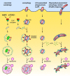
Figure 1.
Figure 1.
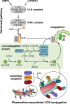
Overview of different autophagy pathways. Hallmark of canonical and selective autophagy—exemplified by xenophagy—is the formation of a double membrane autophagosome. Both pathways are characterized by a hierarchical sequence: initiation via the ULK complex, nucleation, elongation and closure, recycling and degradation by lysosomal enzymes. Canonical autophagy is activated by AMP-activated protein kinase (AMPK) and inhibited by mammalian target of rapamycin complex 1 (mTORC1). A distinctive feature of selective autophagy is the specific recognition of ubiquitinated cargo, which is recognized by autophagy receptors. The receptors mediate recruitment of the autophagosomal membrane. In LC3-associated phagocytosis (LAP), ATG proteins are directly recruited to a single-membrane vacuole. PI3P and the production of reactive oxygen species (ROS) define the target membrane for the LC3 lipidation machinery. The Plasmodium-associated autophagy-related (PAAR) response in P.berghei shares features of xenophagy and LAP. LC3 is directly incorporated into the parasitophorous vacuolar membrane (PVM) and recruits autophagy receptors and ubiquitin in an inverse order. Although Plasmodium has evolved strategies to avoid acidification of the parasitophorous vacuole (PV), parasites can be eliminated by the PAAR response of the host cell.
Figure 2.
Figure 2.
Comparison of the LC3-conjugation pathway during canonical autophagy and Plasmodium-associated autophagy-related (PAAR) response. Upper panel: Autophagosome formation during canonical autophagy depends on the multiprotein complex ULK and the class III phosphatidylinositol 3-kinase (PI3KC3) complex defining phagophore membranes with PI3P (orange). The lipid signal recruits the PI3P-dependent effector WIPI and the LC3-conjugation system to drive elongation of the LC3-positive phagophore (green). Both pathways share the LC3-conjugation system for LC3 lipidation (green box). Lower panel: PAAR response bypasses the canonical steps involved in the initiation, nucleation and elongation of the autophagosome. The pathway upstream of the LC3-conjugation system, which is important for the initiation of the response, and the recognition of the parasitophorous vacuolar membrane (PVM) is unknown.
Figure 3.
Figure 3.
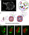
The parasitophorous vacuolar membrane (PVM) and its role during the shedding of host protein. (A) 3D architecture of the PVM and its connected tubovesicular network (TVN). Young P. berghei liver schizont (36 hpi) stained for the PVM protein exported protein 1 (EXP-1) was imaged by confocal laser scanning microscopy (3D-CLSM) with z-increments of 0.22 μm. Morphological features of the TVN represent highly branched tubular structures, large node-like clusters and vesicles. Scale bar, 10 μm. (B) Model of shedding mechanism involved in the removal of host autophagy (LC3) and lysosomal proteins from the PVM of P. berghei during liver stage development. PVM-associated host factors assemble in membrane patches at the PVM, accumulate and become trapped in the TVN, and are finally shed as vesicles into the host cytoplasm. Magnification of the TVN cluster highlights the potential link between the PAAR response and the nourishing capacity of the macroautophagy pathway. Adapted from Agop-Nersesian et al. (2017). (C) Long-term live microscopy of the shedding process. PMV-associated LC3 (green) is progressively removed from the developing P. berghei liver schizonts (24 hpi, red) into the cytoplasm of a HepG2 cell. Time stamp, h:min:s. Scale bar, 10 μm. Adapted from Prado et al. (2015).

Similar articles

Cited by

  • Guidelines for the use and interpretation of assays for monitoring autophagy (4th edition)1.
    Klionsky DJ, Abdel-Aziz AK, Abdelfatah S, Abdellatif M, Abdoli A, Abel S, Abeliovich H, Abildgaard MH, Abudu YP, Acevedo-Arozena A, Adamopoulos IE, Adeli K, Adolph TE, Adornetto A, Aflaki E, Agam G, Agarwal A, Aggarwal BB, Agnello M, Agostinis P, Agrewala JN, Agrotis A, Aguilar PV, Ahmad ST, Ahmed ZM, Ahumada-Castro U, Aits S, Aizawa S, Akkoc Y, Akoumianaki T, Akpinar HA, Al-Abd AM, Al-Akra L, Al-Gharaibeh A, Alaoui-Jamali MA, Alberti S, Alcocer-Gómez E, Alessandri C, Ali M, Alim Al-Bari MA, Aliwaini S, Alizadeh J, Almacellas E, Almasan A, Alonso A, Alonso GD, Altan-Bonnet N, Altieri DC, Álvarez ÉMC, Alves S, Alves da Costa C, Alzaharna MM, Amadio M, Amantini C, Amaral C, Ambrosio S, Amer AO, Ammanathan V, An Z, Andersen SU, Andrabi SA, Andrade-Silva M, Andres AM, Angelini S, Ann D, Anozie UC, Ansari MY, Antas P, Antebi A, Antón Z, Anwar T, Apetoh L, Apostolova N, Araki T, Araki Y, Arasaki K, Araújo WL, Araya J, Arden C, Arévalo MA, Arguelles S, Arias E, Arikkath J, Arimoto H, Ariosa AR, Armstrong-James D, Arnauné-Pelloquin L, Aroca A, Arroyo DS, Arsov I, Artero R, Asaro DML, Aschner M, Ashrafizadeh M, Ashur-Fabian O, Atanasov AG, Au AK, Auberger P, Auner HW, Aurelian L, Autelli R… See abstract for full author list ➔ Klionsky DJ, et al. Autophagy. 2021 Jan;17(1):1-382. doi: 10.1080/15548627.2020.1797280. Epub 2021 Feb 8. Autophagy. 2021. PMID: 33634751 Free PMC article.
  • Hardly Vacuous: The Parasitophorous Vacuolar Membrane of Malaria Parasites.
    Goldberg DE, Zimmerberg J. Goldberg DE, et al. Trends Parasitol. 2020 Feb;36(2):138-146. doi: 10.1016/j.pt.2019.11.006. Epub 2019 Dec 19. Trends Parasitol. 2020. PMID: 31866184 Free PMC article. Review.
  • Plasmodium's fight for survival: escaping elimination while acquiring nutrients.
    Schroeder EA, Chirgwin ME, Derbyshire ER. Schroeder EA, et al. Trends Parasitol. 2022 Jul;38(7):544-557. doi: 10.1016/j.pt.2022.04.004. Epub 2022 May 6. Trends Parasitol. 2022. PMID: 35534377 Free PMC article. Review.
  • Host-pathogen interactions in the Plasmodium-infected mouse liver at spatial and single-cell resolution.
    Hildebrandt F, Iturritza MU, Zwicker C, Vanneste B, Van Hul N, Semle E, Quin J, Pascini T, Saarenpää S, He M, Andersson ER, Scott CL, Vega-Rodriguez J, Lundeberg J, Ankarklev J. Hildebrandt F, et al. Nat Commun. 2024 Aug 19;15(1):7105. doi: 10.1038/s41467-024-51418-2. Nat Commun. 2024. PMID: 39160174 Free PMC article.
  • Mid-Liver Stage Arrest of Plasmodium falciparum Schizonts in Primary Porcine Hepatocytes.
    van der Boor SC, van Gemert GJ, Hanssen AEJ, van Waardenburg YM, McCall MBB, Bousema T, de Wilt JHW, Sauerwein RW, Yang ASP. van der Boor SC, et al. Front Cell Infect Microbiol. 2022 Feb 17;12:834850. doi: 10.3389/fcimb.2022.834850. eCollection 2022. Front Cell Infect Microbiol. 2022. PMID: 35252038 Free PMC article.

References

    1. Agop-Nersesian C, De Niz M, Niklaus L et al. . Shedding of host autophagic proteins from the parasitophorous vacuolar membrane of Plasmodiumberghei. Sci Rep 2017;7:1–14. - PMC - PubMed
    1. Amino R, Thiberge S, Martin B et al. . Quantitative imaging of Plasmodium transmission from mosquito to mammal. Nat Med 2006;12:220–4. - PubMed
    1. Annoura T, van Schaijk BCL, Ploemen IHJ et al. . Two Plasmodium 6-Cys family-related proteins have distinct and critical roles in liver-stage development. FASEB J 2014;28:2158–70. - PubMed
    1. Baer K, Klotz C, Kappe SHI et al. . Release of hepatic Plasmodiumyoelii merozoites into the pulmonary microvasculature. PLoS Pathog 2007;3:1651–68. - PMC - PubMed
    1. Bano N, Romano JD, Jayabalasingham B et al. . Cellular interactions of Plasmodium liver stage with its host mammalian cell. Int J Parasitol 2007;37:1329–41. - PubMed

Publication types