. 2018 May 18;46(9):4752-4770.
doi: 10.1093/nar/gky133.

Protein composition of catalytically active U7-dependent processing complexes assembled on histone pre-mRNA containing biotin and a photo-cleavable linker

Affiliations

Protein composition of catalytically active U7-dependent processing complexes assembled on histone pre-mRNA containing biotin and a photo-cleavable linker

Aleksandra Skrajna et al. Nucleic Acids Res. .

Abstract

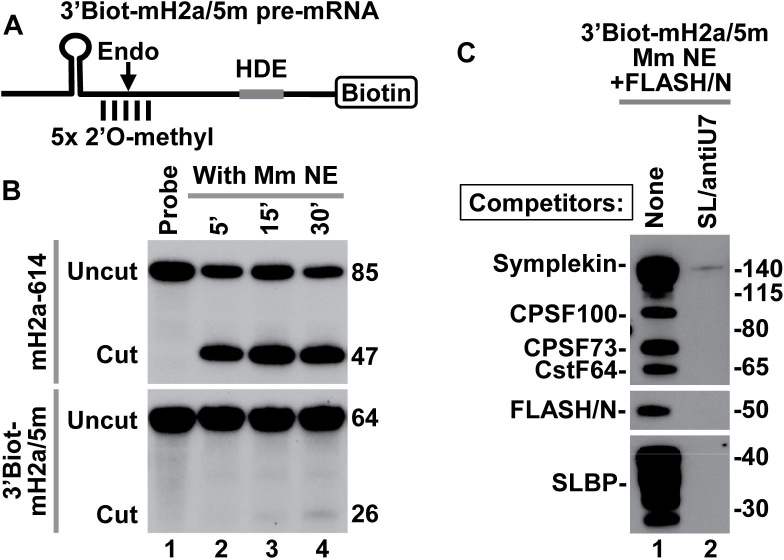
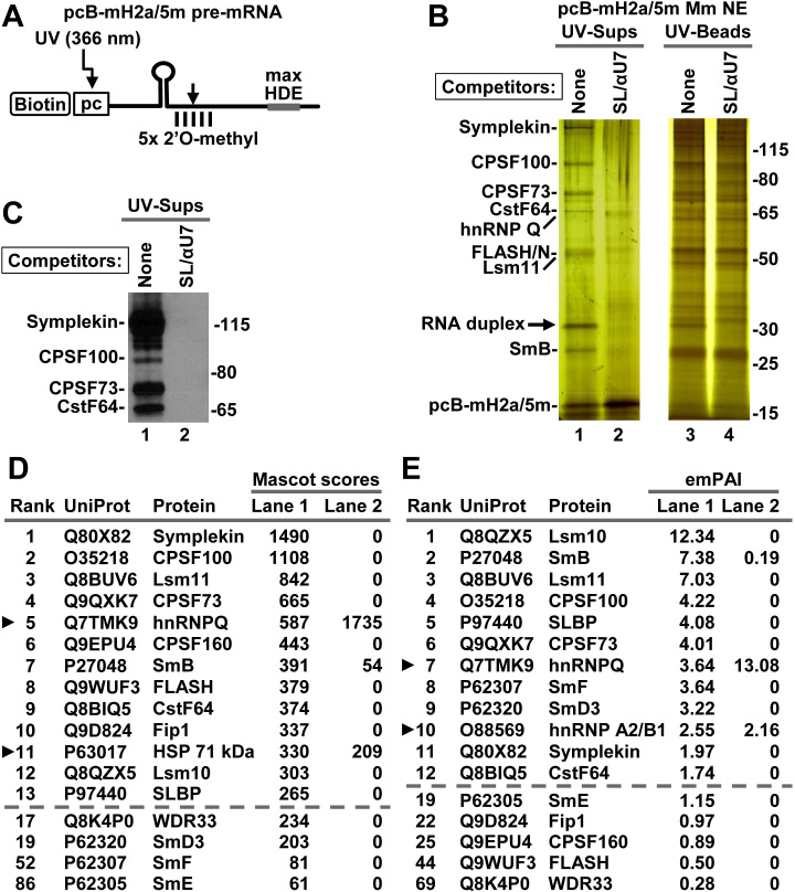
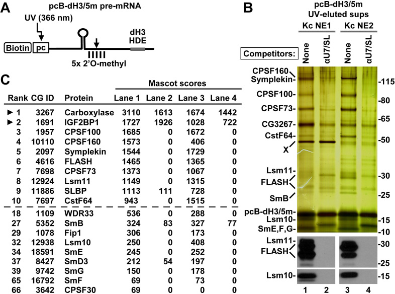
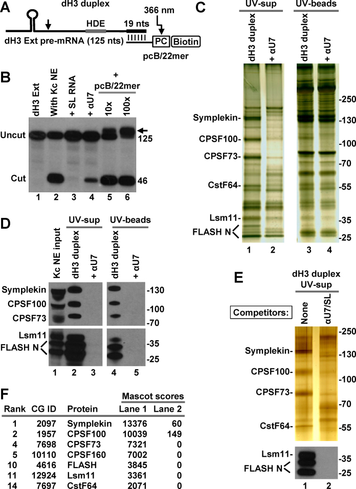
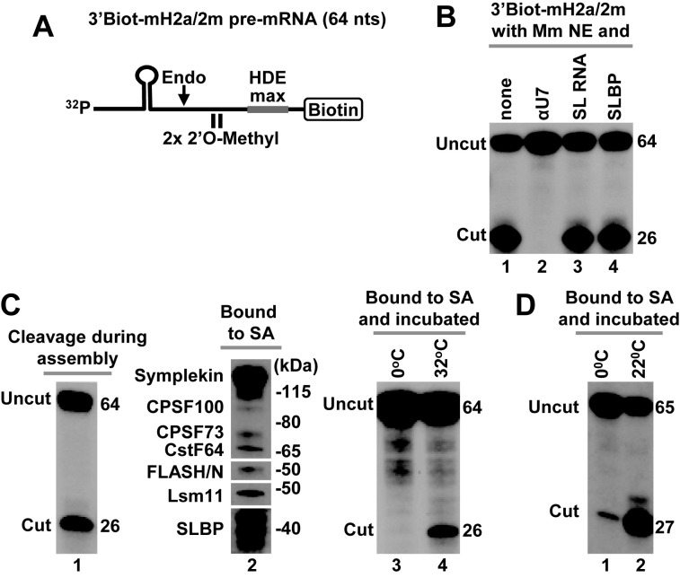
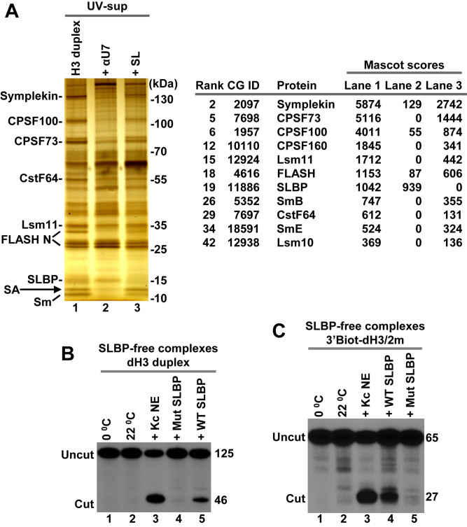
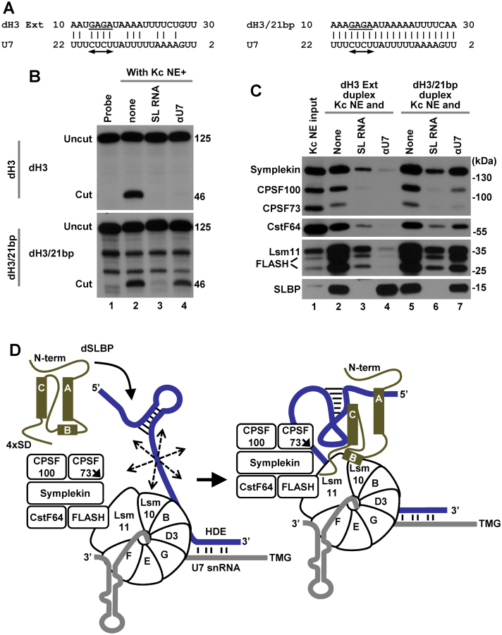
3' end cleavage of metazoan replication-dependent histone pre-mRNAs requires the multi-subunit holo-U7 snRNP and the stem-loop binding protein (SLBP). The exact composition of the U7 snRNP and details of SLBP function in processing remain unclear. To identify components of the U7 snRNP in an unbiased manner, we developed a novel approach for purifying processing complexes from Drosophila and mouse nuclear extracts. In this method, catalytically active processing complexes are assembled in vitro on a cleavage-resistant histone pre-mRNA containing biotin and a photo-sensitive linker, and eluted from streptavidin beads by UV irradiation for direct analysis by mass spectrometry. In the purified processing complexes, Drosophila and mouse U7 snRNP have a remarkably similar composition, always being associated with CPSF73, CPSF100, symplekin and CstF64. Many other proteins previously implicated in the U7-dependent processing are not present. Drosophila U7 snRNP bound to histone pre-mRNA in the absence of SLBP contains the same subset of polyadenylation factors but is catalytically inactive and addition of recombinant SLBP is sufficient to trigger cleavage. This result suggests that Drosophila SLBP promotes a structural rearrangement of the processing complex, resulting in juxtaposition of the CPSF73 endonuclease with the cleavage site in the pre-mRNA substrate.


Figures

Figure 1.
Figure 1.
3′Biot-mH2a/5m pre-mRNA is resistant to cleavage but assembles into processing complexes. (A) A schematic representation of chemically synthesized mouse-specific 3′Biot-mH2a/5m pre-mRNA (64-nt). The major cleavage site (located 5 nt downstream of the stem) and 2 nt on each side of the major cleavage site are modified with a 2′O-methyl group. Biotin is placed at the 3′ end. (B) In vitro processing of 3′Biot-mH2a/5m (bottom) and mH2a-614 (top) pre-mRNAs. mH2a-614 (85 nt) was generated by T7 transcription and contains the same HDE as 3′Biot-mH2a/5m but lacks biotin and modified nucleotides. Each pre-mRNA was labeled at the 5′ end with 32P and incubated in a mouse nuclear extract for 5, 15 and 30 min, as indicated. Probe alone is shown in lane 1. Numbers to the right indicate the length of the input pre-mRNA and the upstream cleavage product. (C) 3′Biot-mH2a/5m was incubated with a mouse myeloma nuclear extract (Mm NE) containing recombinant N-terminal FLASH (FLASH/N, amino acids 53–138) fused to GST. Assembled processing complexes were purified on streptavidin beads and analyzed by western blotting using specific antibodies (lane 1). In lane 2, formation of the processing complexes was blocked by excess SL RNA and αU7 oligonucleotide complementary to the 5′ end of mouse U7 snRNA.
Figure 2.
Figure 2.
Mouse nuclear proteins that bind pcB-mH2a/5m pre-mRNA. (A) A schematic representation of chemically synthesized mouse-specific pcB-mH2a/5m pre-mRNA (61 nt). Five nucleotides around the major cleavage site are modified with a 2′O-methyl group. 5′ biotin (B) is followed by a photo-cleavable (pc) linker sensitive to long wave UV (366 nm). The HDE was altered by two point mutations (max HDE) to increase its base pairing potential with the 5′ end of mouse U7 snRNA. (B) pcB-mH2a/5m was incubated with a mouse myeloma nuclear extract containing recombinant N-terminal FLASH to assemble processing complexes. In negative control, their formation was blocked by addition of SL RNA and αU7 oligonucleotide to the nuclear extract. Proteins bound to pcB-mH2a/5m pre-mRNA were immobilized on streptavidin beads and eluted by irradiation with long wave UV. Same fractions (15%) of the UV-eluted material (UV-sups) and the beads following UV-elution (UV-beads) were analyzed by silver staining. (C) A fraction of the UV-eluted material (15%) was analyzed by western blotting using selected antibodies. (D and E) A fraction (15%) of the UV-eluted material was directly analyzed by mass spectrometry and identified proteins were ranked based on their Mascot scores (panel D) or emPAI values (panel E). The top consecutive 13 and 12 hits are listed in panels D and E, respectively (shown above the dashed lines). Arrowheads indicate proteins that interacted with histone pre-mRNA in the presence of the SL RNA and αU7 oligonucleotide processing competitors. Below the dashed lines shown are the other proteins that fail to interact with histone pre-mRNA in the presence of the two competitors. Note that the emPAI value for FLASH is low since only a short fragment of the protein (amino acids 53–138) was added to the extract.
Figure 3.
Figure 3.
Drosophila nuclear proteins that bind pcB-dH3/5m pre-mRNA. (A) A schematic representation of chemically synthesized Drosophila-specific pcB-dH3/5m pre-mRNA (63 nt). (B) pcB-dH3/5m pre-mRNA was incubated with two different batches of Drosophila Kc nuclear extract in the absence or in the presence of SL RNA and αU7 oligonucleotide complementary to the 5′ end of Drosophila U7 snRNA. Proteins bound to pcB-dH3/5m pre-mRNA were immobilized on streptavidin beads and eluted by irradiation with long wave UV. A fraction of each UV-eluted supernatant was analyzed by silver staining (top panel) or western blotting (bottom panels). (C) The same fraction was also analyzed by mass spectrometry. Proteins with the 10 highest Mascot scores for Kc NE1 are shown above the dashed line, with arrowheads indicating proteins that interact with histone pre-mRNA in the presence of SL RNA and αU7 oligonucleotide. The remaining components of the U7 snRNP and their overall ranking among all identified proteins (numbering for Kc NE1) are shown below the dashed line. Note that carboxylase results from contamination of the UV eluate with a small amount of streptavidin beads.
Figure 4.
Figure 4.
Purification of Drosophila processing complexes with pre-mRNA attached to the photo-cleavable group in trans. (A) A schematic representation of the dH3 Ext duplex formed by annealing T7-generated dH3 Ext pre-mRNA and chemically synthesized 2′O-methyl pcB/22mer oligonucleotide. In pcB/22mer, biotin (B) is placed at the 5′ end and is followed by the photo-cleavable (pc) linker. The last 19 of 22 nt of pcB/22mer are complementary to the 3′ end of dH3 Ext pre-mRNA. (B) dH3 Ext pre-mRNA was labeled at the 5′ end and incubated at room temperature in a Kc nuclear extract either alone (lane 2) or in the presence of indicated oligonucleotides (lanes 3–6). SL RNA and αU7 oligonucleotide were added to a final concentration of 10 ng/μl, pcB/22mer was at either 10 ng/μl or 100 ng/μl, a 10 and 100 molar excess relative to dH3 Ext pre-mRNA, respectively. The arrow indicates an RNA duplex that survived denaturing conditions of the 7M urea gel. Lane 1 contains input dH3 Ext pre-mRNA. (C and D) dH3 Ext duplex was incubated with a Drosophila Kc nuclear extract to form processing complexes (lanes 1 and 3 in panel C, and lanes 2 and 4 in panel D). As a negative control, formation of the processing complexes was blocked by αU7 oligonucleotide complementary to the 5′ end of Drosophila U7 snRNA (lanes 2 and 4 in panel C, and lanes 3 and 5 in panel D). Proteins bound to the duplex RNA were purified on streptavidin beads and UV-eluted. A fraction of the UV-eluted material (UV-sups, lanes 1 and 2) and the beads following UV-elution (UV-beads, lanes 3 and 4) was analyzed by silver staining (panel C) or western blotting (panel D). (E and F) Drosophila processing complexes were assembled on the duplex RNA and purified, as described above with the difference that negative control contained both SL RNA and αU7 oligonucleotide. A fraction of the UV-eluted samples was analyzed by silver staining and western blotting for selected proteins (panel E). The remainder was concentrated by precipitation with acetone, subjected to a brief electrophoresis sufficient for proteins to enter the gel, in-gel digested with trypsin and analyzed by mass spectrometry (panel F). Only top scoring processing factors are listed.
Figure 5.
Figure 5.
Cleavage activity of immobilized mouse and Drosophila processing complexes. (A) A schematic representation of chemically synthesized mouse-specific 3′Biot-mH2a/2m pre-mRNA (64 nt). The two 2′O-methyl groups placed immediately upstream of the HDE have no effect on endonucleolytic cleavage but prevent subsequent 5′-3′ degradation of the downstream cleavage product. Biotin is placed at the 3′ end and the HDE is modified by two point mutations to maximize its base pair interaction with U7 snRNA (HDE max). (B) In vitro processing of 3′Biot-mH2a/2m pre-mRNA in a mouse nuclear extract alone (lane 1) or containing indicated reagents: αU7 oligonucleotide (10 ng/μl), SL RNA (10 ng/μl) or human baculovirus-expressed SLBP (25 ng/μl). (C) Mouse processing complexes (lanes 1–4) were assembled on ice by incubating 32P-labeled 3′Biot-mH2a/2m pre-mRNA with a mouse nuclear extract containing recombinant N-terminal FLASH. During this step, significant cleavage occurred reducing the amount of fully assembled processing complexes (lane 1). Length in nucleotides of the input 3′Biot-mH2a/2m pre-mRNA and of the upstream cleavage product in nucleotides is shown to the right. The assembled complexes were immobilized on streptavidin beads, thoroughly washed and analyzed by western blotting for the presence of SLBP and selected subunits of the U7 snRNP (lane 2) or incubated with a gentle agitation at indicated temperatures to determine their ability to support cleavage (lanes 3 and 4). As explained above, in mouse nuclear extracts SLBP is partially degraded and in SDS gels migrates as a group of closely spaced bands rather than a single full length species of ∼45 kDa. (D) Drosophila processing complexes were assembled on Drosophila-specific 3′Biot-dH3/2m pre-mRNA. The complexes were immobilized on streptavidin beads, washed and incubated with a gentle agitation at 0°C (lane 1) or 22°C (lane 2) to determine their ability to support cleavage.
Figure 6.
Figure 6.
Cleavage activity of Drosophila processing complexes assembled in the absence of SLBP. (A) dH3 Ext duplex was incubated in a Kc nuclear extract in the absence or in the presence of indicated competitors and the UV-eluted samples were visualized by silver staining (left) and analyzed by mass spectrometry following a brief electrophoresis into 4–12 gel SDS/polyacrylamide gel (right). Only SLBP and components of the U7 snRNP are listed. They all fail to interact with dH3 pre-mRNA in the presence of SL RNA or αU7 oligonucleotide. A small amount of contaminating streptavidin (SA) in lanes 1 and 3 is indicated with an arrow. (B and C) SLBP-free Drosophila processing complexes assembled on dH3 Ext duplex (panel B) or 3′Biot-dH3/2m (panel C) in a nuclear extract containing the SL RNA (as in lane 3 of panel A) were immobilized on streptavidin beads, washed and tested for the ability to support cleavage at 22°C (lanes 2–5 in both panels) either alone or in the presence of Kc nuclear extract or indicated SLBP variants. The aliquots incubated on ice are shown in lane 1 in both panels.
Figure 7.
Figure 7.
Effects of increasing the base pairing potential between histone pre-mRNA and Drosophila U7 snRNA. (A) A proposed base pair alignment between the HDE of dH3 Ext (left) and dH3/21bp (right) pre-mRNAs and Drosophila U7 snRNA. The HDE starts 10 nt 3′ of the cleavage site and contains the GAGA sequence (underlined) that is essential for processing and is predicted to form an obligatory duplex with the UCUC motif (indicated with a double-headed arrow) conserved in most U7 snRNAs. (B) Processing of 5′-labeled dH3 Ext (top) and dH3/21bp (bottom) pre-mRNAs in a Kc nuclear extract either lacking (lane 2) or containing indicated competitors (lanes 3 and 4). The input RNA is shown in lane 1. Length of the uncut pre-mRNAs and the upstream cleavage products (cut) in nucleotides is shown to the right (C). dH3 Ext and dH3/21bp pre-mRNAs were annealed to pcB/21mer and the resultant duplexes incubated with a Kc nuclear extract in the absence or presence of oligonucleotide competitors, as indicated. The assembled processing complexes were immobilized on streptavidin beads and analyzed by western blotting for the presence of SLBP and major subunits of the U7 snRNP. (D) A hypothetical model explaining essential role of Drosophila in processing. The three α helices of the RBD of Drosophila SLBP are depicted as rectangles and the repeated SD motif is shown at the C-terminal region. In the absence of Drosophila SLBP, U7 snRNP inefficiently binds histone pre-mRNA (blue line) via limited base paring between U7 snRNA (gray line) and the HDE. The bound U7 snRNP in spite of containing all necessary subunits is unable to carry out the cleavage reaction, possibly due to the excessive flexibility of the pre-mRNA substrate (illustrated by multiple arrows). SLBP bound to the stem-loop uses helix B and the C-terminal region to promote the recruitment of the U7 snRNP to the HDE, likely by contacting Lsm11 bound to FLASH and possibly Lsm10. The same network of interactions may result in a major structural rearrangement of the processing complex and stable alignment of the active site of CPSF73 (indicated with an arrow) with the pre-mRNA.

Similar articles

Cited by

References

    1. Dominski Z., Marzluff W.F.. Three-hybrid screens for RNA-binding proteins: proteins binding the 3′ end of histone mRNA. Methods Mol. Biol. 2001; 177:291–318. - PubMed
    1. Marzluff W.F. Metazoan replication-dependent histone mRNAs: a distinct set of RNA polymerase II transcripts. Curr. Opin. Cell Biol. 2005; 17:274–280. - PubMed
    1. Dominski Z., Carpousis A.J., Clouet-d’Orval B.. Emergence of the beta-CASP ribonucleases: highly conserved and ubiquitous metallo-enzymes involved in messenger RNA maturation and degradation. Biochim. Biophys. Acta. 2013; 1829:532–551. - PubMed
    1. Birchmeier C., Schümperli D., Sconzo G., Birnstiel M.L.. 3′ editing of mRNAs: sequence requirements and involvement of a 60-nucleotide RNA in maturation of histone mRNA precursors. Proc. Natl. Acad. Sci. U.S.A. 1984; 81:1057–1061. - PMC - PubMed
    1. Galli G., Hofstetter H., Stunnenberg H.G., Birnstiel M.L.. Biochemical complementation with RNA in the Xenopus oocyte: a small RNA is required for the generation of 3′ histone mRNA termini. Cell. 1983; 34:823–828. - PubMed

Publication types

MeSH terms