Comparative Study
. 2018 Jul 1;35(7):1588-1598.
doi: 10.1093/molbev/msy021.

Revising the Structural Diversity of Ribosomal Proteins Across the Three Domains of Life

Affiliations
Comparative Study

Revising the Structural Diversity of Ribosomal Proteins Across the Three Domains of Life

Sergey Melnikov et al. Mol Biol Evol. .

Abstract

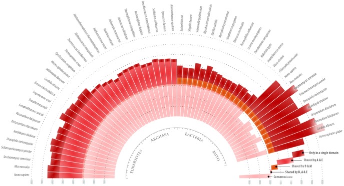
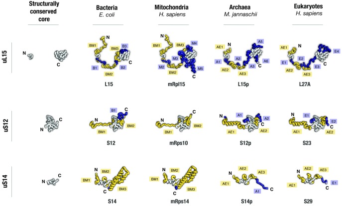
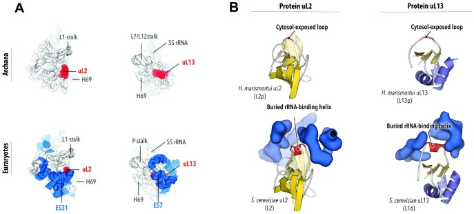
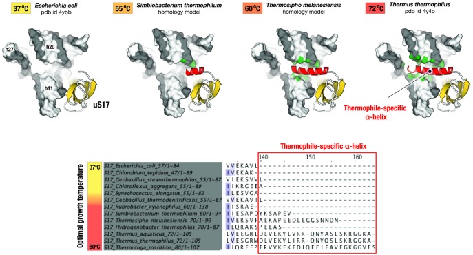
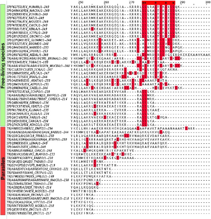
Ribosomal proteins are indispensable components of a living cell, and yet their structures are remarkably diverse in different species. Here we use manually curated structural alignments to provide a comprehensive catalog of structural variations in homologous ribosomal proteins from bacteria, archaea, eukaryotes, and eukaryotic organelles. By resolving numerous ambiguities and errors of automated structural and sequence alignments, we uncover a whole new class of structural variations that reside within seemingly conserved segments of ribosomal proteins. We then illustrate that these variations reflect an apparent adaptation of ribosomal proteins to the specific environments and lifestyles of living species. Finally, we show that most of these structural variations reside within nonglobular extensions of ribosomal proteins-protein segments that are thought to promote ribosome biogenesis by stabilizing the proper folding of ribosomal RNA. We show that although the extensions are thought to be the most ancient peptides on our planet, they are in fact the most rapidly evolving and most structurally and functionally diverse segments of ribosomal proteins. Overall, our work illustrates that, despite being long considered as slowly evolving and highly conserved, ribosomal proteins are more complex and more specialized than is generally recognized.


Figures

<sc>Fig</sc>. 1.
Fig. 1.
Homologous ribosomal proteins have highly diversified structure across the three domains of life. The diagram provides an overview of structural variability in 33 conserved proteins from 45 different species in the three domains of life. Each bar indicates the total number of amino acids in the 33 proteins in each species. The bars are colored to show the number of residues that form either the structurally invariable core (pink) or the variable protein segments (red). As the diagram shows, conserved ribosomal proteins carry nearly as many residues in structurally conserved protein segments as they carry in protein segments with distinct structure in different domains of life, suggesting a high degree of functional specialization of ribosomal proteins across the three domains of life.
<sc>Fig</sc>. 2.
Fig. 2.
Homologous ribosomal proteins have largely conserved globular domains but highly divergent nonglobular extensions. Aligned structures of homologous ribosomal proteins are shown as they appear in bacterial, mitochondrial, archaeal, and eukaryotic ribosomes (pdb ids 4y4b, 3j9m, 4v4n, and 4v6x, respectively). Proteins are colored according to structural conservation: segments that have identical tertiary structure in all four protein homologs are shown in gray; protein segments that have unique tertiary structure or occurrence only in one of the four homologs are shown in blue; protein segments that have unique tertiary structure or occurrence only in bacterial and mitochondrial proteins (labeled as BM) or only in archaeal and eukaryotic proteins are shown in yellow (labeled as AE). The segments are labeled with “B,” “M,” “A,” and “E” to indicate that a protein segment have unique structure or occurrence in bacterial, mitochondrial, archaeal, and eukaryotic proteins, respectively; the segments are numbered as they appear in each protein from its N- to the C-terminus. Apart from common protein names, each protein is named according to its name in bacterial, archaeal, and eukaryotic species. The panel illustrates that, despite homologous ribosomal proteins having comparable size across species, many segments in these proteins have dissimilar secondary and tertiary structure in different domains of life.
<sc>Fig</sc>. 3.
Fig. 3.
Variations in protein globules in an apparent adaptation to ribosomal RNA (rRNA) expansion. The figure compares structures of Haloarcula marismortui and Sacromyces cerevisiae ribosomes. It illustrates how transition from prokaryotes to eukaryotes was accompanied with the formation of a novel secondary structure in cytosol-exposed ribosomal proteins. (A) Views on the large ribosomal subunits illustrate that upon the transition from prokaryotes to eukaryotes, ribosomes have markedly increased in size, and ribosomal proteins uL2 and uL13 (in red) that were exposed on the surface of prokaryotic ribosome became buried in the interior of the eukaryotic ribosome. In eukaryotes, uL2 and uL13 are associated with eukaryote-specific rRNA expansion segments, ES31 and ES7 (in blue). (B) Close-up views on uL13 and uL2 show that the prokaryote-to-eukaryote transition was accompanied with secondary structure transformation in which surface-exposed protein loops of uL2 and uL13 were remodeled into rRNA-binding helices. Names in parenthesis show protein names in H. marismortui and S. cerevisiae.
<sc>Fig</sc>. 4.
Fig. 4.
Variations in protein structures within one domain of life as a possible adaptation to extreme environments. The figure illustrates structural variations in homologous ribosomal proteins upon transition from mesophilic to thermophilic species. The species used for comparison are arranged according to their optimal growth temperature. Fragments of Escherichia coli and Thermus thermophilus ribosome structures and homology models of Symbiobacterium thermophilum and Thermosipho melanesiensis ribosomes illustrate that, in thermophilic species, ribosomal protein uS17 develops an additional C-terminal helix (in red). This helix creates a new RNA–protein interface and stabilizes the RNA fold in the three-way helical junction in the 16S rRNA. Remarkably, as the optimal growth temperature for a given species gets higher, this helix gets progressively longer, and its apparent contacts with rRNA get more extensive (highlighted in green). This example shows that structure of some ribosomal proteins appear to evolutionary respond to higher temperatures by increasing the size of nonglobular extensions to establish new protein–rRNA or protein–protein contacts within the ribosome.
<sc>Fig</sc>. 5.
Fig. 5.
Variations in protein eS6 as a possible adaptation to specific lifestyles. The figure shows a multiple sequence alignment for eukaryotic ribosomal protein eS6 (C-terminal fragment). The C-terminal eS6 segment endows ribosomes with sensitivity to nutrients: it harbors serine residues (highlighted by asterisks) that are phosphorylated in response to hormones and nutrient availability to readjust the overall rate of protein synthesis in a eukaryotic cell. The figure shows that the phosphorylation sites remain conserved in free-living species but are degenerated in parasites, suggesting the lack of a nutrient sensor within parasitic ribosomes.

References

    1. Agrawal RK, Sharma MR, Yassin A, Lahiri I, Spremulli LL (2011). Structure and function of organellar ribosomes as revealed by cryo-EM In: Rodnina MV, Wintermeyer W, Green R, editors. Ribosomes: structure, function, and dynamics. Springer Link.
    1. Ahmed T, Shi J, Bhushan S.. 2017. Unique localization of the plastid-specific ribosomal proteins in the chloroplast ribosome small subunit provides mechanistic insights into the chloroplastic translation. Nucleic Acids Res. 4514:8581–8595. - PMC - PubMed
    1. Amunts A, Brown A, Toots J, Scheres SHW, Ramakrishnan V.. 2015. The structure of the human mitochondrial ribosome. Science 3486230:95–98. - PMC - PubMed
    1. Anger AM, Armache JP, Berninghausen O, Habeck M, Subklewe M, Wilson DN, Beckmann R.. 2013. Structures of the human and Drosophila 80S ribosome. Nature 4977447:80–85. - PubMed
    1. Armache JP, Anger AM, Marquez V, Franckenberg S, Frohlich T, Villa E, Berninghausen O, Thomm M, Arnold GJ, Beckmann R.. 2013. Promiscuous behaviour of archaeal ribosomal proteins: implications for eukaryotic ribosome evolution. Nucleic Acids Res. 412:1284–1293. - PMC - PubMed

Publication types

Substances

LinkOut - more resources