

Can Navigation-assisted Surgery Help Achieve Negative Margins in Resection of Pelvic and Sacral Tumors?
- PMID: 29529631
- PMCID: PMC6260048
- DOI: 10.1007/s11999.0000000000000064
Can Navigation-assisted Surgery Help Achieve Negative Margins in Resection of Pelvic and Sacral Tumors?
Abstract
Background: Navigation-assisted resection has been proposed as a useful adjunct to resection of malignant tumors in difficult anatomic sites such as the pelvis and sacrum where it is difficult to achieve tumor-free margins. Most of these studies are case reports or small case series, but these reports have been extremely promising. Very few reports, however, have documented benefits of navigation-assisted resection in series of pelvic and sacral primary tumors. Because this technology may add time and expense to the surgical procedure, it is important to determine whether navigation provides any such benefits or simply adds cost and time to an already complex procedure.
Questions/purposes: (1) What proportion of pelvic and sacral bone sarcoma resections utilizing a computer-assisted resection technique achieves negative margins? (2) What are the oncologic outcomes associated with computer-assisted resection of pelvic and sacral bone sarcomas? (3) What complications are associated with navigation-assisted resection?
Methods: Between 2009 and 2015 we performed 24 navigation-assisted resections of primary tumors of the pelvis or sacrum. Of those, four were lost to followup after the 2-year postoperative visit. In one patient, however, there was a failure of navigation as a result of inadequate imaging, so nonnavigated resection was performed; the remaining 23 were accounted for and were studied here at a mean of 27 months after surgery (range, 12-52 months). During this period, we performed navigation-assisted resections in all patients presenting with a pelvis or sacral tumor; there was no selection process. No patients were treated for primary tumors in these locations without navigation during this time with the exception of the single patient in whom the navigation system failed. We retrospectively evaluated the records of these 23 patients and evaluated the margin status of these resections. We calculated the proportion of patients with local recurrence, development of metastases, and overall survival at an average 27-month followup (range, 12-52 months). We queried a longitudinally maintained surgical database for any complications and noted which, if any, could have been directly related to the use of the navigation-assisted technique.
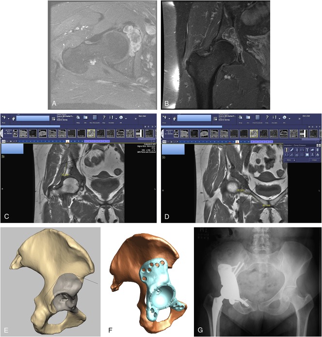
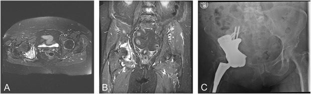
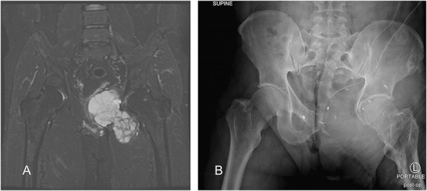
Results: In our series, 21 of 23 patients had a negative margin resection. In all patients the bone margin was negative, but two with sacral resections had positive soft tissue margins. Six of 23 patients experienced local recurrence within the study period. Three patients died during the study period. Seventeen patients demonstrated no evidence of disease at last recorded followup. We noted three intraoperative complications: one dural tear, one iliac vein laceration, and one bladder injury. Eight patients out of 23 had wound complications resulting in operative débridement. Two patients in the series developed transient postoperative femoral nerve palsy, which we believe were caused by stretch of the femoral nerve secondary to the placement of the reference array in the pubic ramus.
Conclusions: Navigation-assisted resection of pelvic and sacral tumors resulted in a high likelihood of negative margin resection in this series, and we observed relatively few complications related specifically to the navigation. We have no comparison group without navigation, and future studies should indeed compare navigated with nonnavigated resection approaches in these anatomic locations. We did identify a potential navigation-related complication of femoral nerve palsy in this series and suggest careful placement and observation of the reference array during the operative procedure to lessen the likelihood of this previously unreported complication. We suggest it is worthwhile to consider the use of navigation-assisted surgery in resection of tumors of the pelvis and sacrum, but further study will be needed to determine its precise impact, if any, on local recurrence and other oncologic outcomes.
Level of evidence: Level IV, therapeutic study.
Conflict of interest statement
Each author certifies that neither he, nor any member of his immediate family, have funding or commercial associations (consultancies, stock ownership, equity interest, patent/licensing arrangements, etc) that might pose a conflict of interest in connection with the submitted article.
All ICMJE Conflict of Interest Forms for authors and
Figures
Comment in
-
CORR Insights®: Can Navigation-assisted Surgery Help Achieve Negative Margins in Resection of Pelvic and Sacral Tumors?Clin Orthop Relat Res. 2018 Mar;476(3):509-510. doi: 10.1007/s11999.0000000000000145. Clin Orthop Relat Res. 2018. PMID: 29529632 Free PMC article. No abstract available.
-
Editorial on "Can navigation-assisted surgery help achieve negative margins in resection of pelvic and sacral tumor?".J Spine Surg. 2018 Sep;4(3):681-683. doi: 10.21037/jss.2018.07.13. J Spine Surg. 2018. PMID: 30547139 Free PMC article. No abstract available.
References
-
- Cartiaux O, Docquier PL, Paul L, Francq BG, Cornu OH, Delloye C, Roucent B, Dehez B, Banse X. Surgical inaccuracy of tumor resection and reconstruction within the pelvis: an experimental study. Acta Orthop. 2008;79:695–702. - PubMed
-
- Cho HS, Kang HG, Kim HS, Han I. Computer-assisted sacral tumor resection. A case report. J Bone Joint Surg Am. 2008;90:1561–1566. - PubMed
-
- Cho HS, Oh JH, Han I, Kim HS. Joint-preserving limb salvage surgery under navigation guidance. J Surg Oncol. 2009;100:227–232. - PubMed
-
- Cho HS, Oh JH, Han I, Kim HS. The outcomes of navigation-assisted bone tumour surgery: minimum three-year follow-up. J Bone Joint Surg Br. 2012;94:1414–1420. - PubMed
-
- Enneking WE, Dunham WK. Resection and reconstruction for primary neoplasms involving the innominate bone. J Bone Joint Surg Am. 1978;60:731–746. - PubMed
MeSH terms
LinkOut - more resources
Full Text Sources
Other Literature Sources
