

Multiple midline defects identified in a litter of golden retrievers following gestational administration of prednisone and doxycycline: a case series
- PMID: 29530019
- PMCID: PMC5848590
- DOI: 10.1186/s12917-018-1419-y
Multiple midline defects identified in a litter of golden retrievers following gestational administration of prednisone and doxycycline: a case series
Abstract
Background: The teratogenic effects of immunomodulatory and certain antimicrobial therapies are described in small rodents and humans. While the described teratogenic effects in small rodents have been extrapolated to make conclusions about its use in the pregnant dam, teratogenic effects of prednisone and doxycycline have not yet been reported in the dog. Here we report and describe midline defects observed in a litter of golden retriever puppies exposed to mid-gestational immunosuppressive and antimicrobial therapy.
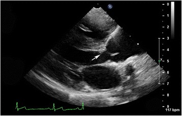
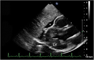
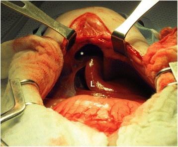
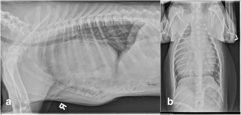
Case presentation: Twenty-one days into gestation, the dam of a litter of eight golden retriever puppies was administered prednisone, doxycycline, and tramadol as treatment for immune-mediated polyarthritis. The individuals in the litter were subsequently diagnosed with a variety of midline defects and congenital cardiac defects. This case series describes the variety of identified defects and presents a descriptive account of complex congenital abnormalities that are likely secondary to teratogenic effects of one or more drugs administered during gestation. The available puppies, dam, and grand dam underwent thorough physical examination, complete echocardiogram, and where indicated, advanced imaging with various surgical corrections when possible. Numerous midline congenital defects and congenital heart disease were identified in the puppies evaluated. Ultimately 5 of 8 puppies born to the dam were presented for thorough evaluation. The midline defects include: gastroschisis (1), peritoneopericardial diaphragmatic hernias (4, PPDH), umbilical hernia (4), unilateral cryptorchidism (1 of 4 males), cleft palate (1), renal agenesis (1), renal abnormalities (1), sternal and vertebral abnormalities (3), remnant liver lobe (1) and malformations consistent with ductal plate malformations with congenital hepatic fibrosis (1). The congenital cardiac defects include: ventricular septal defect (4, VSD) and subaortic stenosis (4, SAS). The presence of greater than one congenital defect was noted in all 5 of the dogs evaluated. Surgical correction was necessary for PPDH in 4 puppies. Medical intervention was recommended for congenital cardiac disease in 1 puppy.
Conclusion: This case report is the first to describe midline defects in dogs that have been exposed to immunomodulatory therapy during gestation. A causative relationship between mid-gestational immunomodulatory exposure and midline defects cannot be proven, however, this case supports a clear association and provides case-based evidence to support its avoidance when possible.
Keywords: Congenital heart defect; Gastroschisis; PPDH; Peritoneopericardial diaphragmatic hernia; Teratogen.
Conflict of interest statement
Ethics approval
Informed client consent for all participants in this case series was obtained upon admission to the hospital and documented in accordance with the Institutional Animal Care and Use Committee of the University of California Davis. Medical care was provided in accordance with the best practice guidelines for veterinary care.
Consent for publication
Informed client consent for publication was obtained upon admission to the hospital.
Competing interests
The authors declare that they have no competing interests.
Publisher’s Note
Springer Nature remains neutral with regard to jurisdictional claims in published maps and institutional affiliations.
Figures
References
-
- Radlinsky MG. Surgery of the digestive system. In: Fossum TW, Dewey CW, Horn CV, Johnson AL, MacPhail CM, Radlinsky MG, Schulz KS, Willard MD, editors. Small animal surgery. 4. Philadelphia: Elsevier; 2013. pp. 502–504.
-
- Evans SM, Biery DN. Congenital peritoneopericardial diaphragmatic hernia in the dog and cat: a literature review and 17 additional case histories. Vet Radiol. 1980;21:108–116. doi: 10.1111/j.1740-8261.1980.tb00589.x. - DOI
Publication types
MeSH terms
Substances
LinkOut - more resources
Full Text Sources
Other Literature Sources
Medical
