

Stearoyl-CoA desaturase-1 promotes colorectal cancer metastasis in response to glucose by suppressing PTEN
- PMID: 29530061
- PMCID: PMC5848567
- DOI: 10.1186/s13046-018-0711-9
Stearoyl-CoA desaturase-1 promotes colorectal cancer metastasis in response to glucose by suppressing PTEN
Erratum in
-
Correction: Stearoyl-CoA desaturase-1 promotes colorectal cancer metastasis in response to glucose by suppressing PTEN.J Exp Clin Cancer Res. 2025 Feb 1;44(1):34. doi: 10.1186/s13046-025-03300-2. J Exp Clin Cancer Res. 2025. PMID: 39891133 Free PMC article. No abstract available.
Abstract
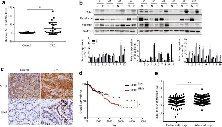
Background: Diabetic patients have a higher risk factor for colorectal cancer (CRC) metastasis. Stearoyl-CoA desaturase 1 (SCD1), the main enzyme responsible for producing monounsaturated fatty acids(MUFA) from saturated fatty acids, is frequently deregulated in both diabetes and CRC. The function and mechanism of SCD1 in metastasis of CRC and its relevance to glucose remains largely unknown.
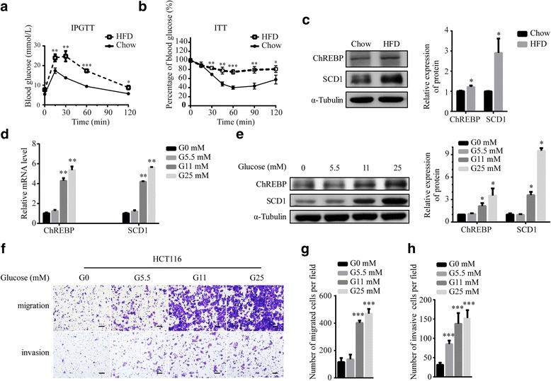
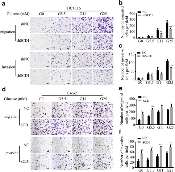
Methods: SCD1 expression levels were analyzed in human CRC tissues and the Cancer Browser database ( https://genome-cancer.ucsc.edu/ ). CRC cell lines stably transfected with SCD1 shRNAs or vector were established to investigate the role of SCD1 in modulating migration and invasion of CRC cells. A glucose concentration gradient was set to investigate regulation of SCD1 in CRC relevant to diabetic conditions.
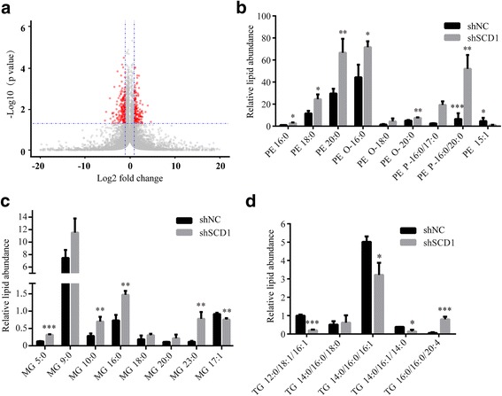
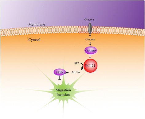
Results: The clinical data analysis showed high expression of SCD1 in CRC tissues with a negative correlation with the prognosis of CRC. In vitro experiments revealed that SCD1 increased CRC progression through promoting epithelial-mesenchymal transition (EMT). Lipidomic analysis demonstrated that SCD1 increased MUFA levels and MUFA administration could rescue migration and invasion defect of CRC cells induced by SCD1 knockdown. Furthermore, SCD1-mediated progression of CRC was promoted by carbohydrate response-element binding protein (ChREBP) in response to high glucose. Mechanistically, hyperglycemia-SCD1-MUFA induced CRC cell migration and invasion by regulating PTEN.
Conclusions: Our findings show that SCD1 promotes metastasis of CRC cells through MUFA production and suppressing PTEN in response to glucose, which may be a novel mechanism for diabetes-induced CRC metastasis.
Keywords: ChREBP; Colorectal cancer; High glucose; Metastasis; PTEN; SCD1.
Conflict of interest statement
Competing interests
The authors declare that they have no competing interests.
Publisher’s Note
Springer Nature remains neutral with regard to jurisdictional claims in published maps and institutional affiliations.
Figures


Similar articles
-
Stearoyl-CoA desaturase 1 inhibition impairs triacylglycerol accumulation and lipid droplet formation in colorectal cancer cells.J Cell Physiol. 2023 Dec;238(12):2888-2903. doi: 10.1002/jcp.31137. Epub 2023 Oct 10. J Cell Physiol. 2023. PMID: 37814830
-
Loss of mitochondrial aconitase promotes colorectal cancer progression via SCD1-mediated lipid remodeling.Mol Metab. 2021 Jun;48:101203. doi: 10.1016/j.molmet.2021.101203. Epub 2021 Mar 3. Mol Metab. 2021. PMID: 33676027 Free PMC article.
-
Clinical and biochemical relevance of monounsaturated fatty acid metabolism targeting strategy for cancer stem cell elimination in colon cancer.Biochem Biophys Res Commun. 2019 Oct 29;519(1):100-105. doi: 10.1016/j.bbrc.2019.08.137. Epub 2019 Aug 31. Biochem Biophys Res Commun. 2019. PMID: 31481234
-
Stearoyl-CoA desaturase 1 as a therapeutic target for cancer: a focus on hepatocellular carcinoma.Mol Biol Rep. 2022 Sep;49(9):8871-8882. doi: 10.1007/s11033-021-07094-2. Epub 2022 Jul 29. Mol Biol Rep. 2022. PMID: 35906508 Review.
-
Stearoyl-CoA desaturase 5 (SCD5), a Δ-9 fatty acyl desaturase in search of a function.Biochim Biophys Acta Mol Cell Biol Lipids. 2021 Jan;1866(1):158840. doi: 10.1016/j.bbalip.2020.158840. Epub 2020 Oct 10. Biochim Biophys Acta Mol Cell Biol Lipids. 2021. PMID: 33049404 Free PMC article. Review.
Cited by
-
Correlation between Expression Profiles of Key Signaling Genes in Colorectal Cancer Samples from Type 2 Diabetic and Non-Diabetic Patients.Life (Basel). 2020 Sep 22;10(9):216. doi: 10.3390/life10090216. Life (Basel). 2020. PMID: 32971867 Free PMC article.
-
A more physiological approach to lipid metabolism alterations in cancer: CRC-like organoids assessment.PLoS One. 2019 Jul 24;14(7):e0219944. doi: 10.1371/journal.pone.0219944. eCollection 2019. PLoS One. 2019. PMID: 31339921 Free PMC article.
-
Crosstalk between fatty acid metabolism and tumour-associated macrophages in cancer progression.Biomedicine (Taipei). 2022 Dec 1;12(4):9-19. doi: 10.37796/2211-8039.1381. eCollection 2022. Biomedicine (Taipei). 2022. PMID: 36816174 Free PMC article. Review.
-
Lipid mechanisms in hallmarks of cancer.Mol Omics. 2020 Feb 17;16(1):6-18. doi: 10.1039/c9mo00128j. Mol Omics. 2020. PMID: 31755509 Free PMC article. Review.
-
Immunoendocrine Peripheral Effects Induced by Atypical Antipsychotics.Front Endocrinol (Lausanne). 2020 Apr 21;11:195. doi: 10.3389/fendo.2020.00195. eCollection 2020. Front Endocrinol (Lausanne). 2020. PMID: 32373066 Free PMC article. Review.
References
MeSH terms
Substances
Grants and funding
LinkOut - more resources
Full Text Sources
Other Literature Sources
Medical
Research Materials
