Review
. 2018 Oct 1;8(10):a031781.
doi: 10.1101/cshperspect.a031781.

Hepatitis A Virus Codon Usage: Implications for Translation Kinetics and Capsid Folding

Affiliations
Review

Hepatitis A Virus Codon Usage: Implications for Translation Kinetics and Capsid Folding

Rosa M Pintó et al. Cold Spring Harb Perspect Med. .

Abstract

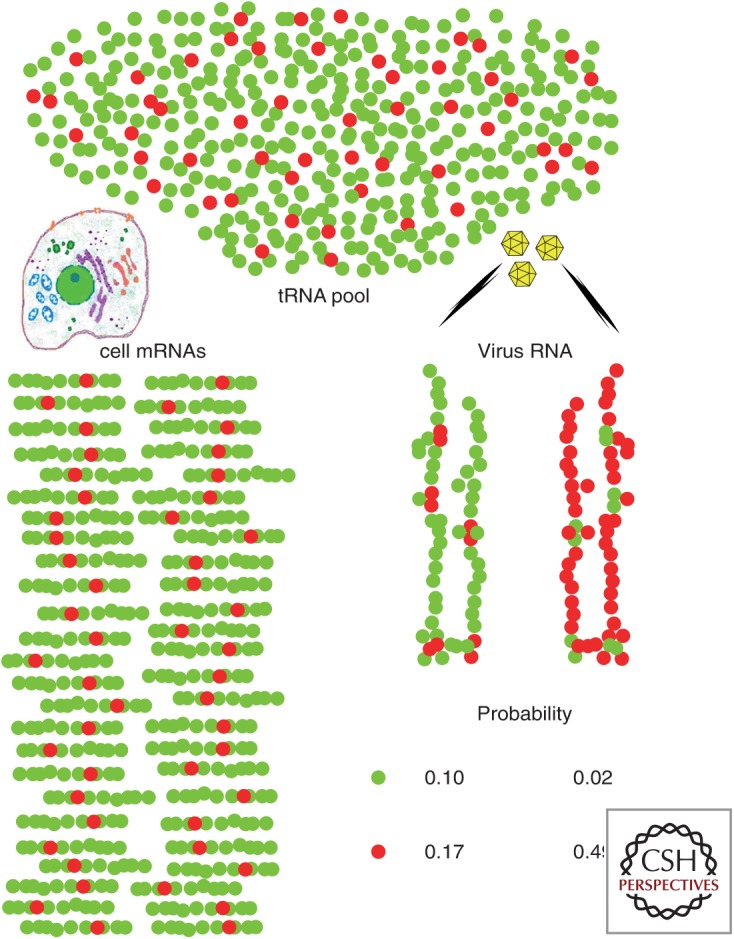
Codon usage bias is universal to all genomes. Hepatitis A virus (HAV) codon usage is highly biased and deoptimized with respect to its host. Accordingly, HAV is unable to induce cellular translational shutoff and its internal ribosome entry site (IRES) is inefficient. Codon usage deoptimization may be seen as a hawk (host cell) versus dove (HAV) game strategy for accessing transfer RNA (tRNA). HAV avoids use of abundant host cell codons and thereby eludes competition for the corresponding tRNAs. Instead, codons that are abundant or rare in cellular messenger RNAs (mRNAs) are used relatively rarely in its genome, although intermediately abundant host cell codons are abundant in the viral genome. Rare codons in the capsid coding region slow down the translation elongation rate, and in doing so intrinsically modulate capsid folding, which is critical to the stability of a virus transmitted through the fecal-oral route. HAV is a paradigmatic example of what has been proposed as a codon usage "code" for protein structure.


Figures

Figure 1.
Figure 1.
Probability that hepatitis A virus (HAV) RNA will pair successfully with the abundant or nonabundant transfer RNAs (tRNAs) required for its translation under two different codon usage simulations, considered within the framework of a very simplistic model for cellular codon usage involving only a single amino acid encoded by either a major abundant codon (green balls) or a minor rare codon (red balls). Three assumptions are made: (1) HAV is a very slowly replicating virus (inefficient internal ribosome entry site [IRES]) with few, but very lengthy translating RNAs compared to cellular messenger RNAs (mRNAs) (the model assumes one translating HAV RNA for every 50 cellular mRNAs per cell, and that it is 6× longer than the average cellular mRNA), (2) HAV is unable to shut down cellular protein synthesis, and (3) tRNA pools are limiting and adapted to the cellular mRNAs. In a hypothetical situation in which HAV adopts the same codon usage as the cell (optimized: viral RNA on the left), the probabilities of tRNA pairing with the most abundant codon and tRNA pairing with the less abundant codon are 0.1 and 0.17, respectively. In contrast, with codon usage opposite with respect to the cell (deoptimized: viral RNA on the right), these probabilities are 0.02 and 0.49. The latter scenario provides an overall advantageous outcome for the virus, with a higher probability of getting tRNAs pairing with its most abundant codons, while accomplishing the goal of using incidentally very low abundance tRNAs to slow down translation at certain positions.
Figure 2.
Figure 2.
Codon usage and its relationship to the rate of translation and replication capacities of two variants derived from the HM175 strain of hepatitis A virus (HAV) (HM175-43c: L0 parental type; HM175-HP: HP fast-growing type) that differ in their level of codon optimization. (A) Relative codon deoptimization index (RCDI) of the L0 and HP strains with respect to the human codon usage. RCDI values shown correspond to overlapping 100 codon segments of the capsid-coding RNA with 15 codon-sliding windows. The higher the RCDI value, the higher the deviation of viral codon usage from host cell codon usage. The HP strain shows a remarkable decrease in RCDI values in a region extending 50% of the VP1 length, compared to the L0 strain, which denotes more optimized codon usage relative to the cell. (B) Rate of translation elongation of the VP1 fragment showing differences in the RCDI between L0 and HP strains. The elongation rate is measured as the relative FLuc/RLuc activity, using a bicistronic vector in which Renilla luciferase (RLuc) translation is cap dependent and Firefly luciferase (FLuc) translation is HAV internal ribosome entry site (IRES) dependent. The VP1 fragment is cloned under control of the IRES just upstream of and in frame with FLuc. Translation elongation was assayed using two bicistronic vectors representing the parental-type IRES (L0-type IRES: two first points of the kinetics) and the mutated-active-type internal ribosome entry site (IRES) (HP-type IRES: two last points of the kinetics), under conditions of no shutoff (–) or shutoff (+). Results are expressed as fold change of the elongation rate relative to the L0 population with the parental-type IRES and in the absence of shutoff. (C) Box plots of the virus yields obtained in FRhK-4 cells. (D) Box plots of virus yields obtained in HuH7-AI cells. In both C and D, the multiplicity of infection was 1. Dotted lines represent mean titer.
Figure 3.
Figure 3.
Cellular shutoff induces a ripple effect on codon usage optimization and the acquisition of internal ribosome entry site (IRES) mutations. In the absence of cellular shutoff with high cellular messenger RNA (mRNA) synthesis (top part of the figure), hepatitis A virus (HAV) shows a very inefficient IRES, a deoptimized codon usage, and very low yields of highly stable capsids. Under conditions of cellular shutoff with a decreased cellular mRNA synthesis (bottom part of the figure), HAV shows an optimized codon usage, a more active IRES, and produces high yields of capsids with subtle folding changes.
Figure 4.
Figure 4.
Hepatitis A virus (HAV) protomer (Wang et al. 2015) showing residues encoded by rare codons (white), and residues replaced in VP3 and VP1 in quasispecies placed under the immune pressure of monoclonal antibodies (mAbs) (yellow). Although residues undergoing replacement are located very close to residues encoded by rare codons, there is a general lack of coincidence with only few matching positions (clear blue). (A) Selection with H7C27 mAb. (B) Selection with K34C8 mAb.

References

    1. Abad FX, Pintó RM, Bosch A. 1994. Survival of enteric viruses on environmental fomites. Appl Environ Microbiol 60: 3704–3710. - PMC - PubMed
    1. Ali IK, McKendrick L, Morley SJ, Jackson RJ. 2001. Activity of the hepatitis A virus IRES requires association between the cap-binding translation initiation factor (eIF4E) and eIF4G. J Virol 75: 7854–7863. - PMC - PubMed
    1. Anthony SJ, St Leger JA, Liang E, Hicks AL, Sanchez-Leon MD, Jain K, Lefkowitch JH, Navarrete-Macias I, Knowles N, Goldstein T, et al. 2015. Discovery of a novel hepatovirus (Phopivirus of seals) related to human hepatitis A virus. mBio 6: e01180–15. - PMC - PubMed
    1. Aragonès L, Bosch A, Pintó RM. 2008. Hepatitis A virus mutant spectra under the selective pressure of monoclonal antibodies: Codon usage constraints limit capsid variability. J Virol 82: 1688–1700. - PMC - PubMed
    1. Aragonès L, Guix S, Ribes E, Bosch A, Pintó RM. 2010. Fine-tuning translation kinetics selection as the driving force of codon usage bias in the hepatitis A virus capsid. PLoS Pathog 6: e1000797. - PMC - PubMed

Publication types

LinkOut - more resources