Review
. 2018 Sep 4;8(9):a031880.
doi: 10.1101/cshperspect.a031880.

Classification and Genomic Diversity of Enterically Transmitted Hepatitis Viruses

Affiliations
Review

Classification and Genomic Diversity of Enterically Transmitted Hepatitis Viruses

Donald B Smith et al. Cold Spring Harb Perspect Med. .

Abstract

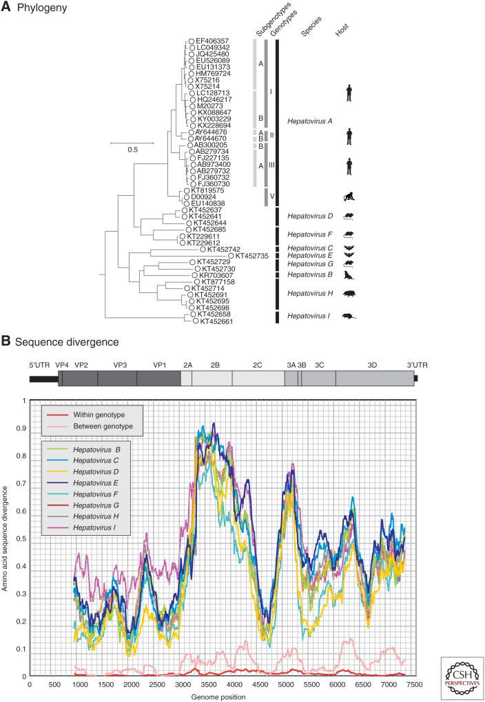
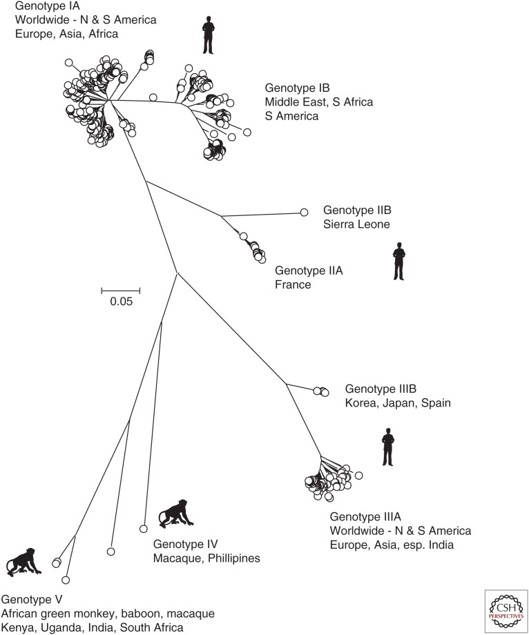
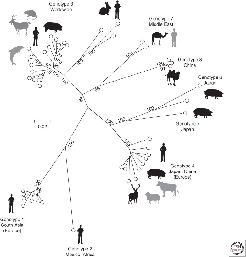
Hepatitis A virus (HAV) and hepatitis E virus (HEV) are significant human pathogens and are responsible for a substantial proportion of cases of severe acute hepatitis worldwide. Genetically, both viruses are heterogeneous and are classified into several genotypes that differ in their geographical distribution and risk group association. There is, however, little evidence that variants of HAV or HEV differ antigenically or in their propensity to cause severe disease. Genetically more divergent but primarily hepatotropic variants of both HAV and HEV have been found in several mammalian species, those of HAV being classified into eight species within the genus Hepatovirus in the virus family Picornaviridae. HEV is classified as a member of the species Orthohepevirus A in the virus family Hepeviridae, a species that additionally contains viruses infecting pigs, rabbits, and a variety of other mammalian species. Other species (Orthohepevirus B-D) infect a wide range of other mammalian species including rodents and bats.


Figures

Figure 1.
Figure 1.
Phylogeny and sequence divergence of hepatitis A viruses (HAVs). (A) Phylogenetic analysis of the complete coding sequences of representative variants of HAV (species A) and of all available sequences of other nonhuman hepatoviruses infecting other mammalian species, labeled as in the key. (The coding region spans positions 723–7407 in the HAV-MBB sequences, M20273 [Paul et al. 1987].) The tree was constructed by maximum likelihood using an optimal substitution model (general time reversible and γ distribution). Robustness of branches was indicated by bootstrap resampling supported by ≥70 from 100 replicate samples. The tree was rooted using sequences from the most closely similar genus of picornaviruses (Tremovirus: KT880668, AY275539, KF979338, AJ225173, and AY517471). The host origins of the different species are indicated diagrammatically and correspond to the following species: Hepatovirus B, seals (Phoca vitulina vitulina); Hepatovirus C, bats (Miniopterus cf. manavi); Hepatovirus D, rodents (Microtus arvalis, Myodes glareolus); Hepatovirus E, bats (Lophuromys sikapusi); Hepatovirus F, rodents (Marmota himalayana, Sigmodon mascotensis); Hepatovirus G, bats (Rhinolophus landeri, Coleura afra); Hepatovirus H, hedgehogs, tupias, and bats (Erinaceus europaeus, Tupaia belangeri chinensis, Eidolon helvum); and Hepatovirus I, shrews (Sorex araneus). (B) Scan of amino acid sequence divergence between HAV and hepatovirus species B–I, and comparison with between and among genotype divergence of HAV (red and pink lines). The distances represent mean values for available sequences of each species and were calculated for sequential fragments spanning >90 codons, incrementing by nine codons between data points.
Figure 2.
Figure 2.
Unrooted phylogenetic analysis of the 625 available hepatitis A virus (HAV) sequences in the VP1 region. Positions 2220–3234 numbered as in Fig. 1. The tree was constructed by neighbor joining of maximum composite likelihood distances as implemented in the program, MEGA6 (Tamura et al. 2013). Bootstrap resampling was performed as described in Figure 1. Sequences were selected for analysis based on being >90% complete in the VP1 coding region and lacking internal stop codons.
Figure 3.
Figure 3.
Phylogeny and sequence divergence of Orthohepevirus species. (A) Phylogenetic relationships of Orthohepevirus species. A conserved region of the capsid protein (nucleotide positions 5485–6497, numbered according to M73218) was aligned for representative isolates and the amino acid sequence used to generate a maximum likelihood tree based on the LG model with a γ distribution of evolutionary rates among sites as implemented in the program MEGA6 (Tamura et al. 2013). Branches supported in >70% of bootstrap replicates are indicated. Genotypes, virus species, and host species are indicated. (B) Scan of amino acid diversity across the Orthohepevirus genome. The sequences used to produce Figure 3A were analyzed as described in Figure 1B.
Figure 4.
Figure 4.
Unrooted phylogenetic analysis of coding sequences of representative members of Orthohepevirus A. Concatenated open reading frame (ORF)1 and ORF2 coding sequences (excluding the hypervariable region) were aligned and used to produce a maximum likelihood tree using a JTT model (with frequencies) and a γ distribution of rate differences among sites, including invariant sites as implemented in the program MEGA6 (Tamura et al. 2013). Branches supported in >70% of bootstrap replicates are indicated. Sequences used were 1a_M73218, 1b_D11092, 1c_X98292, 1d_AY230202, 1e_AY204877, 1f_JF443721, 1_FJ457024, 2a_M74506, 3a_AF082843, 3b_AP003430, 3c_FJ705359, 3e_AB248521, 3_EU360977, 3f_AB369687, 3_EU723513, 3_KJ873911, 3g_AF455784, 3h_JQ013794, 3i_FJ998008, 3j_AY115488, 3_AB290312, 3_AB369689, 3_JQ953664, 3_AB290313, 3ra_FJ906895, 3_JQ013791, 3_KJ013415, 4a_AB197673, 4b_DQ279091, 4c_AB074915, 4d_AJ272108, 4e_AY723745, 4f_AB220974, 4g_AB108537, 4h_GU119961, 4i_DQ450072, 4_AB369688, 5a_AB573435, 6a_AB602441, 6_AB856243, 7a_KJ496143, 7_KJ496144, 8_KX387865, 8_KX387866, and 8_KX387867. Host species are indicated by icons, with less frequent or uncertain species shaded in gray.

Similar articles

Cited by

References

    1. Abravanel F, Lhomme S, El Costa H, Schvartz B, Peron JM, Kamar N, Izopet J. 2017. Rabbit hepatitis E virus infections in humans, France. Emerg Infect Dis 23: 1191–1193. - PMC - PubMed
    1. Anthony SJ, St Leger JA, Liang E, Hicks AL, Sanchez-Leon MD, Jain K, Lefkowitch JH, Navarrete-Macias I, Knowles N, Goldstein T, et al. 2015. Discovery of a novel hepatovirus (Phopivirus of Seals) related to human hepatitis A virus. mBio 6: e01180. - PMC - PubMed
    1. Arankalle VA, Ramakrishnan J. 2009. Simian hepatitis A virus derived from a captive rhesus monkey in India is similar to the strain isolated from wild African green monkeys in Kenya. J Viral Hepat 16: 214–218. - PubMed
    1. Arankalle VA, Chobe LP, Joshi MV, Chadha MS, Kundu B, Walimbe AM. 2002. Human and swine hepatitis E viruses from Western India belong to different genotypes. J Hepatol 36: 417–425. - PubMed
    1. Batts W, Yun S, Hedrick R, Winton J. 2011. A novel member of the family Hepeviridae from cutthroat trout (Oncorhynchus clarkii). Virus Res 158: 116–123. - PubMed

LinkOut - more resources