Crystal structure of a SFPQ/PSPC1 heterodimer provides insights into preferential heterodimerization of human DBHS family proteins
- PMID: 29530979
- PMCID: PMC5925804
- DOI: 10.1074/jbc.RA117.001451
Crystal structure of a SFPQ/PSPC1 heterodimer provides insights into preferential heterodimerization of human DBHS family proteins
Abstract
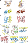
Members of the Drosophila behavior human splicing (DBHS) protein family are nuclear proteins implicated in many layers of nuclear functions, including RNA biogenesis as well as DNA repair. Definitive of the DBHS protein family, the conserved DBHS domain provides a dimerization platform that is critical for the structural integrity and function of these proteins. The three human DBHS proteins, splicing factor proline- and glutamine-rich (SFPQ), paraspeckle component 1 (PSPC1), and non-POU domain-containing octamer-binding protein (NONO), form either homo- or heterodimers; however, the relative affinity and mechanistic details of preferential heterodimerization are yet to be deciphered. Here we report the crystal structure of a SFPQ/PSPC1 heterodimer to 2.3-Å resolution and analyzed the subtle structural differences between the SFPQ/PSPC1 heterodimer and the previously characterized SFPQ homodimer. Analytical ultracentrifugation to estimate the dimerization equilibrium of the SFPQ-containing dimers revealed that the SFPQ-containing dimers dissociate at low micromolar concentrations and that the heterodimers have higher affinities than the homodimer. Moreover, we observed that the apparent dissociation constant for the SFPQ/PSPC1 heterodimer was over 6-fold lower than that of the SFPQ/NONO heterodimer. We propose that these differences in dimerization affinity may represent a potential mechanism by which PSPC1 at a lower relative cellular abundance can outcompete NONO to heterodimerize with SFPQ.
Keywords: DBHS protein family; NONO; PSPC1; RNA binding protein; SFPQ; analytical ultracentrifugation; crystal structure; dimerization; multifunctional protein.
© 2018 by The American Society for Biochemistry and Molecular Biology, Inc.
Conflict of interest statement
The authors declare that they have no conflicts of interest with the contents of this article
Figures


Similar articles
-
A new crystal structure and small-angle X-ray scattering analysis of the homodimer of human SFPQ.Acta Crystallogr F Struct Biol Commun. 2019 Jun 1;75(Pt 6):439-449. doi: 10.1107/S2053230X19006599. Epub 2019 May 21. Acta Crystallogr F Struct Biol Commun. 2019. PMID: 31204691 Free PMC article.
-
Structural plasticity of the coiled-coil interactions in human SFPQ.Nucleic Acids Res. 2025 Jan 11;53(2):gkae1198. doi: 10.1093/nar/gkae1198. Nucleic Acids Res. 2025. PMID: 39698821 Free PMC article.
-
Structure of the heterodimer of human NONO and paraspeckle protein component 1 and analysis of its role in subnuclear body formation.Proc Natl Acad Sci U S A. 2012 Mar 27;109(13):4846-50. doi: 10.1073/pnas.1120792109. Epub 2012 Mar 13. Proc Natl Acad Sci U S A. 2012. PMID: 22416126 Free PMC article.
-
Role of RNA binding proteins of the Drosophila behavior and human splicing (DBHS) family in health and cancer.RNA Biol. 2024 Jan;21(1):1-17. doi: 10.1080/15476286.2024.2332855. Epub 2024 Mar 29. RNA Biol. 2024. PMID: 38551131 Free PMC article. Review.
-
Involvement of paraspeckle components in viral infections.Nucleus. 2024 Dec;15(1):2350178. doi: 10.1080/19491034.2024.2350178. Epub 2024 May 8. Nucleus. 2024. PMID: 38717150 Free PMC article. Review.
Cited by
-
NONO promotes hepatocellular carcinoma progression by enhancing fatty acids biosynthesis through interacting with ACLY mRNA.Cancer Cell Int. 2020 Aug 31;20:425. doi: 10.1186/s12935-020-01520-4. eCollection 2020. Cancer Cell Int. 2020. PMID: 32884448 Free PMC article.
-
RNA-Binding Proteins in the Regulation of Adipogenesis and Adipose Function.Cells. 2022 Jul 31;11(15):2357. doi: 10.3390/cells11152357. Cells. 2022. PMID: 35954201 Free PMC article. Review.
-
NONO, SFPQ, and PSPC1 promote telomerase recruitment to the telomere.Nat Commun. 2025 Jul 1;16(1):5769. doi: 10.1038/s41467-025-60924-w. Nat Commun. 2025. PMID: 40593584 Free PMC article.
-
Multilateral Bioinformatics Analyses Reveal the Function-Oriented Target Specificities and Recognition of the RNA-Binding Protein SFPQ.iScience. 2020 Jul 24;23(7):101325. doi: 10.1016/j.isci.2020.101325. Epub 2020 Jun 28. iScience. 2020. PMID: 32659723 Free PMC article.
-
RNA-binding protein NONO contributes to cancer cell growth and confers drug resistance as a theranostic target in TNBC.Theranostics. 2020 Jul 2;10(18):7974-7992. doi: 10.7150/thno.45037. eCollection 2020. Theranostics. 2020. PMID: 32724453 Free PMC article.
References
-
- Lee M., Sadowska A., Bekere I., Ho D., Gully B. S., Lu Y., Iyer K. S., Trewhella J., Fox A. H., and Bond C. S. (2015) The structure of human SFPQ reveals a coiled-coil mediated polymer essential for functional aggregation in gene regulation. Nucleic Acids Res. 43, 3826–3840 10.1093/nar/gkv156 - DOI - PMC - PubMed
Publication types
MeSH terms
Substances
Associated data
- Actions
- Actions
- Actions
- Actions
LinkOut - more resources
Full Text Sources
Other Literature Sources
Miscellaneous

