. 2018 Apr 27;293(17):6337-6348.
doi: 10.1074/jbc.RA117.000605. Epub 2018 Mar 12.

Impact of altered phosphorylation on loss of function of juvenile Parkinsonism-associated genetic variants of the E3 ligase parkin

Affiliations

Impact of altered phosphorylation on loss of function of juvenile Parkinsonism-associated genetic variants of the E3 ligase parkin

Jacob D Aguirre et al. J Biol Chem. .

Abstract

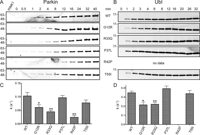
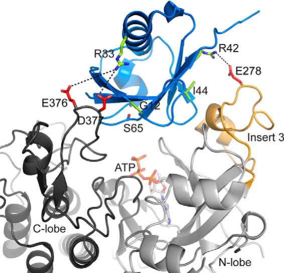
Autosomal recessive juvenile Parkinsonism (ARJP) is an inherited neurodegenerative disease in which 50% of affected individuals harbor mutations in the gene encoding the E3 ligase parkin. Parkin regulates the mitochondrial recycling pathway, which is induced by oxidative stress. In its native state, parkin is auto-inhibited by its N-terminal ubiquitin-like (Ubl) domain, which blocks the binding site for an incoming E2∼ubiquitin conjugate, needed for parkin's ubiquitination activity. Parkin is activated via phosphorylation of Ser-65 in its Ubl domain by PTEN-induced putative kinase 1 (PINK1) and a ubiquitin molecule phosphorylated at a position equivalent to Ser-65 in parkin. Here we have examined the underlying molecular mechanism of phosphorylation of parkin's Ubl domain carrying ARJP-associated substitutions and how altered phosphorylation modulates parkin activation and ubiquitination. We found that three substitutions in the Ubl domain (G12R, R33Q, and R42P) significantly decrease PINK1's ability to phosphorylate the Ubl domain. We noted that two basic loss-of-function substitutions (R33Q and R42P) are close to acidic patches in the proposed PINK1-parkin interface, indicating that ionic interactions at this site may be important for efficient parkin phosphorylation. Increased auto-ubiquitination with unique ubiquitin chain patterns was observed for two other Ubl domain substitutions (G12R and T55I), suggesting that these substitutions, along with phosphorylation, increase parkin degradation. Moreover, Ubl domain phosphorylation decreased its affinity for the potential effector protein ataxin-3, which edits ubiquitin chain building by parkin. Overall, our work provides a framework for the mechanisms of parkin's loss-of-function, indicating an interplay between ARJP-associated substitutions and phosphorylation of its Ubl domain.

Keywords: PTEN-induced putative kinase 1 (PINK1); Parkinson's disease (autosomal recessive, early onset) 7 (PARK7); parkin; protein folding; protein structure; protein–protein interaction; ubiquitylation (ubiquitination).


Conflict of interest statement

The authors declare that they have no conflicts of interest with the contents of this article

Figures

Figure 1.
Figure 1.
Schematic of the parkin Ser-65–phosphorylated Ubl domain. The secondary structure and the locations of substituted residues (blue) are shown. The position of phosphoserine 65 is also highlighted (red). This figure was produced using PDB coordinates from 5TR5.
Figure 2.
Figure 2.
PINK1-mediated phosphorylation of full-length parkin and Ubl domain monitored by Western blotting. A–D, each reaction monitored 1 μm parkin (A and C) or Ubl domain (B and D) using either 50 nm (Ubl) or 500 nm (parkin) PINK1 for the specified times. Detection utilized a pSer-65–specific antibody with DyLight680-conjugated secondary antibody. Fluorescence was imaged and quantified at 700 nm and plotted to determine relative rate constants (kobs) of phosphorylation.
Figure 3.
Figure 3.
A and B, time course phosphorylation assays for full-length parkin (A) and the Ubl domain (B) carrying early-onset Parkinson's disease substitutions. Relative rates of phosphorylation were determined as shown in Fig. 2 and as described under “Experimental procedures.” C and D, rates were calculated from global fits of three independent experiments ± S.E. (represented by error bars) and plotted for parkin (C) and the Ubl domain (D). Statistically significant deviations for each substituted protein with respect to WT protein are indicated (*, p ≤ 0.05; **, p ≤ 0.01), as determined by a two-tailed t test. The UblR42P protein was unfolded and insoluble and could not be examined.
Figure 4.
Figure 4.
Phosphorylation at Ser-65 uniformly decreases the stability of the parkin Ubl domain carrying PD substitutions. A–F, denaturation curves derived from urea-mediated unfolding experiments measured at 218 nm by CD spectropolarimetry. Shown are data for Ubl (●), pUbl (■), and UblS65E (□) (A); UblG12R (●) and pUblG12R (■) (B); UblR33Q (●) and pUblR33Q (■) (C); UblP37L (●) and pUblP37L (■) (D); UblT55I (●) and pUblT55I (■) (E); and UblI44A (●) (F). All data were fit as described under “Experimental procedures.”
Figure 5.
Figure 5.
Substitutions in the Ubl domain alter parkin autoubiquitination. The assay shows ubiquitination products monitored using fluorescently labeled Ub (Ub800). Each ubiquitination series shows parkin and phosphoparkin taken at the 0, 15, and 30-min time points for the WT and four different substitutions. All other ubiquitination conditions are described under “Experimental procedures.”
Figure 6.
Figure 6.
ARJP substitutions and phosphorylation of Ubl alter the affinity with ataxin-3. A series of 1H-15N HSQC spectra were used to monitor the interactions between the 15N-labeled Ubl variants and the unlabeled tandem UIM region from ataxin-3 (ataxin194–361). Regions of the spectra for Ubl (A, top), pUbl (center), and UblS65E (bottom) and UblP37L and pUblP37L (B) are shown with common residues that shift upon ataxin194–361 addition (colored contours) compared with the Ubl domain alone (black contours). Residues used for affinity calculations are indicated by arrows and are plotted as change in chemical shift for either 1H or 15N for Ile-44 (●), Lys-48 (■), Glu-49 (▴), Gln-63 (♦), Gln-64 (▾), and Ser-65/pSer-65/S65E (●). The curves were obtained from a global fit for a particular dataset to obtain a best fit value for Kd. Titration experiments used 150 μm Ubl domain (75 μm pUBLP37L) in 25 mm HEPES buffer and 100 mm NaCl (pH 7.0) at 25 °C.
Figure 7.
Figure 7.
Model of the parkin Ubl interaction with PINK1. The model was constructed using the coordinates for PINK1 (PDB code 6EQI) and superposition of the Ubl domain structure with the bound ubiquitin in the structure. The ATP molecule from the cAMP-dependent protein kinase (PDB code 1ATP) was added as described previously (31). The model shows the activation loop of the C lobe (gray) and Insert3 of the N lobe of PINK1 against the Ubl domain (blue) and ATP (orange). The side chains of Glu-278, Glu-376, and Asp-377 were modeled in, as these were not visible in the crystal structure. PD substitutions for R33Q, R42P, and G12R are shown with potential ionic contacts to PINK1.

References

    1. Ishikawa A., and Tsuji S. (1996) Clinical analysis of 17 patients in 12 Japanese families with autosomal-recessive type juvenile parkinsonism. Neurology 47, 160–166 10.1212/WNL.47.1.160 - DOI - PubMed
    1. Chan N. C., Salazar A. M., Pham A. H., Sweredoski M. J., Kolawa N. J., Graham R. L., Hess S., and Chan D. C. (2011) Broad activation of the ubiquitin–proteasome system by Parkin is critical for mitophagy. Hum. Mol. Genet. 20, 1726–1737 10.1093/hmg/ddr048 - DOI - PMC - PubMed
    1. Yoshii S. R., Kishi C., Ishihara N., and Mizushima N. (2011) Parkin mediates proteasome-dependent protein degradation and rupture of the outer mitochondrial membrane. J. Biol. Chem. 286, 19630–19640 10.1074/jbc.M110.209338 - DOI - PMC - PubMed
    1. Guo C., Sun L., Chen X., and Zhang D. (2013) Oxidative stress, mitochondrial damage and neurodegenerative diseases. Neural Regen. Res. 8, 2003–2014 - PMC - PubMed
    1. Schwarz K., Siddiqi N., Singh S., Neil C. J., Dawson D. K., and Frenneaux M. P. (2014) The breathing heart: mitochondrial respiratory chain dysfunction in cardiac disease. Int. J. Cardiol. 171, 134–143 10.1016/j.ijcard.2013.12.014 - DOI - PubMed

Publication types

Grants and funding

LinkOut - more resources