. 2018 May;29(5):1475-1489.
doi: 10.1681/ASN.2017080932. Epub 2018 Mar 12.

Gpr97 Exacerbates AKI by Mediating Sema3A Signaling

Affiliations

Gpr97 Exacerbates AKI by Mediating Sema3A Signaling

Wei Fang et al. J Am Soc Nephrol. 2018 May.

Abstract

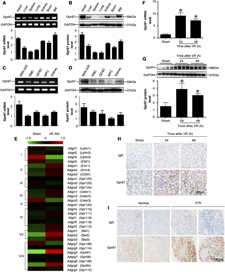
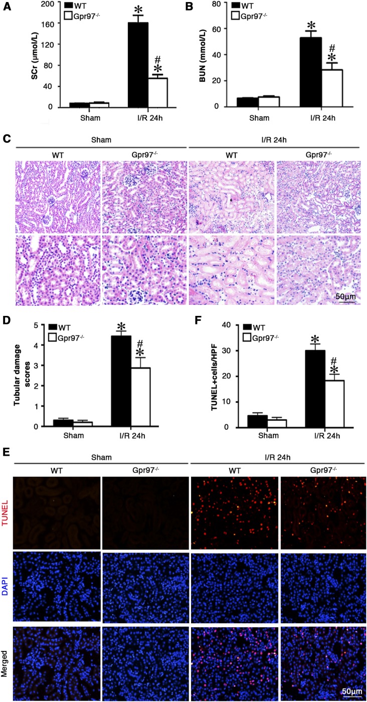
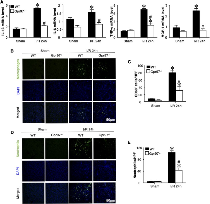
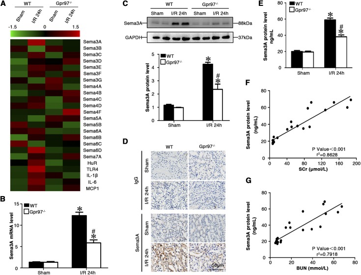
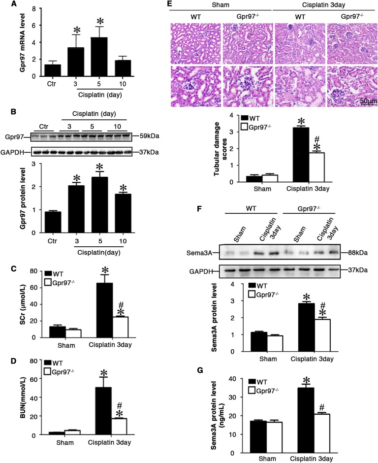
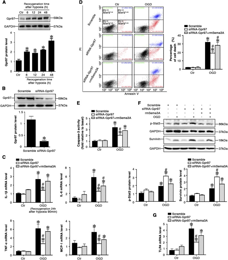
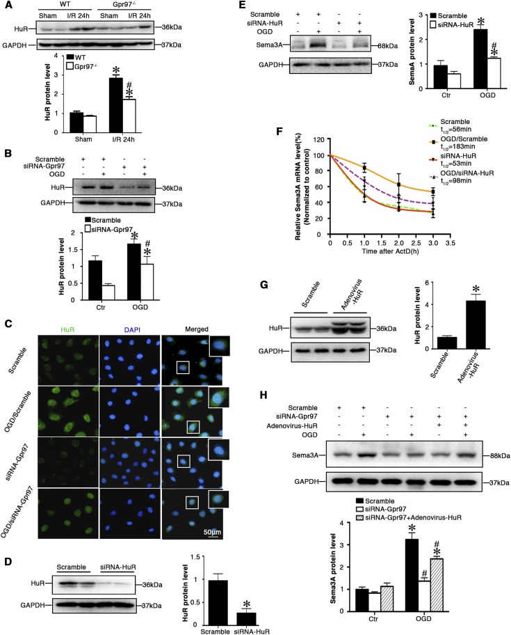
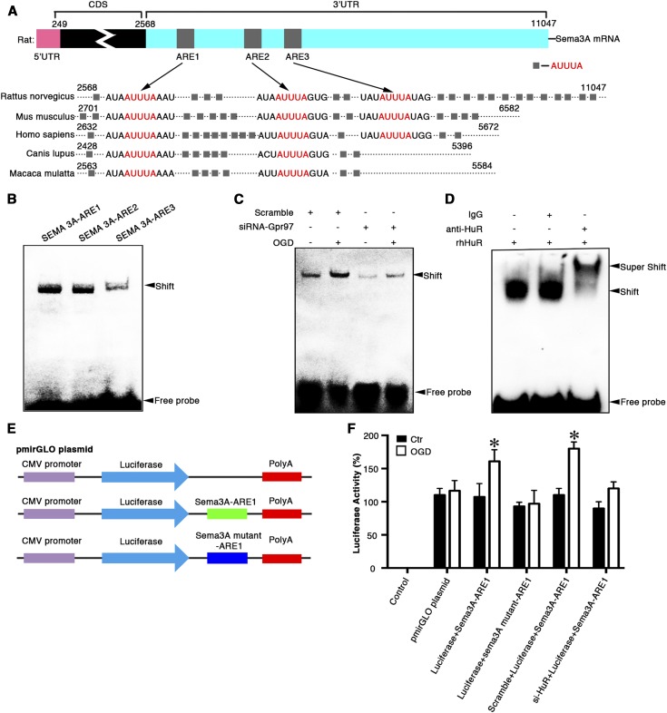
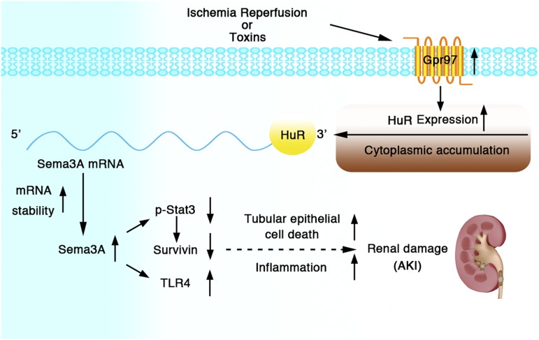
Background G protein-coupled receptors (GPCRs) participate in a variety of physiologic functions, and several GPCRs have critical physiologic and pathophysiologic roles in the regulation of renal function. We investigated the role of Gpr97, a newly identified member of the adhesion GPCR family, in AKI.Methods AKI was induced by ischemia-reperfusion or cisplatin treatment in Gpr97-deficient mice. We assessed renal injury in these models and in patients with acute tubular necrosis by histologic examination, and we conducted microarray analysis and in vitro assays to determine the molecular mechanisms of Gpr97 function.Results Gpr97 was upregulated in the kidneys from mice with AKI and patients with biopsy-proven acute tubular necrosis compared with healthy controls. In AKI models, Gpr97-deficient mice had significantly less renal injury and inflammation than wild-type mice. Gpr97 deficiency also attenuated the AKI-induced expression of semaphorin 3A (Sema3A), a potential early diagnostic biomarker of renal injury. In NRK-52E cells subjected to oxygen-glucose deprivation, siRNA-mediated knockdown of Gpr97 further increased the expression of survivin and phosphorylated STAT3 and reduced toll-like receptor 4 expression. Cotreatment with recombinant murine Sema3A protein counteracted these effects. Finally, additional in vivo and in vitro studies, including electrophoretic mobility shift assays and luciferase reporter assays, showed that Gpr97 deficiency attenuates ischemia-reperfusion-induced expression of the RNA-binding protein human antigen R, which post-transcriptionally regulates Sema3A expression.Conclusions Gpr97 is an important mediator of AKI, and pharmacologic targeting of Gpr97-mediated Sema3A signaling at multiple levels may provide a novel approach for the treatment of AKI.

Keywords: acute renal failure; cisplatin nephrotoxicity; ischemia-reperfusion; renal cell biology; renal tubular epithelial cells; tubular epithelium.


Figures

Figure 1.
Figure 1.
Gpr97 was significantly induced in the kidneys of mice with renal IRI. (A) RT-PCR analysis of Gpr97 mRNA levels in selected murine tissues, including heart, liver, spleen, lung, kidney, brain, and bone marrow (BM). (B) Western blot analysis of the relative protein levels of Gpr97 in selected murine tissues. (C) RT-PCR analysis of Gpr97 mRNA levels in renal cells including rat proximal tubule epithelial cells (NRK-52E), rat glomerular mesangial cells, rat glomerular endothelial cells, and murine podocytes. (D) Western blot analysis of the relative protein levels of Gpr97 in renal cells. (E) Representative heatmap of gene expression levels of adhesion GPCRs in the kidneys of mice with IRI by multiplex qRT-PCR array. (F) Relative mRNA levels of Gpr97 in the kidneys of mice with renal IRI. (G) Representative Western blot gel documents and summarized data showing the protein levels of Gpr97 in the kidneys of mice with renal IRI. (H) Representative photomicrographs of 3,3'-diaminobenzidine (DAB) immunohistochemical staining of Gpr97 in the kidneys of mice with IRI. (I) Representative photomicrographs of 3,3'-diaminobenzidine (DAB) immunohistochemical staining of Gpr97 in the kidneys of patients with biopsy-proven acute tubular necrosis (ATN). Normal kidney tissues were obtained from the healthy kidney poles of individuals who underwent tumor nephrectomies without renal disease. *P<0.05 versus sham-operated wild-type mice (n=8).
Figure 2.
Figure 2.
Gpr97 deficiency ameliorated renal IRI in mice. (A) Serum creatinine (SCr) concentration in different groups of mice. (B) BUN levels in different groups of mice. (C) Representative micrographs showing the morphology by hematoxylin and eosin (H&E) staining of the kidney from different groups of mice. (D) Quantitative assessment of tubular damage. (E) In situ terminal deoxynucleotidyl transferase–mediated UTP nick-end labeling (TUNEL) assays were performed to assess renal cell death. Nuclei were revealed using 4’,6-diamidino-2-phenylindole (DAPI) staining. (F) Quantitative assessment of the number of cell death (numbers per high-power field [HPF]). *P<0.05 versus sham-operated wild-type mice (n=8); #P<0.05 versus ischemic wild-type mice at the same experimental conditions.
Figure 3.
Figure 3.
Gpr97 deficiency reduced inflammatory responses in the kidney after renal IRI in mice. (A) The mRNA levels of proinflammatory mediators including IL-1β, IL-6, TNF-α, and monocyte chemoattractant protein-1 (MCP-1) in the kidney from different groups of mice. (B) Representative sections of kidney stained for macrophages from different groups of mice. (C) Data analysis of macrophage infiltrates in the kidney (numbers per high-power field [HPF]) from different groups of mice. (D) Representative sections of kidney stained for neutrophils from different groups of mice. (E) Data analysis of neutrophil infiltrates in the kidney (numbers per HPF) from different groups of mice. *P<0.05 versus sham-operated wild-type mice; #P<0.05 versus ischemic wild-type mice at the same experimental conditions (n=8).
Figure 4.
Figure 4.
Gpr97 mediated Sema3A expression. (A) Representative heatmap of gene expression levels in the kidneys of mice with IRI by multiplex qRT-PCR array. (B) Summarized data showing the mRNA levels of Sema3A in the kidneys of mice with renal IRI. (C) Representative Western blot gel documents and summarized data showing the protein levels of Sema3A in the kidneys of mice with renal IRI. (D) Representative photomicrographs of Sema3A 3,3'-diaminobenzidine (DAB) immunohistochemical staining in the kidneys of mice with renal IRI. (E) Summarized data showing the serum levels of Sema3A in mice with renal IRI. (F) Positive correlation between the serum levels of Sema3A and serum creatinine (SCr) in all mice. (G) Positive correlation between the serum levels of Sema3A and BUN in all mice. *P<0.05 versus sham-operated wild-type mice; #P<0.05 versus ischemic wild-type mice at the same experimental conditions (n=8).
Figure 5.
Figure 5.
Gpr97 deficiency protected against AKI induced by cisplatin. (A) Relative mRNA levels of Gpr97 in the kidney from different groups of mice. (B) Representative Western blot gel documents and summarized data showing the levels of Gpr97 in the kidney from different groups of mice. (C) Serum creatinine (SCr) concentration in different groups of mice. (D) BUN levels in different groups of mice. (E) Representative micrographs showing the morphology by hematoxylin and eosin (H&E) stainingand and quantitative assessment of tubular damage from different groups of mice. (F) Summarized data showing the expression levels of Sema3A in the kidney from different groups of mice. (G) Summarized data showing the serum levels of Sema3A in different groups of mice. *P<0.05 versus sham-operated wild-type mice; #P<0.05 versus wild-type mice with cisplatin treatment (n=8).
Figure 6.
Figure 6.
Gpr97 contributed to hypoxia-induced production of proinflammatory mediators and cell death in NRK-52E cells via Sema3A. (A) Representative Western blot gel documents and summarized data showing the protein levels of Gpr97 in NRK-52E cells under OGD conditions (cultured in a hypoxic environment for 90 minutes at 0.1% O2, followed by reoxygenation at different time points [6, 12, 24, or 48 hours]). (B) Representative Western blot gel documents and summarized data showing the gene silencing efficiency of Gpr97 by siRNA-Gpr97 transfection. (C) The effect of Gpr97 on the mRNA levels of proinflammatory mediators in NRK-52E cells under OGD conditions (cultured in a hypoxic environment for 90 minutes at 0.1% O2, followed by reoxygenation at 24 hours). (D) Summarized data showing the overall percentage of cell death, including the amount of apoptotic and necrotic cells determined by flow cytometric analysis in NRK-52E cells with different treatments. (E) Summarized data showing the caspase-3 activity in NRK-52E cells with different treatments. (F) Representative Western blot gel documents and summarized data showing the effect of Gpr97 on the levels of STAT3 phosphorylation and survivin in NRK-52E cells with different treatments. (G) Summarized data showing the mRNA levels of TLR4 in NRK-52E cells with different treatments. *P<0.05 versus control; #P<0.05 versus vehicle of NRK-52E cells under OGD conditions (n=6).
Figure 7.
Figure 7.
Gpr97 induced Sema3A expression by mediating HuR expression and activation. (A) Representative Western blot gel documents and summarized data showing the protein levels of HuR in the kidneys of mice with renal IRI. *P<0.05 versus sham-operated wild-type mice; #P<0.05 versus ischemic wild-type mice (n=8). (B) Representative Western blot gel documents and summarized data showing the protein levels of HuR in NRK-52E cells under OGD conditions. *P<0.05 versus control; #P<0.05 versus vehicle of NRK-52E cells under OGD conditions (n=6). (C) Representative microscopic images for immunofluorescence staining showing the distribution of HuR in NRK-52E cells under OGD conditions. (D) Representative Western blot gel documents and summarized data showing the gene silencing efficiency of HuR by siRNA-HuR transfection. (E) Representative Western blot gel documents and summarized data showing the effect of HuR on the Sema3A protein in NRK-52E cells under OGD conditions. *P<0.05 versus control; #P<0.05 versus vehicle of NRK-52E cells under OGD conditions (n=6). (F) Summarized data showing the effect of HuR on the regulation of Sema3A mRNA stability. After NRK-52E cells were transfected with siRNA-HuR or scramble, the cells were treated without (control) or with OGD conditions for 12 hours before addition of actinomycin D (ActD). (G) Representative Western blot gel documents and summarized data showing the overexpression of HuR by adenovirus transfection (ORF of ELAVL1 [HuR] in adenoviral vector pAd with C-terminal Flag and His tag; Adenovirus-HuR) in NRK-52E cells. *P<0.05 versus control (n=6). (H) Representative Western blot gel documents and summarized data showing that HuR overexpression counteracted Gpr97 deficiency-reduced Sema3A expression. *P<0.05 versus control; #P<0.05 versus vehicle of NRK-52E cells under OGD conditions (n=6).
Figure 8.
Figure 8.
HuR interacted with the 3′-UTR of Sema3A mRNA and was involved in hypoxia-induced Sema3A mRNA stability. (A) Sequence homology analysis of 3′UTR of Sema3A mRNA among different species. In rats, 22 canonical ARE sites were labeled. (B) The cytoplasmic extracts from NRK-52E cells were prepared for RNA-EMSA analysis. Complexes were formed when Sema3A-ARE1, Sema3A-ARE2, and Sema3A-ARE3 were applied for the binding reaction. (C) RNA-EMSA assay showing that the binding ability was strongly increased in extracts from NRE-52E cells under OGD conditions, which was abolished by gene silencing of Gpr97 using Sema3A-ARE1. (D) Recombinant human HuR proteins (rhHuR) were used as positive controls to confirm the interaction between HuR and Sema3A-ARE1 by RNA-EMSA. (E) A schematic representation of the construction of various plasmids containing luciferase and 3′-UTR of Sema3A-ARE1 mRNA. (F) The luciferase activity was quantified by in NRE-52E cells transfected with different plasmids. *P<0.05 versus control (n=6).
Figure 9.
Figure 9.
Schematic representation showing that Gpr97 exacerbates AKI via mediating Sema3A signaling. In this model, Gpr97 contributed to the development of postischemic renal inflammation and promoted tubular epithelial cell death, which was associated with the regulation of Sema3A. Gpr97-induced Sema3A expression and mRNA stability were regulated by HuR.

References

    1. Jang HR, Rabb H: Immune cells in experimental acute kidney injury. Nat Rev Nephrol 11: 88–101, 2015 - PubMed
    1. Rewa O, Bagshaw SM: Acute kidney injury-epidemiology, outcomes and economics. Nat Rev Nephrol 10: 193–207, 2014 - PubMed
    1. Paavola KJ, Sidik H, Zuchero JB, Eckart M, Talbot WS: Type IV collagen is an activating ligand for the adhesion G protein-coupled receptor GPR126. Sci Signal 7: ra76, 2014 - PMC - PubMed
    1. Hamann J, Aust G, Araç D, Engel FB, Formstone C, Fredriksson R, et al. .: International Union of Basic and Clinical Pharmacology. XCIV. Adhesion G protein-coupled receptors. Pharmacol Rev 67: 338–367, 2015 - PMC - PubMed
    1. Kishore A, Hall RA: Versatile signaling activity of adhesion GPCRs. Handb Exp Pharmacol 234: 127–146, 2016 - PubMed

Publication types

MeSH terms