. 2018 Mar 12;8(1):4345.
doi: 10.1038/s41598-018-22748-1.

Shelf Life Evaluation of Clinical Grade Chondrogenic Induced Aged Adult Stem Cells for Cartilage Regeneration

Affiliations

Shelf Life Evaluation of Clinical Grade Chondrogenic Induced Aged Adult Stem Cells for Cartilage Regeneration

C C Ude et al. Sci Rep. .

Abstract

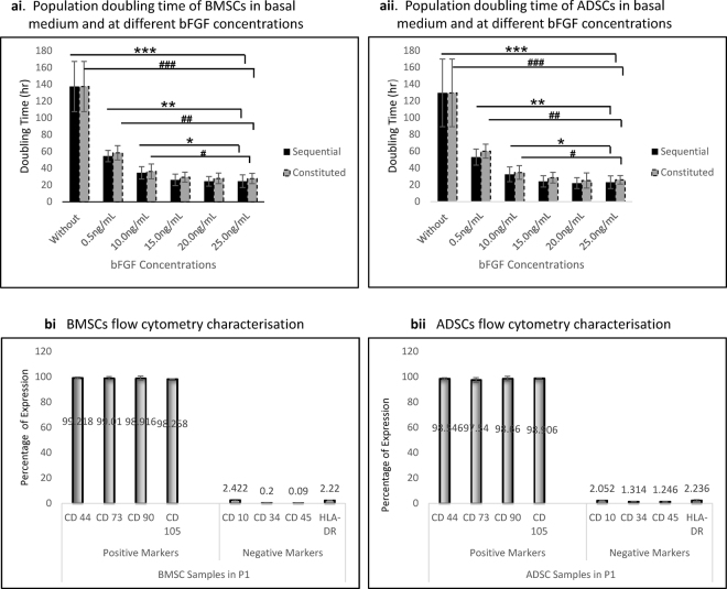
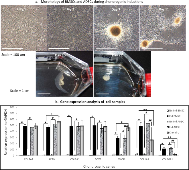
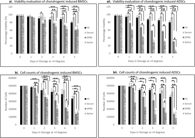
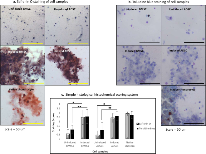
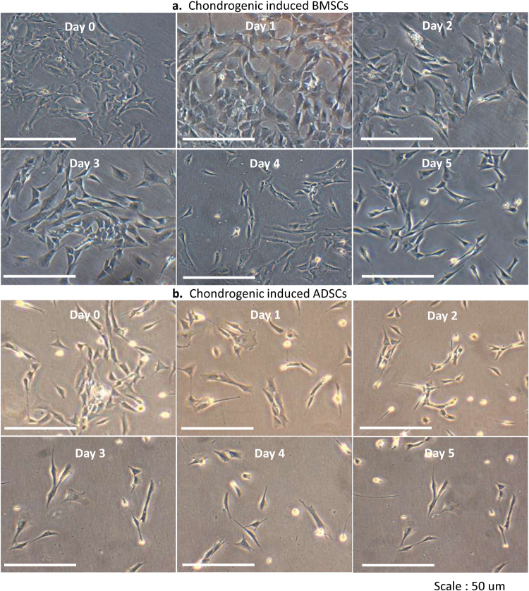
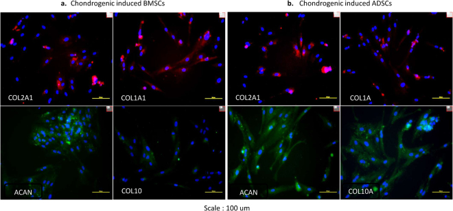
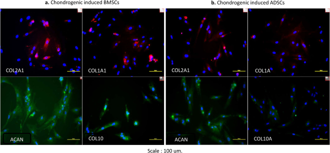
The study objectives include, enhancing the proliferations of aged bone marrow stem cells (BMSCs) and adipose stem cells (ADSCs); and evaluating the shelf lives of clinical grade chondrogenically induced cells from both samples. ADSCs and BMSCs from 56 patients (76 ± 8 yrs) were proliferated using basal medium (FD) and at (5, 10, 15, 20 and 25) ng/ml of basal fibroblast growth factor (bFGF). They were induced to chondrogenic lineage and stored for more than 120 hrs in FD, serum, Dulbecco's phosphate buffered saline (DPBS) and saline at 4 °C. In FD, cells stagnated and BMSCs' population doubling time (PDT) was 137 ± 30 hrs, while ADSCs' was 129.7 ± 40 hrs. bFGF caused PDT's decrease to 24.5 ± 5.8 hrs in BMSCs and 22.0 ± 6.5 hrs in ADSCs (p = 0.0001). Both cells were positive to stem cell markers before inductions and thereafter, expressed significantly high chondrogenic genes (p = 0.0001). On shelf life, both cells maintained viabilities and counts above 70% in FD and serum after 120 hrs. BMSCs' viabilities in DPBS fell below 70% after 96 hrs and saline after 72 hrs. ADSCs' viability fell below 70% in DPBS after 24 hrs and saline within 24 hrs. Concentrations between 20 ng/ml bFGF is ideal for aged adult cells' proliferation and delivery time of induced BMSCs and ADSCs can be 120 hrs in 4 °C serum.


Conflict of interest statement

The authors declare no competing interests.

Figures

Figure 1
Figure 1
Growth Kinetics and Monolayer phenotype characterisation of cell samples. (ai) Population doubling time of BMSCs in basal medium and at different concentrations of bFGF. PDT without bFGF was significantly higher compare to every addition of bFGF, (***)p = 0.0001. 5 ng/ml was significantly higher to other concentrations in both sequential and constitution addition, (***)p = 0.0001. The same was found at 10 ng/ml, (*)p = 0.005. At 15, 20 and 25 ng/ml, there was no significance difference. (aii) Population doubling time of ADSCs at different concentrations of bFGF. Without addition of bFGF, PDT was significantly higher to every addition of bFGF, (***)p = 0.0001. PDT at 5 ng/ml was significantly higher to other increased concentrations (***)p = 0.0001. The same at 10  ng/ml, (*)p = 0.005. There was no significance difference between PDTs at (15, 20 and 25) ng/ml. (bi) BMSCs flow cytometry. BMSCs were highly positive to the markers CD 44, CD73, CD90 and CD105 associated with positive markers of stem cells. They were also negative to CD 10, CD34, CD45 and HLA-DR associated with negative markers of stem cells. (bii) ADSCs flow cytometry. ADSCs were highly negative to CD 44, CD73, CD90 and CD105 associated with positive markers of stem cells. They were also negative to CD 10, CD34, CD45 and HLA-DR associated with negative markers of stem cells respectively.
Figure 2
Figure 2
Chondrogenic Inductions. (a) Morphologies of BMSCs and ADSCs during chondrogenic inductions. The characteristic morphologies of both BMSCs and ADSCs as they underwent chondrogenesis were shown. The spindle morphologies of uninduced BMSCs and ADSCs (day 1), on interaction with induction media, changed to polygonal shapes (day 2), formed aggregates of cells and matrixes (days 7 to 11), and later become whitish cartilage structure (day 15 to 21) (b) RTPCR gene expression analysis. COL2A1, ACAN, COL9A1, SOX9, FMOD, COL1A1 and COL10A1, revealed that chondrogenically induced BMSCs and ADSCs had significantly higher expressions compare to their uninduced forms (*, #)p = 0.0001. The native chondrocytes as positive control had increased expressions in all the hyaline genes and were significantly higher than induced BMSCs at ACAN and FMOD (a), and induced ADSCs at FMOD (a a)p = 0.005. Both BMSCs and ADSCs had significantly higher expressions compare to the native chondrocytes at COL1A1 and COL10A1 respectively (**, ##)p = 0.005.
Figure 3
Figure 3
Shelf life evaluation of revived chondrogenic induced cell samples in different media (ai) Viability evaluation of chondrogenic induced BMSCs. On day 1, viability decreased slightly in all media, but was not significant. On the second day, viability of cells in DPBS and Saline decrease more. Serum had higher viability to DPBS only (*)p = 0.003. From the third day through fifth, further decreases occurred in DPBS and Saline. Serum and FD remained highly comparable with day 1 values. Both were significantly higher to DPBS and Saline (*, **) (∞, ∞∞)p = 0.001 respectively. (aii) Viability evaluation of chondrogenic induced ADSCs. From day 1, viabilities decreased drastically in both DPBS and Saline. FD and Serum maintained significantly higher values (*, **), (∞, ∞∞)p = 0.001. From the second day through fifth, more decreases were observed in DPBS and Saline. Both Serum and FD were significantly higher to DPBS and saline (*, **), (∞, ∞∞) respectively. DPBS also had significantly higher viabilities to saline (#)p = 0.0001. (bi) Cell counts of chondrogenic induced BMSCs. On day 1, a significant decrease was observed with Saline compare to FD, Serum and DPBS, (∞∞), (**), (#) respectively, P = 0.001. On the second day, FD and Serum remained significantly higher to DPBS and Saline (∞, ∞∞), (*, **) respectively, p = 0.0001. From the third and fourth day, counts in FD and Serum were significantly higher to both DPBS and Saline, while DPBS was also higher to Saline (∞, ∞∞), (*, **), (#) respectively, p = 0.0001. On the fifth day, FD and Serum remained significantly higher (∞, ∞∞) (*, **)p = 0.0001. (bii) Cell counts of chondrogenic induced ADSCs. There was a significant decrease with cells in DPBS and Saline compare to FD and Serum from day 1 (∞, ∞∞), (*, **)p = 0.001; DPBS also had significantly higher counts to saline (#)p = 0.001. The same trend was seen on days 2, 3 and 4 (∞, ∞∞), (*, **), (#)p = 0.001 respectively. On the fifth day, FD and Serum retained significantly higher difference to DPBS and Saline, but DPBS was not difference to Saline (∞, ∞∞), (*, **)p = 0.001 respectively.
Figure 4
Figure 4
Histological evaluations of cell samples. (a) Safranin O staining of cell samples. Chondrogenic induced ADSCs and BMSCs stained positive. The appearances were in accordance with the presence of accumulated glycosaminoglycans. The two tests samples and the positive control (chondrocytes) were comparable, while uninduced cells were negative to the stain. (b) Toluidine blue staining of cell samples. Chondrogenic induced ADSCs and BMSCs stained positive. Cells and matrixes of test samples exhibited the presence of accumulated proteoglycans revealed via Toluidine blue. The two tests samples and the positive control (chondrocytes) were comparable, while uninduced cells were negative has slight positive stain. (c) The simple histological scoring Evaluation. Both chondrogenic induced BMSCs and ADSCs had significantly higher stains on both Safranin O and Toluidine blue compare to their negative control samples (*, **), (#, ##) respectively, p = 0.001. Both induced cells and the chondrocytes had no significant differences.
Figure 5
Figure 5
Attachment and proliferation of revived chondrogenic induced cell samples. (a) BMSCs. There were attachments and proliferations of the cells after storage. It provided evidence that the stored induced BMSCs were viable and proliferating. (b) ADSCs. There were attachments and proliferations of the cells after storage. It provided evidence that the stored induced ADSCs were viable and proliferating.
Figure 6
Figure 6
Immunohistochemistry of revived chondrogenic induced cell samples in serum (day 1). (a) BMSCs. The positive expression of collagen type I alpha 1, collagen type II alpha 1, aggrecan, and collagen type X alpha 1, evaluated on stored chondrogenic induced BMSCs in serum, on day 1 after cultures (b) ADSCs. The positive expression of collagen type I alpha 1, collagen type II alpha 1, aggrecan, and collagen type X alpha 1, evaluated on stored chondrogenic induced ADSCs in serum, on day 1 after cultures.
Figure 7
Figure 7
Immunohistochemistry of revived chondrogenic induced cell samples in serum (day 5). (a) BMSCs. The positive expression of collagen type I alpha 1, collagen type II alpha 1, aggrecan, and collagen type X alpha 1, evaluated on stored chondrogenic induced BMSCs in serum, on day 5 after cultures (b) ADSCs. The positive expression of collagen type I alpha 1, collagen type II alpha 1, aggrecan, and collagen type X alpha 1, evaluated on stored chondrogenic induced BMSCs in serum, on day 5 after cultures.

Similar articles

Cited by

References

    1. Saim AB, et al. Engineering autogenous cartilage in the shape of a helix using an injectable hydrogel scaffold. The Laryngoscope. 2000;110:1694–1697. doi: 10.1097/00005537-200010000-00023. - DOI - PubMed
    1. Horas U, Pelinkovic D, Herr G, Aigner T, Schnettler R. Autologous chondrocyte implantation and osteochondral cylinder transplantation in cartilage repair of the knee joint A prospective, comparative trial. The Journal of Bone and Joint Surgery (American). 2003;85:185–192. doi: 10.2106/00004623-200302000-00001. - DOI - PubMed
    1. Kim HJ, Im GI. Chondrogenic differentiation of adipose tissue-derived mesenchymal stem cells: greater doses of growth factor are necessary. J Orthop Res. 2009;27:612. doi: 10.1002/jor.20766. - DOI - PubMed
    1. Madry H, Cucchiarini M. Clinical potential and challenges of using genetically modified cells for articular cartilage repair. Croat Med J. 2011;52:245. doi: 10.3325/cmj.2011.52.245. - DOI - PMC - PubMed
    1. Murphy JM, Fink DJ, Hunziker EB, Barry FP. Stem cell therapy in a caprine model of osteoarthritis. Arthritis Rheum. 2003;48:3464–3474. doi: 10.1002/art.11365. - DOI - PubMed

Publication types

LinkOut - more resources