

Automated Whole-Body Bone Lesion Detection for Multiple Myeloma on 68Ga-Pentixafor PET/CT Imaging Using Deep Learning Methods
- PMID: 29531504
- PMCID: PMC5817261
- DOI: 10.1155/2018/2391925
Automated Whole-Body Bone Lesion Detection for Multiple Myeloma on 68Ga-Pentixafor PET/CT Imaging Using Deep Learning Methods
Abstract
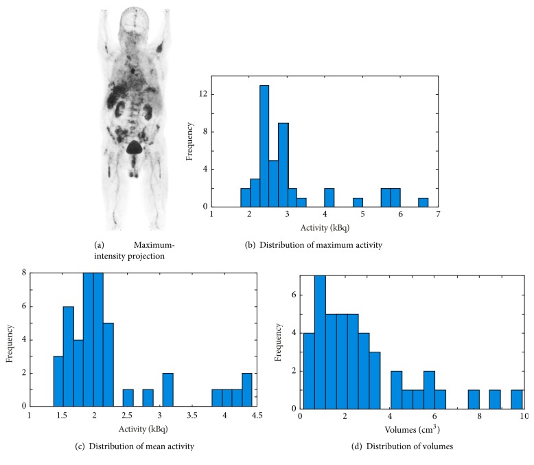
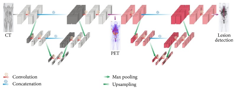
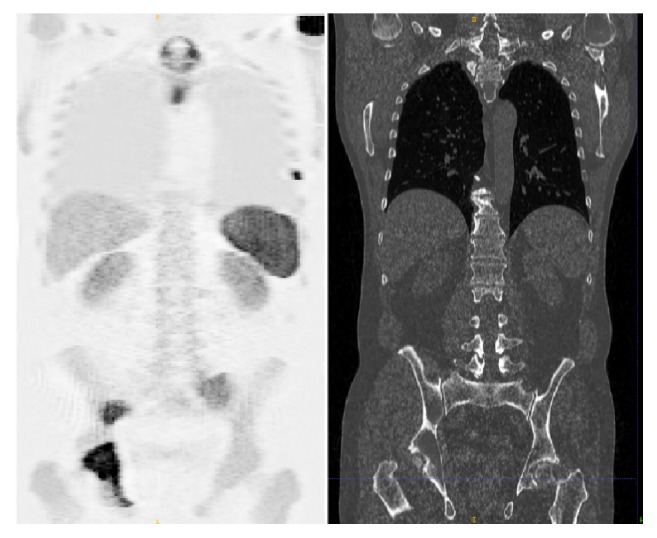
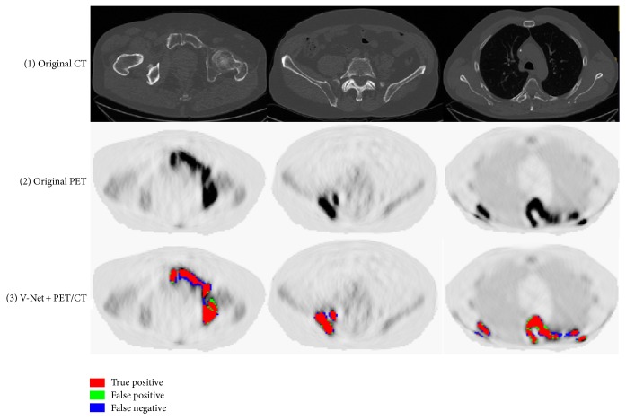
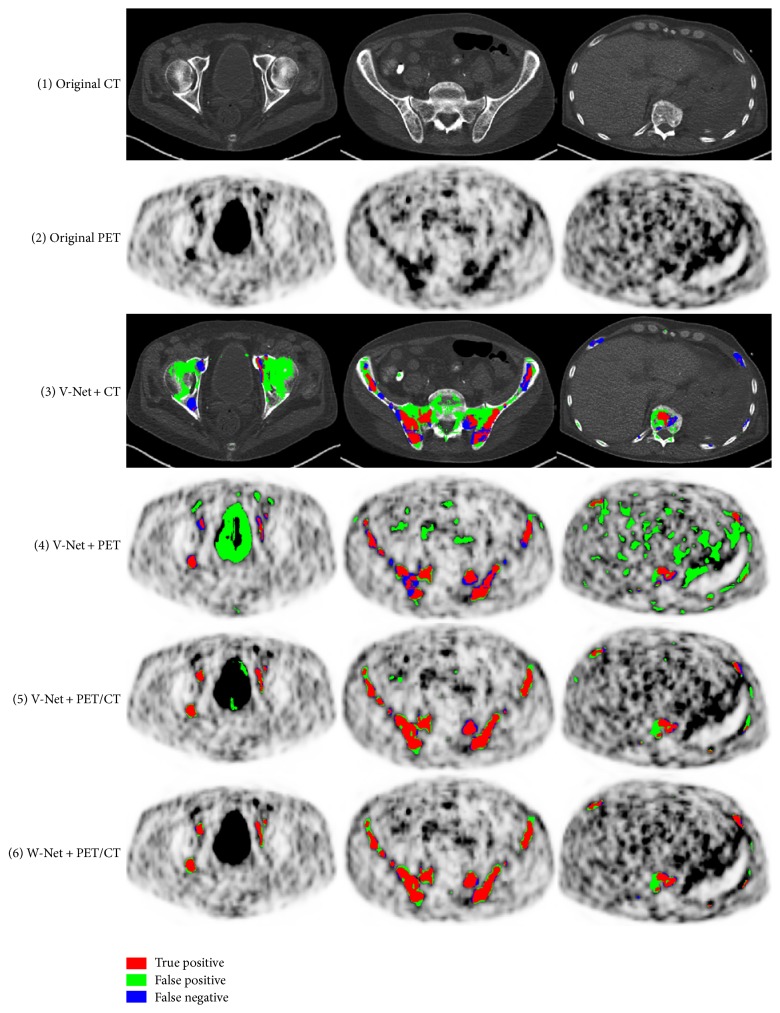
The identification of bone lesions is crucial in the diagnostic assessment of multiple myeloma (MM). 68Ga-Pentixafor PET/CT can capture the abnormal molecular expression of CXCR-4 in addition to anatomical changes. However, whole-body detection of dozens of lesions on hybrid imaging is tedious and error prone. It is even more difficult to identify lesions with a large heterogeneity. This study employed deep learning methods to automatically combine characteristics of PET and CT for whole-body MM bone lesion detection in a 3D manner. Two convolutional neural networks (CNNs), V-Net and W-Net, were adopted to segment and detect the lesions. The feasibility of deep learning for lesion detection on 68Ga-Pentixafor PET/CT was first verified on digital phantoms generated using realistic PET simulation methods. Then the proposed methods were evaluated on real 68Ga-Pentixafor PET/CT scans of MM patients. The preliminary results showed that deep learning method can leverage multimodal information for spatial feature representation, and W-Net obtained the best result for segmentation and lesion detection. It also outperformed traditional machine learning methods such as random forest classifier (RF), k-Nearest Neighbors (k-NN), and support vector machine (SVM). The proof-of-concept study encourages further development of deep learning approach for MM lesion detection in population study.
Figures
Similar articles
-
Imaging CXCR4 receptors expression for staging multiple myeloma by using 68Ga-Pentixafor PET/CT: comparison with 18F-FDG PET/CT.Br J Radiol. 2022 Aug 1;95(1136):20211272. doi: 10.1259/bjr.20211272. Epub 2022 Jun 30. Br J Radiol. 2022. PMID: 35731811 Free PMC article.
-
[68Ga]Pentixafor-PET/CT for imaging of chemokine receptor CXCR4 expression in multiple myeloma - Comparison to [18F]FDG and laboratory values.Theranostics. 2017 Jan 1;7(1):205-212. doi: 10.7150/thno.16576. eCollection 2017. Theranostics. 2017. PMID: 28042328 Free PMC article.
-
Chemokine Receptor 4-Targeted PET/CT with [68Ga]pentixather in Newly Diagnosed Multiple Myeloma: a Comparative Study with [68Ga]pentixafor PET/CT.Mol Imaging Biol. 2024 Dec;26(6):986-994. doi: 10.1007/s11307-024-01953-7. Epub 2024 Sep 20. Mol Imaging Biol. 2024. PMID: 39304574
-
Advances in PET Imaging of the CXCR4 Receptor: [68Ga]Ga-PentixaFor.Semin Nucl Med. 2024 Jan;54(1):163-170. doi: 10.1053/j.semnuclmed.2023.09.002. Epub 2023 Nov 3. Semin Nucl Med. 2024. PMID: 37923671 Free PMC article. Review.
-
Nuclear Medicine Application of Pentixafor/Pentixather Targeting CXCR4 for Imaging and Therapy in Related Disease.Mini Rev Med Chem. 2023;23(7):787-803. doi: 10.2174/1389557523666221216095821. Mini Rev Med Chem. 2023. PMID: 36529918 Review.
Cited by
-
Positron Emission Tomography-Derived Radiomics and Artificial Intelligence in Multiple Myeloma: State-of-the-Art.J Clin Med. 2023 Dec 13;12(24):7669. doi: 10.3390/jcm12247669. J Clin Med. 2023. PMID: 38137738 Free PMC article. Review.
-
Deep learning-based detection of parathyroid adenoma by 99mTc-MIBI scintigraphy in patients with primary hyperparathyroidism.Ann Nucl Med. 2022 May;36(5):468-478. doi: 10.1007/s12149-022-01726-8. Epub 2022 Feb 18. Ann Nucl Med. 2022. PMID: 35182328
-
Deep learning image segmentation approaches for malignant bone lesions: a systematic review and meta-analysis.Front Radiol. 2023 Aug 8;3:1241651. doi: 10.3389/fradi.2023.1241651. eCollection 2023. Front Radiol. 2023. PMID: 37614529 Free PMC article. Review.
-
Deep learning applications in visual data for benign and malignant hematologic conditions: a systematic review and visual glossary.Haematologica. 2023 Aug 1;108(8):1993-2010. doi: 10.3324/haematol.2021.280209. Haematologica. 2023. PMID: 36700396 Free PMC article.
-
Recent advances in imaging and artificial intelligence (AI) for quantitative assessment of multiple myeloma.Am J Nucl Med Mol Imaging. 2024 Aug 25;14(4):208-229. doi: 10.62347/NLLV9295. eCollection 2024. Am J Nucl Med Mol Imaging. 2024. PMID: 39309415 Free PMC article. Review.
References
-
- Terpos E., Kanellias N. Practical Considerations for Bone Health in Multiple MyelomaPersonalized Therapy for Multiple Myeloma. Springer; 2018. - DOI
-
- Lütje S., Rooy J. W. J., Croockewit S., Koedam E., Oyen W. J. G., Raymakers R. A. Role of radiography, MRI and FDG-PET/CT in diagnosing, staging and therapeutical evaluation of patients with multiple myeloma. Annals of Hematology. 2009;88(12):1161–1168. doi: 10.1007/s00277-009-0829-0. - DOI - PMC - PubMed
Publication types
MeSH terms
Substances
LinkOut - more resources
Full Text Sources
Other Literature Sources
Medical
