. 2018 Feb 20:4:33.
doi: 10.1038/s41420-018-0027-8. eCollection 2018 Dec.

Multiscale analysis of a regenerative therapy for treatment of volumetric muscle loss injury

Affiliations

Multiscale analysis of a regenerative therapy for treatment of volumetric muscle loss injury

Carlos A Aguilar et al. Cell Death Discov. .

Erratum in

Abstract

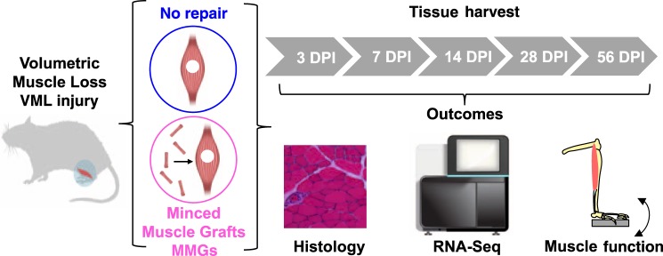
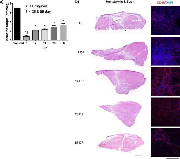
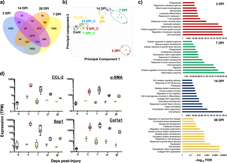
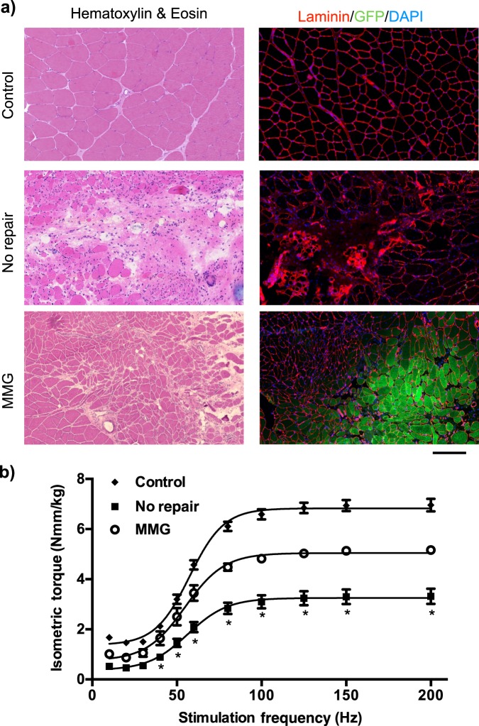
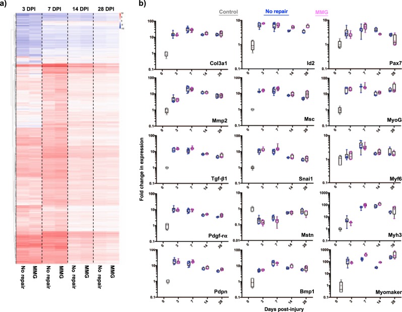
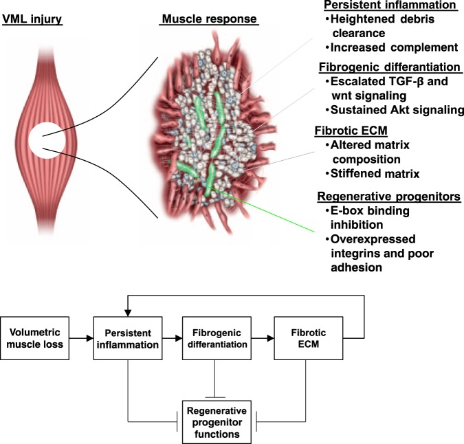
Skeletal muscle possesses a remarkable capacity to regenerate when injured, but when confronted with major traumatic injury resulting in volumetric muscle loss (VML), the regenerative process consistently fails. The loss of muscle tissue and function from VML injury has prompted development of a suite of therapeutic approaches but these strategies have proceeded without a comprehensive understanding of the molecular landscape that drives the injury response. Herein, we administered a VML injury in an established rodent model and monitored the evolution of the healing phenomenology over multiple time points using muscle function testing, histology, and expression profiling by RNA sequencing. The injury response was then compared to a regenerative medicine treatment using orthotopic transplantation of autologous minced muscle grafts (~1 mm3 tissue fragments). A chronic inflammatory and fibrotic response was observed at all time points following VML. These results suggest that the pathological response to VML injury during the acute stage of the healing response overwhelms endogenous and therapeutic regenerative processes. Overall, the data presented delineate key molecular characteristics of the pathobiological response to VML injury that are critical effectors of effective regenerative treatment paradigms.


Conflict of interest statement

The authors declare that they have no competing interests.

Figures

Fig. 1
Fig. 1. Schematic of experimental approach to define the molecular response to volumetric muscle loss (VML) injury and how transplantation of autologous minced muscle graft (MMGs) impacts regenerative trajectory.
Injured and uninjured muscles were extracted at multiple days post injury (DPI) and characterized using histology, high-throughput sequencing of RNA (RNA-Seq), and muscle function
Fig. 2
Fig. 2. VML injury in the rat TA muscle induces chronic strength deficits and prolonged tissue damage.
a TA muscle maximal isometric torque was elicited using common peroneal nerve stimulation in uninjured and VML injured (non-repair) muscle at the specified days post injury (DPI). Values are mean ± sem. *, †: p < 0.05. b Representative TA muscle hematoxylin and eosin (H&E) and macrophage (CD68) probed sections are presented. Scale bars for H&E and CD68 images are 1000 and 150 µm, respectively. TA tibialis anterior, VML volumetric muscle loss
Fig. 3
Fig. 3. Characterization of molecular response to VML injury.
a Venn diagram showing number of unique and overlapping differentially expressed genes for muscle tissues administered VML injury and extracted at different days post injury (DPI). Days 3 and 7 show the most unique differentially expressed genes. b Principal component analysis (PCA) of RNA-Seq data sets from control (cont), injured (example: 3 DPI), and uninjured (example: 3 DPI_C) data sets from different time points harvested show distinct separation between injured and uninjured samples. c Enriched pathways associated with differentially expressed genes from each time point. Enrichment scores are plotted as −log10(FDR), where FDR is the false discovery rate. d Individual graphs of gene expression plotted in TPM (transcripts per million reads) where uninjured samples are plotted first and injured samples are plotted second for each time point. CCL2: C-C Motif chemokine ligand 2, SPP1: secreted phosphoprotein 1, α-SMA: alpha smooth muscle actin, Col1a1: collagen type I alpha chain. VML volumetric muscle loss
Fig. 4
Fig. 4. Autologous MMGs promote partial muscle fiber regeneration and functional recovery.
a TA muscle sections stained with hematoxylin and eosin or probed for laminin, GFP, nuclei from control (uninjured), VML injured no repair and minced graft repaired muscles 56 days post injury are presented. Scale bar is 200 µm. b Maximal TA muscle isometric torque was measured as a function of stimulation frequency in each group at 56 days post injury. Values are mean ± sem. *p < 0.05. MMG minced muscle graft, TA tibialis anterior
Fig. 5
Fig. 5. Transcriptional response of regenerative (MMGs) treatment for VML show marginal effects compared to non-repaired tissue at time points sampled.
a Heatmap of differentially expressed genes (injured vs. uninjured) for each treatment and time point. Data are plotted as union of biological replicates. b Individual box plots of gene expression plotted as fold change (injured vs. uninjured) for four different time points (Sham-gray, No Repair-blue, MMG-purple). DPI days post-injury, MMG minced muscle graft, VML volumetric muscle loss
Fig. 6
Fig. 6. VML induces formation of an inhibitive feedback response loop that modulates the regenerative actions of myogenic progenitors.
The abrupt tissue removal engenders a sustained inflammatory response that invokes fibrogenic cells to differentiate and produce excess matrix. The modified matrix and milieu in turn modifies the regenerative functions of myogenic progenitors and prevents healing. ecm extracellular matrix, VML

Comment in

References

    1. Scharner J, Zammit PS. The muscle satellite cell at 50: the formative years. Skelet. Muscle. 2011;1:28. doi: 10.1186/2044-5040-1-28. - DOI - PMC - PubMed
    1. Grogan BF, Hsu JR. Volumetric muscle loss. J. Am. Acad. Ortho. Surg. 2011;19:S35–S37. doi: 10.5435/00124635-201102001-00007. - DOI - PubMed
    1. Garg K, et al. Volumetric muscle loss: persistent functional deficits beyond frank loss of tissue. J. Ortho. Res. 2015;33:40–46. doi: 10.1002/jor.22730. - DOI - PubMed
    1. Corona BT, Wenke JC, Ward CL. Pathophysiology of volumetric muscle loss injury. Cells Tissues Organs. 2015;202:180–188. doi: 10.1159/000443925. - DOI - PubMed
    1. Corona BT, et al. The promotion of a functional fibrosis in skeletal muscle with volumetric muscle loss injury following the transplantation of muscle-ECM. Biomaterials. 2013;34:3324. doi: 10.1016/j.biomaterials.2013.01.061. - DOI - PubMed