. 2017:1:993-1003.
doi: 10.1038/s41551-017-0167-9. Epub 2017 Dec 12.

Surveillance nanotechnology for multi-organ cancer metastases

Affiliations

Surveillance nanotechnology for multi-organ cancer metastases

Harini Kantamneni et al. Nat Biomed Eng. 2017.

Abstract

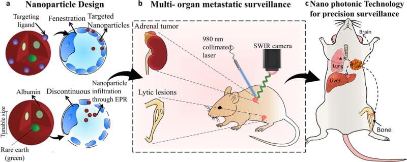
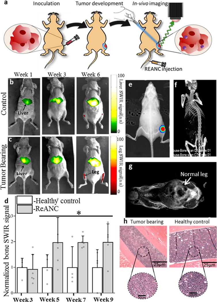
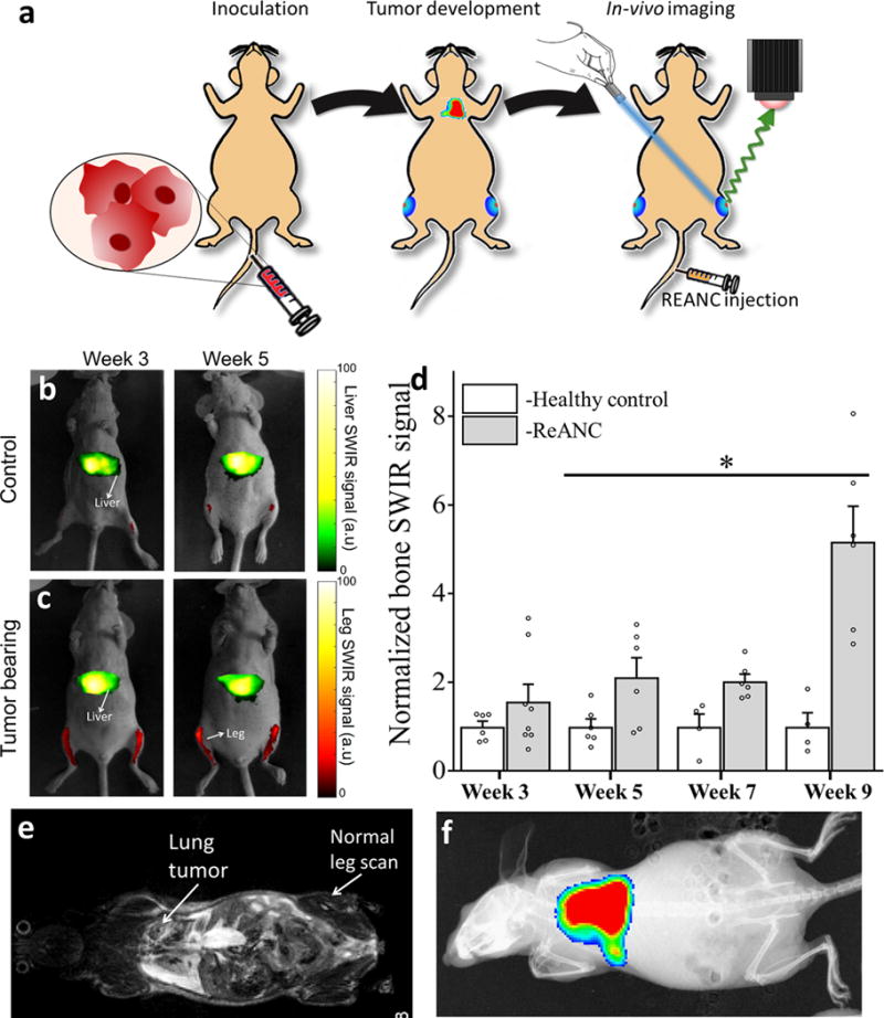
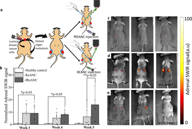
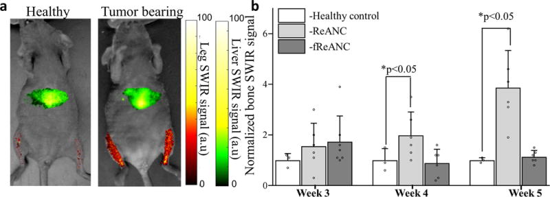
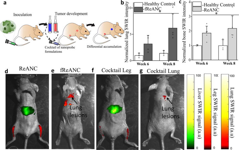
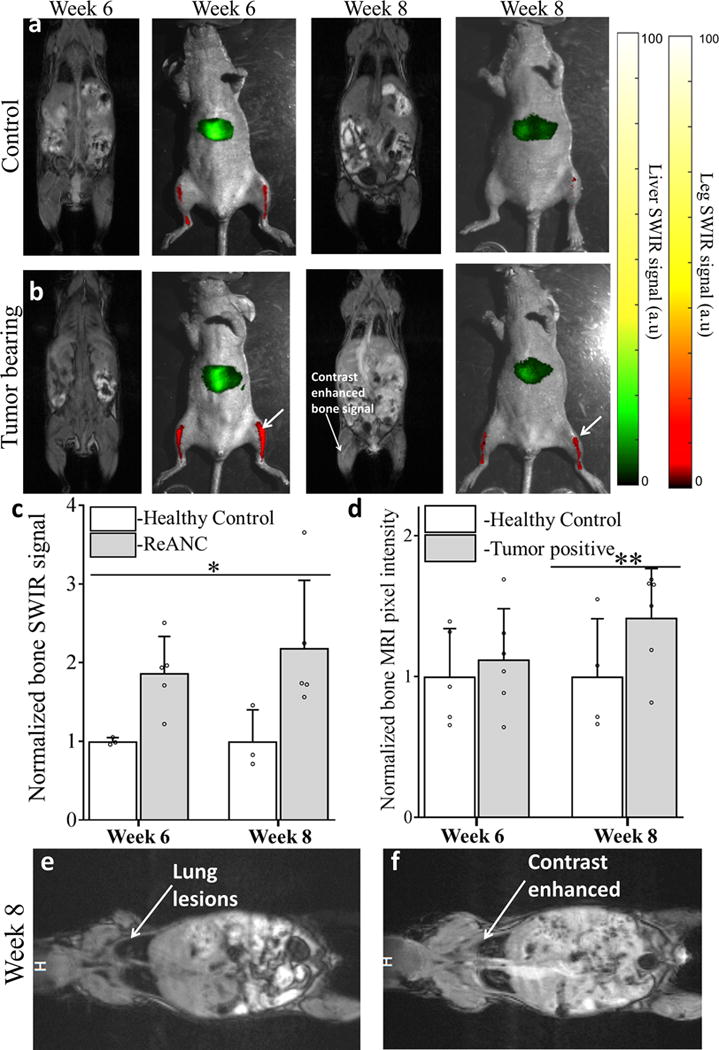
The identification and molecular profiling of early metastases remains a major challenge in cancer diagnostics and therapy. Most in vivo imaging methods fail to detect small cancerous lesions, a problem that is compounded by the distinct physical and biological barriers associated with different metastatic niches. Here, we show that intravenously injected rare-earth-doped albumin-encapsulated nanoparticles emitting short-wave infrared light (SWIR) can detect targeted metastatic lesions in vivo, allowing for the longitudinal tracking of multi-organ metastases. In a murine model of basal human breast cancer, the nanoprobes enabled whole-body SWIR detection of adrenal gland microlesions and bone lesions that were undetectable via contrast-enhanced magnetic resonance imaging (CE-MRI) as early as, respectively, three weeks and five weeks post-inoculation. Whole-body SWIR imaging of nanoprobes functionalized to differentially target distinct metastatic sites and administered to a biomimetic murine model of human breast cancer resolved multi-organ metastases that showed varied molecular profiles at the lungs, adrenal glands and bones. Real-time surveillance of lesions in multiple organs should facilitate pre-therapy and post-therapy monitoring in preclinical settings.

Keywords: cancer metastasis; nanotechnology; rare earths; shortwave infrared imaging.


Conflict of interest statement

Competing interests The authors declare no competing financial interests.

Figures

Figure 1
Figure 1. Design and workflow of photonic nanotechnology for cancer-metastasis detection and profiling
Distinct nanoparticles were designed with rare-earth-doped cores based on differences in tissue microenvironment (a) to enable whole body screening based on deeper-tissue emanating short-wave infrared emissions. When administered in vivo to biomimetic breast cancer models, these nanoparticles are targeted to reach multi-organ metastatic sites across different pharmacologic barriers(b). Metastatic lesions (in the long bones or adrenal glands) can be detected earlier than conventional methods (bioluminescence, MRI, CT) and molecular changes in cancer cell signatures can be obtained, forming the basis for future metastatic-site specific, personalized cancer therapies(c).
Figure 2
Figure 2. SWIR imaging of photonic nanoparticles discerns bone lesions in vivo that are undetectable by conventional MRI and CT
(a) Schematic diagram illustrating intra-tibial inoculation of MDA-MB-231 derived SCP2 cells followed by tail vein administration of ReANCs and in vivo SWIR imaging. Representative SWIR images from (b) healthy (non-tumor-bearing) animals and (c) tumor-bearing mice. (d) Quantification of SWIR signal in the tibiae of tumor-bearing and healthy controls shows a 2-fold increase in signal at 5-weeks post inoculation. Data is expressed as mean±S.D; n=6 for tumor-bearing group and n=3 for healthy control group. *P<0.1, determined by Welch’s t-test. Data in (d) is represented as a fold increase compared to healthy control. (e) Validation of tumor presence by BLI. (f) End point CT and (g) MRI do not indicate bone deformities while (h) ex vivo histopathology shows possible hypo-cellularization, potentially indicating lytic activity due to the presence of tumors in tumor-bearing legs compared to healthy controls. SWIR intensities were normalized to those of the healthy control groups for the region of interest at each time point.
Figure 3
Figure 3. Distal bone lesions can be detected with SWIR imaging earlier than by MRI and CT in a biomimetic metastasis model
(a) Schematic diagram illustrating intravenous inoculation of MDA-MB-231 derived SCP28 cells followed by tail vein administration of ReANCs and in vivo SWIR imaging. Representative images from (b) non-tumor-bearing control animals and (c) tumor-bearing animals at weeks 3 and 5 post-inoculation. (d) Quantification of SWIR intensity shows at least a 2-fold increase in signal over healthy controls from week 5 onwards. Data is expressed as mean±S.D; n=4 for tumor-bearing group and n=3 for healthy control group. *two-tailed P<0.06, determined by Welch’s t-test. Data in (d) is represented as a fold increase compared to healthy control. (e) MRI and (f) BLI show no visible bone abnormalities at the study end point. SWIR intensities were normalized to those of the healthy control groups for the region of interest at each time point.
Figure 4
Figure 4. Differential niche-based accumulation of ReANC and fReANC formulations leads to multi-organ detection of metastases in a luminal breast cancer model
(a) Athymic nude mice were injected with MCF7 derived cells via the left ventricle to form metastases in adrenals and bones; (b) Weekly intravenous administration of fReANCs show earlier detection of adrenal metastases compared to untargeted ReANCs. Quantitative analysis of SWIR intensity shows significantly higher accumulation of fREANCs compared to ReANCs at 5 weeks post inoculation whereas comparison between healthy controls and tumor-bearing animals injected with fReANC shows higher SWIR signal from adrenal lesions as early as 3 weeks post inoculation. Volumetric MRI analysis found the smallest tumor volume detected 5-weeks post inoculation with fReANCs to be 7.8 mm3, compared to a volume of 15.7 mm3 with REANCs. Representative dorsal (prone) images showing SWIR signal from adrenal lesions of healthy controls (c) and tumor-bearing animals injected with fReANC (d) and ReANC(e) shows earlier detection of adrenal metastases with active targeting. Data in (b) is expressed as mean±S.D; n=6 for tumor-bearing group and n=3 for healthy control group. *two-tailed P<0.05, determined by Welch’s t-test. ** two-tailed P<0.05, determined by Welch’s t-test. SWIR intensities were normalized to those of the healthy control groups for the region of interest at each time point.
Figure 5
Figure 5. Passive accumulation of ReANCs, via the discontinuous fenestration architecture of bone tissue, in a metastatic luminal breast cancer model
Athymic nude mice were injected with MCF7 derived cells via the left ventricle to form metastases in bones followed by weekly injections of ReANC nanoprobes. (a) Representative ventral (supine) images showing SWIR signal from the bone space of tumor-bearing and healthy control animals show significant accumulation of ReANCs in the bone lesions 5 weeks post inoculation. (b) There is significant increase in SWIR emission intensity in bone starting 4 weeks post inoculation in tumor-bearing and healthy controls injected with ReANCs. There was no significant increase in accumulation of fReANC in bones of tumor-bearing and healthy control animals. Data in (b) is expressed as mean±S.D; n=6 for tumor-bearing group and n=3 for healthy control group represented as a fold increase compared to healthy control group.*two-tailed P<0.05 determined using a Welch’s t-test. SWIR intensities were normalized to those of the healthy control groups for the region of interest at each time point.
Figure 6
Figure 6. Differential accumulation of ReANCs in leg lesions and fReANCs in lung lesions in a multi-organ metastatic model
(a) Schematic illustrating intravenous inoculation of MDA-MB-231 cells followed by intravenous administration of a cocktail of nanoprobe formulations and SWIR imaging. Quantitative analysis of SWIR signal shows significantly higher lung intensity compared to healthy controls (b). Representative images show localization of ReANC localization to leg lesions (c); and fReANC localization to lung lesions (d). Representative animal injected with a cocktail of ReANC and fReANC probes showed leg signal (e) and lung signal (f). Data is expressed as mean ± S.D; n=4 for tumor-bearing group and n=3 for healthy control group represented as fold increase compared to healthy control group. *p<0.07, determined by Welch’s t test. SWIR intensities were normalized to those of the healthy control groups for the region of interest at each time point.
Figure 7
Figure 7. Distal bone lesions can be detected with SWIR imaging earlier than by contrast-enhanced MRI in a biomimetic metastasis model
Representative images from non-tumor control animals (a) and Tumor-bearing animals (b) shows enhanced SWIR signal 6 weeks’ post inoculation with no significant changes in CE-MRI. At 8 weeks’ post inoculation both SWIR and CE-MRI shows enhanced signal from the bone space. Quantitative comparison of SWIR emission intensity (c) in bone lesions shows significant increase starting week 6 post inoculation compared to non-tumor control animals. Quantitative analysis of increase in pixel intensity pre- and post-contrast injection in MR imaging does not show significant enhancement until week 8 (d). Representative image of lung metastases prior to (e) and post (f) contrast injection at week 8, used as positive control, to show enhanced uptake of MRI contrast by tumor lesions. Data is expressed as mean± S.D; n=5 for tumor-bearing group and n=3 for healthy control group. *two-tailed P<0.05, determined by Welch’s t-test; n=5 **two tailed P<0.05 determined by Welch’s t-test; n=6. Data in c and d is represented as a fold increase compared to healthy control. SWIR intensities were normalized to those of the healthy control groups for the region of interest at each time point.

Similar articles

Cited by

References

    1. Kennecke H, et al. Metastatic behavior of breast cancer subtypes. Journal of clinical oncology : official journal of the American Society of Clinical Oncology. 2010;28:3271–3277. doi: 10.1200/JCO.2009.25.9820. - DOI - PubMed
    1. Chaffer CL, Weinberg RA. A perspective on cancer cell metastasis. Science. 2011;331:1559–1564. doi: 10.1126/science.1203543. - DOI - PubMed
    1. Dawood S, Broglio K, Ensor J, Hortobagyi GN, Giordano SH. Survival differences among women with de novo stage IV and relapsed breast cancer. Annals of oncology : official journal of the European Society for Medical Oncology / ESMO. 2010;21:2169–2174. doi: 10.1093/annonc/mdq220. - DOI - PMC - PubMed
    1. Dawood S, et al. Differences in survival among women with stage III inflammatory and noninflammatory locally advanced breast cancer appear early: a large population-based study. Cancer. 2011;117:1819–1826. doi: 10.1002/cncr.25682. - DOI - PubMed
    1. Manders K, et al. Clinical management of women with metastatic breast cancer: a descriptive study according to age group. BMC cancer. 2006;6:179. doi: 10.1186/1471-2407-6-179. - DOI - PMC - PubMed

LinkOut - more resources