. 2018 Feb 28;4(2):246-259.
doi: 10.1021/acscentsci.7b00556. Epub 2018 Jan 17.

Ube2V2 Is a Rosetta Stone Bridging Redox and Ubiquitin Codes, Coordinating DNA Damage Responses

Affiliations

Ube2V2 Is a Rosetta Stone Bridging Redox and Ubiquitin Codes, Coordinating DNA Damage Responses

Yi Zhao et al. ACS Cent Sci. .

Abstract

Posttranslational modifications (PTMs) are the lingua franca of cellular communication. Most PTMs are enzyme-orchestrated. However, the reemergence of electrophilic drugs has ushered mining of unconventional/non-enzyme-catalyzed electrophile-signaling pathways. Despite the latest impetus toward harnessing kinetically and functionally privileged cysteines for electrophilic drug design, identifying these sensors remains challenging. Herein, we designed "G-REX"-a technique that allows controlled release of reactive electrophiles in vivo. Mitigating toxicity/off-target effects associated with uncontrolled bolus exposure, G-REX tagged first-responding innate cysteines that bind electrophiles under true kcat/Km conditions. G-REX identified two allosteric ubiquitin-conjugating proteins-Ube2V1/Ube2V2-sharing a novel privileged-sensor-cysteine. This non-enzyme-catalyzed-PTM triggered responses specific to each protein. Thus, G-REX is an unbiased method to identify novel functional cysteines. Contrasting conventional active-site/off-active-site cysteine-modifications that regulate target activity, modification of Ube2V2 allosterically hyperactivated its enzymatically active binding-partner Ube2N, promoting K63-linked client ubiquitination and stimulating H2AX-dependent DNA damage response. This work establishes Ube2V2 as a Rosetta-stone bridging redox and ubiquitin codes to guard genome integrity.


Conflict of interest statement

The authors declare the following competing financial interest(s): Provisional patent for the G-REX method in filing stages (by Cornell University).

Figures
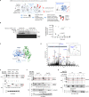
Figure 1
Figure 1
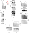
G-REX identifies endogenous privileged first responder (PFR)-cysteines, including two novel sensors, Ube2V1 and Ube2V2, through its capability to genome-wide target-ID endogenous PFRs under electrophile-limited conditions. (A) General setup for G-REX. Treatment of HEK293T cells ectopically expressing HaloTag results in specific binding of the inert photocaged RES-precursor [e.g., Ht-PreHNE(alkyne)]. (In dotted box is the ribbon model of Halo bound to Ht-PreHNE(alkyne), chemical structure of which is shown in Figure S1B). Any unbound probe is washed out. Upon low-energy light exposure of cells (see methods section), this Halo–Ht-PreHNE(alkyne) complex releases a stoichiometric amount of HNE(alkyne) (t1/2 < 1–2 min) (red dot) within the microenvironment of Halo, enabling substoichiometric covalent tagging of native PFRs to HNE. Indicated established pulldown-proteomics analysis permits HT-target-ID genome-wide. (B) (Left) The indicated recombinant protein (12 μM) was treated with HNE-alkyne (12 μM) at 37 °C and aliquots were removed as a function of indicated time and diluted into Click reaction mixture containing Cy5-azide (reporting on HNEylation) and subsequently analyzed by Sypro-Ruby (total protein). (Right) Plot of total unlabeled protein remaining as a function of time based on band-intensity quantitation. Also see Figure S3A. (C) An illustrative model of N-terminal HaloTagged Ube2V2 complexed to Ube2N with the photocaged-precursor, Ht-PreHNE, bound at Halo. (See Figure S1A for general T-REX setup.) Ribbon structure is adapted from PDB: 1J7D. The newly discovered HNE-responsive C69 (this work) within Ube2V2, and the catalytic cysteine (C87) within Ube2N, are indicated. (D) MS/MS spectrum of a triply charged ion at m/z 795.75653+ identifying an HNE alkyne modified peptide: IYSLKVECGPKYPEAPPSVR in which Cys69 of the UBE2 V2 protein digested by ArgC is modified by HNE alkyne with Xcorr score at 2.20 by Proteome Discoverer database search. The MS/MS spectrum was manually inspected and confirmed. The relative low Xcorr score is apparently due to the labile nature of the HNE alkyne which readily loses the HNE alkyne molecule from Cys69 residue during the CID fragmentation. This observation was supported by the two dominant ions (m/z 745.353+ and m/z 1117.372+) in the MS/MS spectrum representing the native targeted peptide (after loss of HNE alkyne). The inset shows the expanded view of MS survey scan for the precursor ion of the HNE-alkyne-modified peptide (m/z 795.75653+) with observed mass accuracy at 1.04 ppm. Also see Tables S1–S2. (E) HEK293T cells were transfected with the indicated plasmids, treated with Ht-PreHNE, and either exposed to light, or not irradiated. Normalized lysates from these two sets of cells were treated with either TEV-protease or buffer alone, respectively. Lysates were then subject to Click coupling with Cy5-azide, and analyzed by in-gel fluorescence for Cy5 signal. “1, 2, and N” respectively designate Ube2V1, Ube2V2, and Ube2N. Refer to Figure S4A for Cy5 gel and blots in full-view. (F) Similar to E but cells were cotransfected with either empty vector (−) or a plasmid of the same vector expressing HA-Ube2N (+). Region of interest in Cy5 gel is marked by a red rectangle. Refer to Figure S4B for Cy5 gel and blots in full-view. (G) Similar to F. Refer to Figure S4C for Cy5 gel and blots in full-view. M designates molecular weight marker lane in all gels/blots in this and all figures elsewhere.
Figure 2
Figure 2
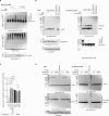
T-REX-assisted Ube2V2(C69)-specific HNEylation elicits K63-linked-polyubiquitination that is lost in the C69S mutant. (A) HNEylation of Ube2V1 (specifically of C94), but not Ube2V2, selectively elicits upregulation in NF-κB-signaling (mean ± s.e.m, n ≥ 2 independent biological replicates). (B) HEK293T cells expressing wt-Halo-(FLAG)-Ube2V2 or the C69S mutant were treated with the DUB-inhibitor PR619 and high-molecular-weight (HMW) polyubiquitylated-Ube2V2 was assessed by western blot. (C) HEK293T cells expressing wt-Halo-(FLAG)-Ube2V2 (or the C69S mutant) and HA-Ubiquitin were exposed to the indicated conditions. Halo-(FLAG)-Ube2V2 from these cells was enriched by FLAG-immunoprecipitation (IP), and resulting samples were analyzed using the indicated antibodies. Left panel = “Input lanes”; Right panel = “IP-samples”. (D) Similar experiment to C, except nonenriched whole cell lysates were analyzed using the indicated antibodies, and an additional sample from the use of no-alkyne-variant of Ht-PreHNE but otherwise treated under identical conditions was also analyzed. (E) Similar experiment to C except global Ub pools were precipitated using HA IP (right panel) (there is no change in Ub since Ub is being IP-ed).
Figure 3
Figure 3
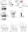
Ube2V2(C69)-specific HNEylation functionally impacts the monoubiquitinated state of Ube2N. (A) HEK293T cells ectopically expressing wt-Halo-(FLAG)-Ube2V2 and HA-Ubiquitin were cotransfected with either empty vector (EV) or a plasmid of the same backbone expressing HA-Ube2N and analyzed for HMW band of HaloUbe2V2 (i.e., Ube2V2-polyUbiquitin) by indicated antibodies. (B) HEK293T cells transfected with the indicated plasmids were subjected to T-REX conditions against controls followed by immunoprecipitation using FLAG resin (Input: top panel; IP: panel on right). Eluates and inputs were analyzed by western blot using indicated antibodies. Levels of Ube2N/Ube2N-monoUb bound to Ube2V2 were analyzed by western blot. See Figure 3C for quantitation. (C) Quantitation of the relative amount of mono-Ub-Ube2N bound to Halo-Ube2V2 enriched from cells subjected to T-REX against controls. See representative blots, for instance, in Figure 3B,D and Figures S5B and S6E (mean ± s.d., n = 3 independent sets of biological replicates at different passages). (D) HEK293T cells transfected with the indicated plasmids were subjected to T-REX conditions against indicated controls. Levels of Ube2N/Ube2N-monoUb bound to Halo-(FLAG)-Ube2V2 were analyzed by western blot subsequent to enrichment using anti-FLAG-beads (IP: right panel). Half of precipitated fractions were treated with NH2OH (conditions known to hydrolyze thioester bonds; detailed in SI methods) and analyzed separately. All samples were exposed to light in this experiment.
Figure 4
Figure 4
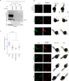
HNEylation of Ube2V2 upregulates γ-H2AX and decreases DNA synthesis: these phenotypes depend on both C69 and Ube2N. (A) Ube2N knockdown lines #16, 17, 18, and control-knockdown line were transfected with Halo-(FLAG)-Ube2V2 and HA-Ubiquitin, then subjected to T-REX against no-“Ht-PreHNE”-controls. HA-Ubiquitin was immunoprecipitated, and inputs (top panels) and elutions (lower panels) were analyzed by western blot using indicated antibodies. See Figure 4B for quantitation [n = 3 independent biological replicates at different passages except shUbe2N-#17 (n = 1)]. (B) Quantitation of data from A. (C) SILAC workflow used to identify proteins that bind preferentially to HNEylated-Ube2V2/Ube2N complex (upper left) and graphical depiction of hits (bottom right). (D) HEK293T cells coexpressing Myc-MCM6 and either wt-Halo-(FLAG)-Ube2V2 or C69S-Halo-(FLAG)-Ube2V2 were subjected to T-REX conditions against “no Ht-PreHNE”-controls. 3-h-Post light exposure, cells were lysed and analyzed by western blot using the indicated antibodies. (E) Similar experiment to D, except cells were transfected with Halo-(Flag)-Ube2V2 (wt- or C69S mutant) and HA-Ubiquitin, and lysates were analyzed for endogenous PCNA-ubquitination using sandwich ELISA [binding: anti-HA(Ub); detection: anti-PCNA (endogenous) antibody] as detailed in SI methods. [mean ± s.d., two independent replicates were performed. N = 3 for each set of cells transfected with either wt- or C69S-Halo-(FLAG)-Ube2V2, under individual experimental conditions as indicated. (F) HEK293T cells were transfected with Halo-(FLAG)-Ube2V2 (or the C69S mutant), then exposed to T-REX conditions. The EdU/BrdU-dual-pulse DNA-labeling (detailed in SI methods) was measured and levels of second pulse (BrdU) from EdU-positive-only cells were quantified and displayed. [mean ± s.e.m., for wt-Halo-(FLAG)-Ube2V2, n = 339 (T-REX), n = 375 (Light alone), n = 300 (Ht-PreHNE alone), n = 465 (DMSO), n = 280 (mitomycin C), for C69S- Halo-(FLAG)-Ube2V2, n = 266 (T-REX), n = 212 (Light alone), n = 283 (Ht-PreHNE alone), n = 312 (DMSO), n = 305 (mitomycin C)]. See Figure S8B for representative images for data in panel F. (G) HEK293T cells were transfected with Halo-(FLAG)-Ube2V2 (or the C69S mutant), then exposed to T-REX conditions. Levels of γ-H2AX were assessed by immunofluorescence (detailed in SI methods) as a function of time post light exposure. [mean ± s.e.m., for wt-Halo-(FLAG)-Ube2V2, n = 648 (0 h), n = 624 (1 h), n = 634 (3 h), n = 571 (6 h), n = 542 (18 h); for C69S- Halo-(FLAG)-Ube2V2, n = 585 (0 h), n = 615 (1 h), n = 644 (3 h), n = 649 (6 h), n = 646 (18 h)]. (H) Ube2N knockdown lines #16 (shUbe2N-16) and control-knockdown line (shLacZ-D11) were transfected with wt-Halo-(FLAG)-Ube2V2, then exposed to T-REX conditions. Levels of γ-H2AX were assessed by immunofluorescence at the similar peak hour (3 h) as in F [mean ± s.e.m., for shUbe2N-#16, n = 379 (T-REX), n = 297 (Light alone), n = 342 (Ht-PreHNE alone), n = 402 (DMSO), n = 241 (Mitomycin C); for shLacZ-D11, n = 434 (T-REX), n = 390 (Light alone), n = 483 (Ht-PreHNE alone), n = 434 (DMSO), n = 445 (Mitomycin C)].
Figure 5
Figure 5
Ube2V2-Specific HNEylation regulates γ-H2AX levels in zebrafish. (A) Casper zebrafish embryos were injected with mRNA-encoding Halo-(FLAG)-Ube2V2 at the 1–4 cell stage. Once injection was complete, eggs were either exposed to Ht-PreHNE or DMSO. After 24-h-incubation, fish were washed and exposed to light. After dechorionation and deyolking at 4 °C, embryos were lysed, biotin was attached using Click chemistry by biotin-azide, and lysates were ethanol precipitated. After resolubilization, biotinylated [i.e., HNE(alkyne)-modified] proteins were pulled down using streptavidin and analyzed by western blot. (See Figure S1A for workflow; Clicking with biotin-azide.) (B) Similar experiment to A, but at 3-h postlight-exposure, fish were fixed, permeabilized, and analyzed by whole-mount immunofluorescence using indicated antibodies. Scale bars, 100 μm. (C) Quantitation of images in B [mean ± s.e.m., n = 69 (T-REX), n = 74 (Light alone), n = 64 (Ht-PreHNE alone), n = 69 (DMSO)].

Similar articles

Cited by

References

    1. Pearlman S. M.; Serber Z.; Ferrell J. E. Jr. A mechanism for the evolution of phosphorylation sites. Cell 2011, 147 (4), 934–46. 10.1016/j.cell.2011.08.052. - DOI - PMC - PubMed
    1. Yau R.; Rape M. The increasing complexity of the ubiquitin code. Nat. Cell Biol. 2016, 18, 579–586. 10.1038/ncb3358. - DOI - PubMed
    1. Wilkinson K. D.; Ventii K. H.; Friedrich K. L.; Mullally J. E. The ubiquitin signal: assembly, recognition and termination. Symposium on ubiquitin and signaling. EMBO Rep. 2005, 6 (9), 815–820. 10.1038/sj.embor.7400506. - DOI - PMC - PubMed
    1. Tarrant M. K.; Cole P. A. The chemical biology of protein phosphorylation. Annu. Rev. Biochem. 2009, 78, 797–825. 10.1146/annurev.biochem.78.070907.103047. - DOI - PMC - PubMed
    1. Love K. R.; Catic A.; Schlieker C.; Ploegh H. L. Mechanisms, biology and inhibitors of deubiquitinating enzymes. Nat. Chem. Biol. 2007, 3 (11), 697–705. 10.1038/nchembio.2007.43. - DOI - PubMed