. 2018 Mar 7;4(3):eaar2631.
doi: 10.1126/sciadv.aar2631. eCollection 2018 Mar.

Mechanisms of KCNQ1 channel dysfunction in long QT syndrome involving voltage sensor domain mutations

Affiliations

Mechanisms of KCNQ1 channel dysfunction in long QT syndrome involving voltage sensor domain mutations

Hui Huang et al. Sci Adv. .

Abstract

Mutations that induce loss of function (LOF) or dysfunction of the human KCNQ1 channel are responsible for susceptibility to a life-threatening heart rhythm disorder, the congenital long QT syndrome (LQTS). Hundreds of KCNQ1 mutations have been identified, but the molecular mechanisms responsible for impaired function are poorly understood. We investigated the impact of 51 KCNQ1 variants with mutations located within the voltage sensor domain (VSD), with an emphasis on elucidating effects on cell surface expression, protein folding, and structure. For each variant, the efficiency of trafficking to the plasma membrane, the impact of proteasome inhibition, and protein stability were assayed. The results of these experiments combined with channel functional data provided the basis for classifying each mutation into one of six mechanistic categories, highlighting heterogeneity in the mechanisms resulting in channel dysfunction or LOF. More than half of the KCNQ1 LOF mutations examined were seen to destabilize the structure of the VSD, generally accompanied by mistrafficking and degradation by the proteasome, an observation that underscores the growing appreciation that mutation-induced destabilization of membrane proteins may be a common human disease mechanism. Finally, we observed that five of the folding-defective LQTS mutant sites are located in the VSD S0 helix, where they interact with a number of other LOF mutation sites in other segments of the VSD. These observations reveal a critical role for the S0 helix as a central scaffold to help organize and stabilize the KCNQ1 VSD and, most likely, the corresponding domain of many other ion channels.


Figures

Fig. 1
Fig. 1. KCNQ1 expression levels and surface trafficking efficiencies.
Data are color-coded: LQTS mutant (red), VUS (blue), or predicted neutral polymorphism (black). For all three panels, reported expression levels are relative to WT results and are expressed as mean ± SEM based on at least three independent experiments. (A) Total expression levels plotted as a scatterplot versus surface expression. The inset boxes illustrate which sets of mutants yield near-zero or WT-like (100%) surface expression levels. (B) Surface trafficking efficiency versus total expression level, where efficiency is defined as [(surface)mutant/(total)mutant]/[(surface)WT/(total)WT] × 100. (C) Cell surface expression levels as measured in this study versus K+ channel peak current density, as originally reported elsewhere (22). The vertical red lines indicate 65% of WT, the effective upper limit cutoff for LOF.
Fig. 2
Fig. 2. Treatment of cells with a proteasome inhibitor (MG132) has modest impact on surface expression levels of the trafficking-deficient KCNQ1 variants (A) but often increases the total expression (B).
Cells expressing WT or mutant KCNQ1 were treated with 25 μM MG132 or vehicle for 20 hours. Cells were fixed and permeabilized (this step was omitted for measuring the surface expression) and then stained with myc-tag mouse monoclonal antibody. Cells were then washed and stained with anti-mouse Alexa Fluor 647 antibody. Immunostaining of 2500 cells was quantitated by flow cytometry. Results are expressed as mean ± SEM for at least three independent experiments. Data are color-coded: LQTS mutant (red), VUS (blue), or predicted neutral polymorphism (black). Data labeled with an asterisk and a horizontal bar indicate those for which the measured KCNQ1 protein level for vehicle-treated cells was statistically different from the level measured in MG132-treated cells (P < 0.05).
Fig. 3
Fig. 3. Effect on KCNQ1 trafficking of coexpression of WT KCNQ1 with mutant KCNQ1.
(A) Total WT + mutant expression levels and (B) surface WT + mutant expression levels. HEK293 cells were transiently transfected with either 0.5 μg WT or mutant plasmid only (results on the left of each panel) or were cotransfected with both 0.25 μg WT and 0.25 μg mutant plasmids (heterozygous conditions, results presented on the right of each panel). See the legend of Fig. 1 for additional details.
Fig. 4
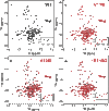
Fig. 4. NMR spectra of WT KCNQ1 and representative mutant forms.
1H/15N-TROSY NMR spectra (900 MHz) of the WT KCNQ1 VSD (residues 100 to 249) (A) and representative mutant forms (B to D). ppm, parts per million. The spectrum of each mutant VSD is shown in red, superimposed on the black spectrum of the WT VSD spectrum. Data were collected at 50°C for WT and mutant forms of VSD in LMPG micelles at pH 5.5. The LMPG concentration for all samples was 50 to 80 mM, and the KCNQ1 VSD concentration was 0.3 mM in all samples. (B) The spectrum from the V110I mutant, for which the only changes relative to the WT spectrum are shifts in peak positions. (C) The spectrum from the H126L mutant that is deemed to be moderately destabilized on the basis of a modest degree of line broadening for a number of peaks relative to the corresponding peaks in the WT spectrum. (D) The spectrum from the E115G mutant, which is deemed to be severely destabilized on the basis of extensive peak broadening and even disappearance of a number of peaks.
Fig. 5
Fig. 5. Structural locations and key interactions involving mutation sites and/or S0 in the KCNQ1 VSD.
(A) Three views of the VSD illustrating the locations of the five sites in S0 (red side chains: Q107, Y111, L114, E115, and P117) subject to LQTS mutations that destabilize the VSD, resulting in mistrafficking and degradation of KCNQ1. The side chains for four residues that interact with these S0 residues and that are also subject to destabilizing VUS or LQTS mutations resulting in channel LOF are shown in magenta. The open-state VSD coordinates from the cryo–electron microscopy (EM) structure of KCNQ1 [Protein Data Bank (PDB) ID: 5VMS] were used to generate this figure (23). The V110 LQTS mutation site is also located in S0, but the mutation does not appear to destabilize the protein. (B to F) Results from the MD simulation of the KCNQ1 VSD. (B) Structural model of the WT open-state human VSD in a DMPC bilayer after 500 ns of MD. The VSD is displayed in cartoon representation, with S1-S4 colored pale green and the S0 helix colored cyan. DMPC molecules are depicted as spheres and colored by atom identity: C, gray; O, red; N, blue; P, orange. (C to F) Nonbonded interactions involving sites in S0 and sites contacting S0 for which LQTS and VUS mutations were characterized in this study (see Results). Amino acid side chains are drawn as sticks. LQTS and VUS mutation sites are colored light red and blue, respectively. Residues for which mutations are neutral or have not been characterized in this work are colored gray and green, respectively. Predicted hydrogen bond interactions are indicated by black dotted lines and atoms. The nature of the nonbonded interactions involving S0 is further described in the main body of the text and in table S2.

References

    1. Abbott G. W., KCNE1 and KCNE3: The yin and yang of voltage-gated K+ channel regulation. Gene 576, 1–13 (2016). - PMC - PubMed
    1. Cui J., Voltage-dependent gating: Novel insights from KCNQ1 channels. Biophys. J. 110, 14–25 (2016). - PMC - PubMed
    1. Jespersen T., Grunnet M., Olesen S.-P., The KCNQ1 potassium channel: From gene to physiological function. Physiology 20, 408–416 (2005). - PubMed
    1. Liin S. I., Barro-Soria R., Larsson H. P., The KCNQ1 channel—Remarkable flexibility in gating allows for functional versatility. J. Physiol. 593, 2605–2615 (2015). - PMC - PubMed
    1. Nakajo K., Kubo Y., KCNQ1 channel modulation by KCNE proteins via the voltage-sensing domain. J. Physiol. 593, 2617–2625 (2015). - PMC - PubMed

Publication types

MeSH terms

Substances