. 2017 Dec:4:87-96.
doi: 10.1016/j.cobme.2017.09.006. Epub 2017 Sep 22.

Progress and Remaining Challenges in the Application of High Frequency Oscillations as Biomarkers of Epileptic Brain

Affiliations

Progress and Remaining Challenges in the Application of High Frequency Oscillations as Biomarkers of Epileptic Brain

Fatemeh Khadjevand et al. Curr Opin Biomed Eng. 2017 Dec.

Abstract

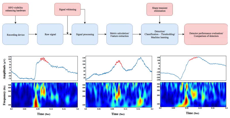
High-frequency oscillations (HFOs: 100 - 600 Hz) have been widely proposed as biomarkers of epileptic brain tissue. In addition, HFOs over a broader range of frequencies spanning 30 - 2000 Hz are potential biomarkers of both physiological and pathological brain processes. The majority of the results from humans with focal epilepsy have focused on HFOs recorded directly from the brain with intracranial EEG (iEEG) in the high gamma (65 - 100 Hz), ripple (100 - 250 Hz), and fast ripple (250 - 600 Hz) frequency ranges. These results are supplemented by reports of HFOs recorded with iEEG in the low gamma (30 - 65Hz) and very high frequency (500 - 2000 Hz) ranges. Visual detection of HFOs is laborious and limited by poor inter-rater agreement; and the need for accurate, reproducible automated HFOs detection is well recognized. In particular, the clinical translation of HFOs as a biomarker of the epileptogenic brain has been limited by the ability to reliably detect and accurately classify HFOs as physiological or pathological. Despite these challenges, there has been significant progress in the field, which is the subject of this review. Furthermore, we provide data and corresponding analytic code in an effort to promote reproducible research and accelerate clinical translation.


Figures

Figure 1
Figure 1. Generic algorithm flow chart of automated HFOs detection
A) Schematic of automatic HFOs detection. The brain activity is recorded with a wide bandwidth acquisition system. Then, the raw broadband electrophysiologic recording is “whitened” to better highlight the low amplitude HFOs transients. A wide range of features can be extracted to aid classification. After the feature extraction through different methods, the candidate events can be classified using a wide range of classification approaches, like machine learning. The output of these detectors should be evaluated and have comparable outputs from other detectors and laboratories. Red boxes correspond to the advances in HFOs detection and blue boxes are the established flow of HFOs detection. B) Three HFOs detected in a wide bandwidth recording from human brain. Each figure is composed, from top to bottom, of the raw signal (in blue) with the automatically detected HFOs (red) in the signal, and the continuous time frequency image.
Figure 2
Figure 2. A) Cognitive task paradigm for studying physiological nHFOs
Patients with epilepsy who are implanted with subdural and depth electrodes for seizure monitoring as part of the clinical evaluation for drug-resistant epilepsy provide a unique opportunity for neuroscientists to perform cognitive tasks and record the electrophysiological signals directly from the human brain. Areas of active research include investigation of the neural correlates of cognition. These cognitive tasks may help to improve the discrimination of nHFOs from pHFOs. In our study, we used iEEG recorded during a verbal memory task in 11 epilepsy patients to analyze gamma frequency events within and outside the seizure generating brain regions. A. a) Diagram of free recall verbal memory task, A. b) Epileptic patients participating in the study during second phase monitoring doing verbal memory tasks on a laptop, A. c) brain surface of an example patient with implanted grid and strip electrodes the iEEG signal is recording from (unpublished data). B) From top to bottom, B. a) shows the trial-averaged ERP signal for a whole session (300 words presented), plots below are raw data from individual example trial during same session, that subject subsequently recalled the presented word. From top to bottom, the unfiltered EEG (fig 2. B. b), the band-pass filtered signal for high gamma activity (HGA) (fig 2. B. c), and the continuous time frequency image with detected HGAs highlighted with black rectangles (fig 2. B. d). Red lines are stimulus (word presentation) onset and offset (F Khadjevand et al., unpublished).
Figure 3
Figure 3. Brain mapping using physiologic task-induced HFOs and pathological HFOs Top, left)
Word lists are presented one-by-one for encoding & subsequent recall. Presentation of words induces high gamma activities (60–120 Hz) in specific brain areas. In this figure subsequently recalled words printed in red and forgotten words presented in blue. Top, right) Spectrogram of local field power during encoding epochs aligned to the presented word. Bottom) Left panel, brain surface maps of high physiological gamma power interpolated over 4×6 temporal cortex grid (White arrow focus of high activation) and in right panel the pathological HFOs over the epileptic region of the brain.

Similar articles

Cited by

References

    1. Buzsáki G, da Silva FL. High frequency oscillations in the intact brain. Progress in neurobiology. 2012;98(3):241–249. - PMC - PubMed
    1. Engel J, Jr, Bragin A, Staba R, Mody I. High-frequency oscillations: What is normal and what is not? Epilepsia. 2009;50(4):598–604. - PubMed
    1. Kucewicz MT, Cimbalnik J, Matsumoto JY, Brinkmann BH, Bower MR, Vasoli V, Sulc V, Meyer F, Marsh WR, Stead SM, Worrell GA. High frequency oscillations are associated with cognitive processing in human recognition memory. Brain. 2014;137(Pt 8):2231–2244. - PMC - PubMed
    1. Worrell GA, Gardner AB, Stead SM, Hu S, Goerss S, Cascino GJ, Meyer FB, Marsh R, Litt B. High-frequency oscillations in human temporal lobe: Simultaneous microwire and clinical macroelectrode recordings. Brain. 2008;131(4):928–937. - PMC - PubMed
    1. Bragin A, Mody I, Wilson CL, Engel J. Local generation of fast ripples in epileptic brain. Journal of Neuroscience. 2002;22(5):2012–2021. - PMC - PubMed

LinkOut - more resources