

Physiology of Cardiac Development: From Genetics to Signaling to Therapeutic Strategies
- PMID: 29532042
- PMCID: PMC5844510
- DOI: 10.1016/j.cophys.2017.09.002
Physiology of Cardiac Development: From Genetics to Signaling to Therapeutic Strategies
Abstract
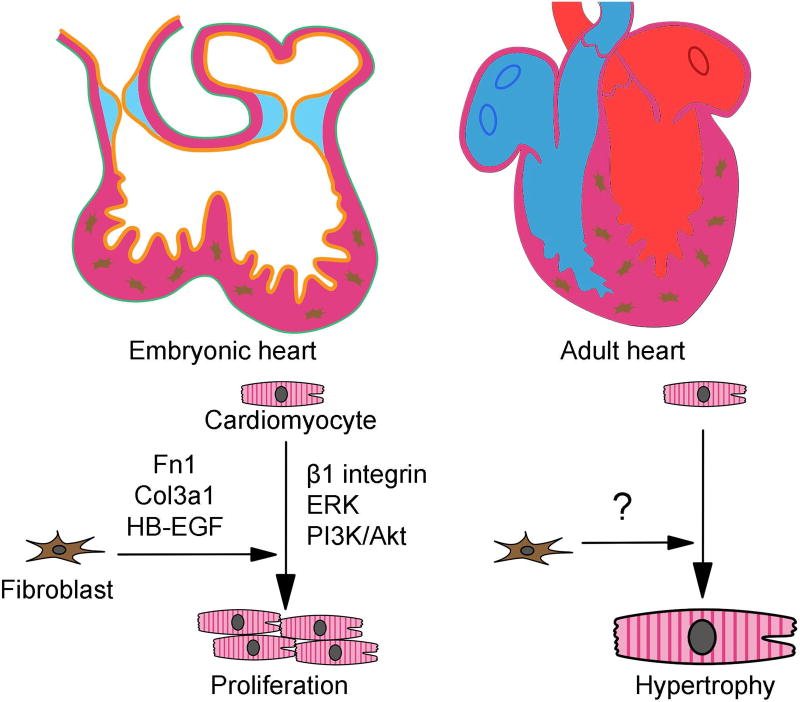
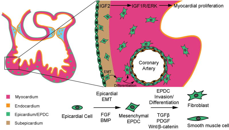
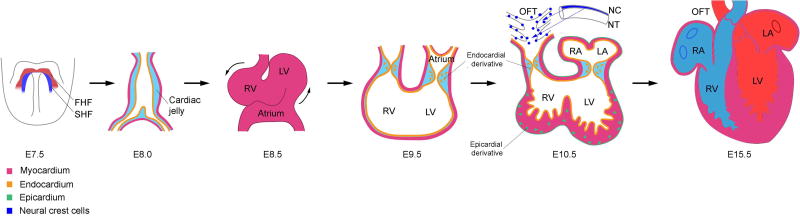
The heart is one of the first organs to form and function during embryonic development. It is comprised of multiple cell lineages, each integral for proper cardiac development, and include cardiomyocytes, endothelial cells, epicardial cells and neural crest cells. The molecular mechanisms regulating cardiac development and morphogenesis are dependent on signaling crosstalk between multiple lineages through paracrine interactions, cell-ECM interactions, and cell-cell interactions, which together, help facilitate survival, growth, proliferation, differentiation and migration of cardiac tissue. Aberrant regulation of any of these processes can induce developmental disorders and pathological phenotypes. Here, we will discuss each of these processes, the genetic factors that contribute to each step of cardiac development, as well as the current and future therapeutic targets and mechanisms of heart development and disease. Understanding the complex interactions that regulate cardiac development, proliferation and differentiation is not only vital to understanding the causes of congenital heart defects, but to also finding new therapeutics that can treat both pediatric and adult cardiac disease in the near future.
Keywords: congenital; development; disease; genetics; heart; signaling.
Conflict of interest statement
The authors wish to disclose that there are no conflicts of interest.
Figures


Similar articles
-
Endocardial-Myocardial Interactions During Early Cardiac Differentiation and Trabeculation.Front Cardiovasc Med. 2022 May 4;9:857581. doi: 10.3389/fcvm.2022.857581. eCollection 2022. Front Cardiovasc Med. 2022. PMID: 35600483 Free PMC article. Review.
-
Role of the vascular endothelial growth factor isoforms in retinal angiogenesis and DiGeorge syndrome.Verh K Acad Geneeskd Belg. 2005;67(4):229-76. Verh K Acad Geneeskd Belg. 2005. PMID: 16334858 Review.
-
Cardiac outflow tract defects in mice lacking ALK2 in neural crest cells.Development. 2004 Jul;131(14):3481-90. doi: 10.1242/dev.01214. Development. 2004. PMID: 15226263
-
Misregulation of SDF1-CXCR4 signaling impairs early cardiac neural crest cell migration leading to conotruncal defects.Circ Res. 2013 Aug 16;113(5):505-16. doi: 10.1161/CIRCRESAHA.113.301333. Epub 2013 Jul 9. Circ Res. 2013. PMID: 23838132
-
Cardiac neural crest.Semin Cell Dev Biol. 2005 Dec;16(6):704-15. doi: 10.1016/j.semcdb.2005.06.004. Epub 2005 Jul 27. Semin Cell Dev Biol. 2005. PMID: 16054405 Review.
Cited by
-
Single-cell reconstruction and mutation enrichment analysis identifies dysregulated cardiomyocyte and endothelial cells in congenital heart disease.Physiol Genomics. 2023 Dec 1;55(12):634-646. doi: 10.1152/physiolgenomics.00070.2023. Epub 2023 Oct 9. Physiol Genomics. 2023. PMID: 37811720 Free PMC article.
-
scaRNA1 Levels Alter Pseudouridylation in Spliceosomal RNA U2 Affecting Alternative mRNA Splicing and Embryonic Development.Pediatr Cardiol. 2020 Feb;41(2):341-349. doi: 10.1007/s00246-019-02263-4. Epub 2020 Jan 17. Pediatr Cardiol. 2020. PMID: 31953571
-
Telomeres and Telomerase in Heart Ontogenesis, Aging and Regeneration.Cells. 2020 Feb 22;9(2):503. doi: 10.3390/cells9020503. Cells. 2020. PMID: 32098394 Free PMC article. Review.
-
Cardiomyogenesis Modeling Using Pluripotent Stem Cells: The Role of Microenvironmental Signaling.Front Cell Dev Biol. 2019 Aug 9;7:164. doi: 10.3389/fcell.2019.00164. eCollection 2019. Front Cell Dev Biol. 2019. PMID: 31448277 Free PMC article. Review.
-
A genome-wide association study of obstructive heart defects among participants in the National Birth Defects Prevention Study.Am J Med Genet A. 2022 Aug;188(8):2303-2314. doi: 10.1002/ajmg.a.62759. Epub 2022 Apr 22. Am J Med Genet A. 2022. PMID: 35451555 Free PMC article.
References
-
- Chong JJ, Forte E, Harvey RP. Developmental origins and lineage descendants of endogenous adult cardiac progenitor cells. Stem Cell Res. 2014;13:592–614. - PubMed
-
- Katz AM. Physiology of the Heart. fifth. Lippincott Williams & Wilkins; Philaddelphia: 2011.
Grants and funding
LinkOut - more resources
Full Text Sources
Other Literature Sources