

Method and reliability of measuring midurethral area and echogenicity, and changes during and after pregnancy
- PMID: 29532128
- PMCID: PMC6132674
- DOI: 10.1007/s00192-018-3580-z
Method and reliability of measuring midurethral area and echogenicity, and changes during and after pregnancy
Abstract
Introduction and hypothesis: Internal closure of the urethral sphincter is one of the mechanisms in maintaining continence. Little is known about changes in the urethral sphincter during pregnancy. We designed this study to develop a reliable method to measure the area and mean echogenicity of the midurethra during and after pregnancy and to assess changes over time.
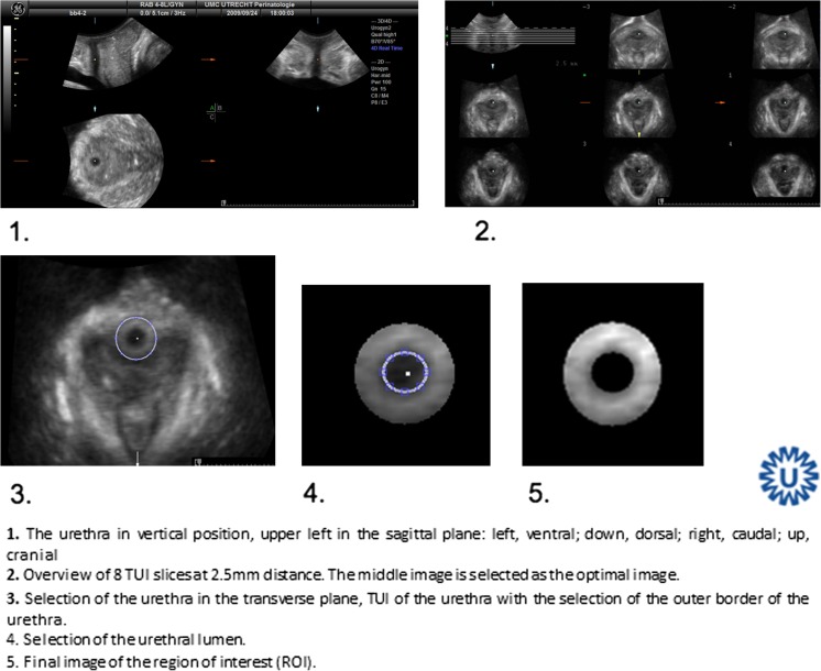
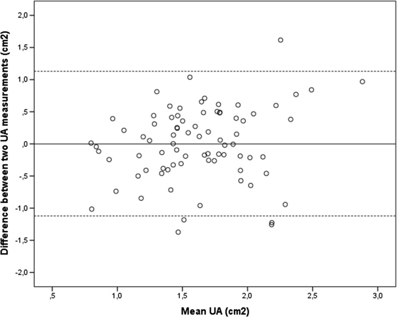
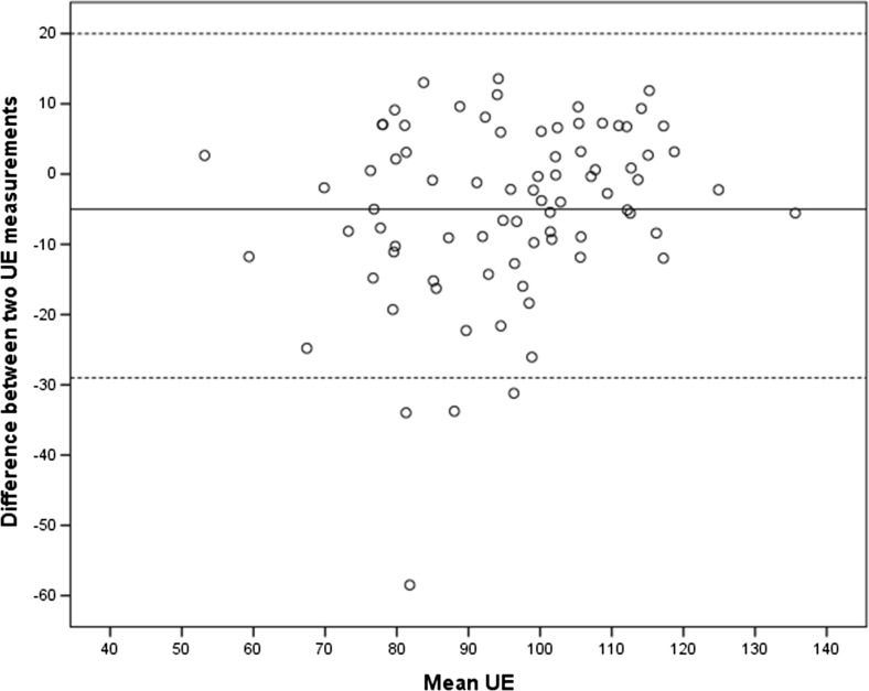
Methods: Two observers independently segmented the urethra as follows: in the sagittal plane, the urethra was positioned vertically, the marker was placed in the middle section of the lumen of the urethra, and eight tomographic US images of 2.5 -mm slices were obtained. The central image was selected, and area and mean echogenicity were calculated automatically. Intra- and interobserver reliability were determined by intraclass correlation coefficients (ICC) and their 95% confidence intervals (CI). Two hundred and eighty women underwent TPUS at 12 weeks and 36 weeks of gestation and 6 months postpartum, and 3D/4D transperineal ultrasound (TPUS) images of 40 pregnant nulliparous women were used for the reliability study. Paired t tests were used to assess changes in echogenicity and area.
Results: The ICC for measuring the area was substantial, at 0.77 and for measuring mean echogenicity was almost perfect, at 0.86. In the total study group (n = 280), midurethral area and mean echogenicity were significantly lower 6 months after delivery compared with 12 and 36 weeks of gestation.
Conclusions: Our protocol for measuring area and mean echogenicity of the midurethra is reliable. This study indicates that structural changes in the midurethraoccur during pregnancy.
Keywords: 3D/4D; Area; Echogenicity; Transperineal ultrasound; Urethra.
Conflict of interest statement
None.
Figures

Similar articles
-
Measuring echogenicity and area of the puborectalis muscle: method and reliability.Ultrasound Obstet Gynecol. 2014 Oct;44(4):481-5. doi: 10.1002/uog.13409. Ultrasound Obstet Gynecol. 2014. PMID: 24817256
-
Reliability of pelvic floor measurements on three- and four-dimensional ultrasound during and after first pregnancy: implications for training.Ultrasound Obstet Gynecol. 2013 Nov;42(5):590-5. doi: 10.1002/uog.12523. Ultrasound Obstet Gynecol. 2013. PMID: 23729398
-
Reliability of a new method for assessing urethral compression following midurethral tape procedures using four-dimensional ultrasound.Ultrasound Obstet Gynecol. 2011 Aug;38(2):210-6. doi: 10.1002/uog.9003. Ultrasound Obstet Gynecol. 2011. PMID: 21425199
-
Intraobserver and interobserver reliability of the three-dimensional ultrasound imaging of female urethral sphincter using a translabial technique.Int Urogynecol J. 2012 Aug;23(8):1063-8. doi: 10.1007/s00192-012-1669-3. Epub 2012 Jan 21. Int Urogynecol J. 2012. PMID: 22270730
-
Three-dimensional ultrasound: a novel technique for investigating the urethral sphincter in the third trimester of pregnancy.Ultrasound Obstet Gynecol. 2001 May;17(5):421-4. doi: 10.1046/j.1469-0705.2001.00354.x. Ultrasound Obstet Gynecol. 2001. PMID: 11380967
References
-
- Dietz HP. Ultrasound imaging of the pelvic floor. Part II: three-dimensional or volume imaging. Ultrasound Obstet Gynecol. 2004; 10.1002/uog.1072. - PubMed
-
- Cosimato C, Cipullo LM, Troisi J, Di Spiezio SA, Tommaselli GA, Oro RR, et al. Ultrasonographic evaluation of urethrovesical junction mobility: correlation with type of delivery and stress urinary incontinence. Int Urogynecol J. 2015; 10.1007/s00192-015-2736-3. - PubMed
-
- van Veelen GA, Schweitzer KJ, van der Vaart CH. Ultrasound imaging of the pelvic floor: changes in anatomy during and after first pregnancy. Ultrasound Obstet Gynecol. 2014; 10.1002/uog.13301. - PubMed
-
- Falkert A, Willmann A, Endress E, Meint P, Seelbach-Gobel B. Three-dimensional ultrasound of pelvic floor: is there a correlation with delivery mode and persisting pelvic floor disorders 18-24 months after first delivery? Ultrasound Obstet Gynecol. 2013; 10.1002/uog.11214. - PubMed
-
- Wijma J, Weis Potters AE, de Wolf BT, Tinga DJ, Aarnoudse JG. Anatomical and functional changes in the lower urinary tract during pregnancy. BJOG. 2001. - PubMed
MeSH terms
LinkOut - more resources
Full Text Sources
Other Literature Sources
