. 2018 Jun;247(6):818-831.
doi: 10.1002/dvdy.24627. Epub 2018 Apr 10.

A novel role for cilia-dependent sonic hedgehog signaling during submandibular gland development

Affiliations

A novel role for cilia-dependent sonic hedgehog signaling during submandibular gland development

Kelsey H Elliott et al. Dev Dyn. 2018 Jun.

Abstract

Background: Submandibular glands (SMGs) are specialized epithelial structures which generate saliva necessary for mastication and digestion. Loss of SMGs can lead to inflammation, oral lesions, fungal infections, problems with chewing/swallowing, and tooth decay. Understanding the development of the SMG is important for developing therapeutic options for patients with impaired SMG function. Recent studies have suggested Sonic hedgehog (Shh) signaling in the epithelium plays an integral role in SMG development; however, the mechanism by which Shh influences gland development remains nebulous.

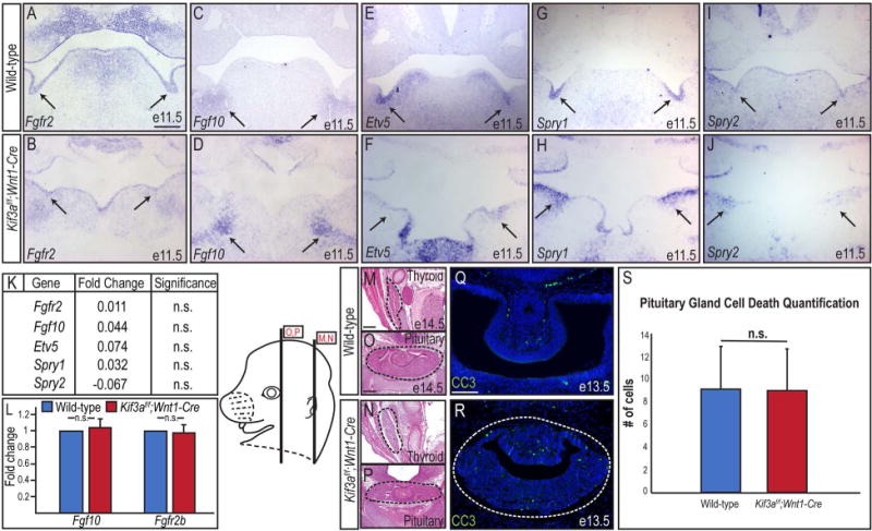
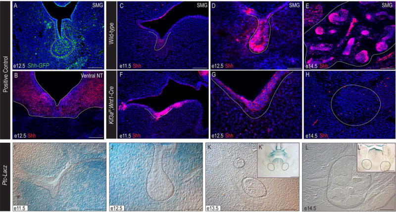
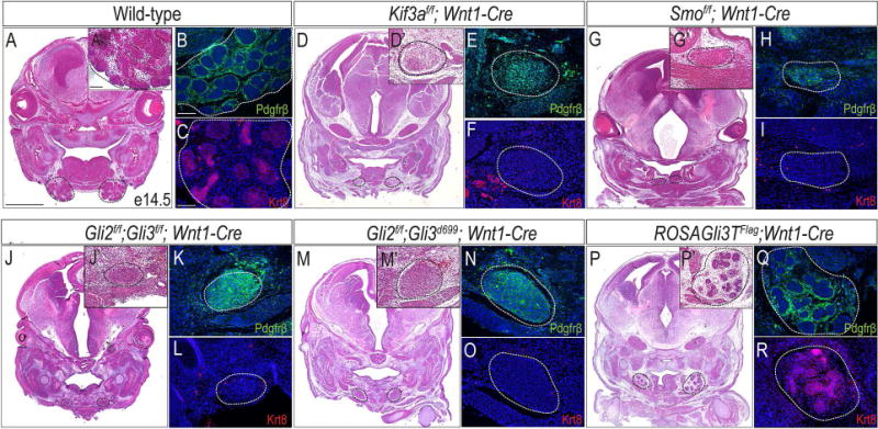
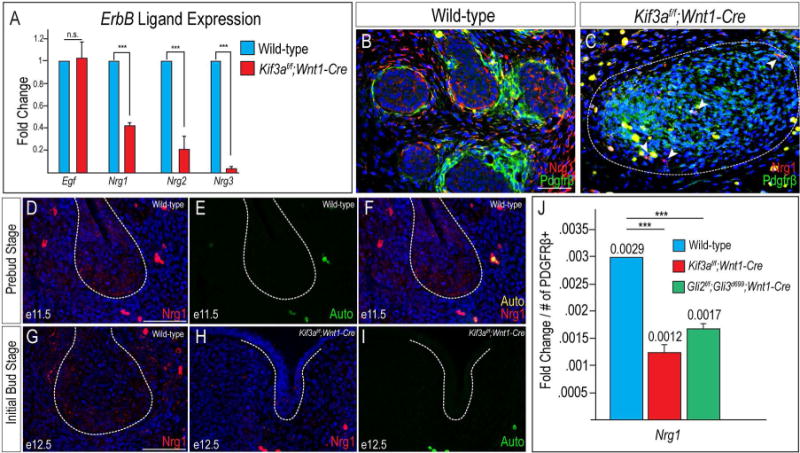
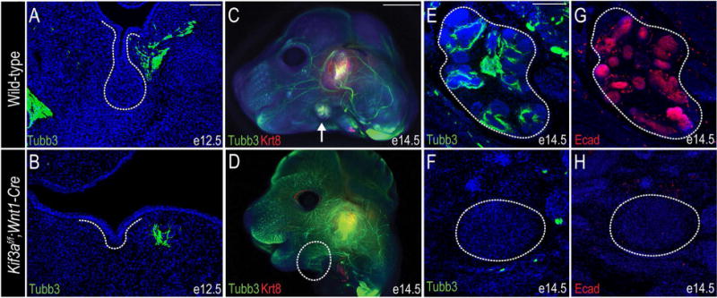
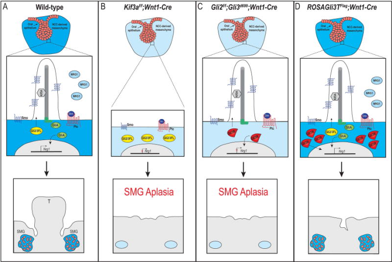
Results: Using the Kif3af/f ;Wnt1-Cre ciliopathic mouse model to prevent Shh signal transduction by means of the loss of primary cilia in neural crest cells, we report that mesenchymal Shh activity is necessary for gland development. Furthermore, using a variety of murine transgenic lines with aberrant mesenchymal Shh signal transduction, we determine that loss of Shh activity, by means of loss of the Gli activator, rather than gain of Gli repressor, is sufficient to cause the SMG aplasia. Finally, we determine that loss of the SMG correlates with reduced Neuregulin1 (Nrg1) expression and lack of innervation of the SMG epithelium.

Conclusions: Together, these data suggest a novel mechanistic role for mesenchymal Shh signaling during SMG development. Developmental Dynamics 247:818-831, 2018. © 2018 Wiley Periodicals, Inc.

Keywords: Gli; Hedgehog; primary cilia; submandibular salivary glands.


Figures

Figure 1
Figure 1. Conditional loss of primary cilia on NCCs results in aplasia of the SMG
(A, B) H&E on e11.5 frontal sections through wild-type and Kif3af/f;Wnt1-Cre SMG prebud epithelium (dotted black lines and black arrows). (A’, B’) High-magnification images of A, B. (C, D) H&E on e12.5 frontal sections through wild-type and Kif3af/f;Wnt1-Cre initial bud SMG (dotted black lines). (E, F) H&E on e14.5 frontal sections through wild-type and Kif3af/f;Wnt1-Cre mutant embryos showing the presence and absence of SMGs (dotted black lines), respectively. (E’, F’) High-magnification images of E, F. (G-J) Anti-Pdgfrβ (green) and Anti-Krt8 (red) immunostaining on e14.5 wild-type and Kif3af/f;Wnt1-Cre mutant SMGs. The (G, I) SMG or (H, J) mutant mesenchymal capsule are indicated with dotted white lines. Nuclei are counterstained with Hoechst. (K, L) Anti-Sox9 (red) immunostaining on e11.5 wild-type and Kif3af/f;Wnt1-Cre mutant embryos, showing the presence (dotted white lines) or absence (white asterisks) of Sox9 expression in the prebud epithelium. Nuclei are counterstained with Hoechst. (M, N) Anti-Arl13b (green) immunostaining on e11.5 wild-type and Kif3af/f;Wnt1-Cre developing SMGs (dotted white lines). Nuclei are counterstained with Hoechst. (O) Quantification of CC3-positive cells in the e11.5 prebud epithelium and mesenchyme of wild-type and Kif3af/f;Wnt1-Cre mutants. (P) Quantification of PHH3-positive cells in the e11.5 prebud epithelium and mesenchyme of wild-type and Kif3af/f;Wnt1-Cre mutants. (Q) X-gal staining on a frontal section through the developing SMG of an e14.5 R26R;Wnt1-Cre embryo. The SMG is indicated with a dotted black line. (R) Frontal section through the developing SMG of a ROSAmT/mG;Wnt1-Cre embryo at e14.5. NCCs express EGFP (green). Non-NCCs express Td Tomato (red). Scale bars= (A-D, E’, F’, G-J) 100μm; (E, F) 1mm; (K, L) 20μm; (M, N, R) 200μm; (Q) 500μm.
Figure 2
Figure 2. Fgf signaling is maintained in Kif3af/f;Wnt1-Cre mutants
(A-J) Sectioned in situ hybridization for Fgf10, Fgfr2, Etv5, Spry1, and Spry2 in SMG epithelium (black arrows) of e11.5 wild-type and Kif3af/f;Wnt1-Cre mutant embryos. (K) Fold change in expression levels of Fgf10, Fgfr2, Etv5, Spry1, and Spry2 by RNA-Sequencing in e11.5 Kif3af/f;Wnt1-Cre mutant mandibular prominences compared to e11.5 wild-type mandibular prominences. (L) RT-qPCR for Fgf10 and Fgfr2b transcript levels in wild-type SMGs and Kif3af/f;Wnt1-Cre mutant mesenchymal capsules. (M-P) H&E on frontal sections through the thyroid and pituitary glands (dotted black lines) of e14.5 wild-type and Kif3af/f;Wnt1-Cre embryos. (Q, R) Anti-CC3 immunostaining (green) on e13.5 wild-type and Kif3af/f;Wnt1-Cre pituitary glands (dotted white lines). Nuclei counterstained with Hoechst. (S) Quantification of Q and R. Scale bars= (A-J, M-P) 200μm; (Q, R) 100μm.
Figure 3
Figure 3. Shh pathway is active during early SMG development
(A) Anti-GFP immunostaining (green) on e12.5 Shh-GFP SMG (dotted white line). (B) Anti-Shh immunostaining (red) on e12.5 ventral neural tube (dotted white line). (C-H) Anti-Shh immunostaining (red) on e11.5, e12.5, and e14.5 wild-type and Kif3af/f;Wnt1-Cre mutant SMGs (dotted white lines). Nuclei counterstained with Hoechst. (I-L) X-gal staining on frontal sections of Ptc-LacZ embryos. Dotted black lines indicate developing SMGs. (K’, L’) High-magnification images of K and L. (A, B, C, E, F, H, I) 100μm; (D, G, J) 50μm; (K, L) 500μm; (K’, L’) 200μm.
Figure 4
Figure 4. Shh and GliA activity are required in NCCs for SMG development
(A, D, G, J, M, P) H&E staining on frontal sections of wild-type, Kif3af/f;Wnt1-Cre, Smof/f;Wnt1-Cre, Gli2f/f;Gli3f/f;Wnt1-Cre, Gli2f/f;Gli3d699;Wnt1-Cre, and ROSAGli3TFlag;Wnt1-Cre embryos. Dotted black lines indicate developing and mutant SMGs. (A’, D’, G’, J’, M’, P’) High-magnification images of A, D, G, J, M, P. (B, E, H, K, N, Q) Anti-Pdgfrβ immunostaining (green) on wild-type, Kif3af/f;Wnt1-Cre, Smof/f;Wnt1-Cre, Gli2f/f;Gli3f/f;Wnt1-Cre, Gli2f/f;Gli3d699;Wnt1-Cre, and ROSAGli3TFlag;Wnt1-Cre SMGs or mutant mesenchymal capsules (dotted white lines). (C, F, I, L, O, R) Anti-Krt8 immunostaining (red) on wild-type, Kif3af/f;Wnt1-Cre, Smof/f;Wnt1-Cre, Gli2f/f;Gli3f/f;Wnt1-Cre, Gli2f/f;Gli3d699;Wnt1-Cre, and ROSAGli3TFlag;Wnt1-Cre SMGs or mutant mesenchymal capsules (dotted white lines). Nuclei counterstained with Hoechst. Scale bars= (A, D, G, J, M, P) 1mm; (A’, B, C, D’, E, F, G’, H, I, J’, K, L, M’, N, O, P’, Q, R) 100μm.
Figure 5
Figure 5. Nrg1 expression is reduced in ciliopathic and GliA mutants
(A) RT-qPCR for Egf, Nrg1, Nrg2, and Nrg3 transcript levels in wild-type SMGs and Kif3af/f;Wnt1-Cre mutant mesenchymal capsules. (B, C) Anti-Nrg1 (red) and anti-Pdgfrβ (green) co-immunostaining on e14.5 wild-type SMGs and Kif3af/f;Wnt1-Cre mutant mesenchymal capsules (dotted white lines). White arrow heads in C indicate a small number of Nrg1-positive cells in the Kif3af/f;Wnt1-Cre mutant mesenchymal capsules. (D) Anti-Nrg1 immunostaining (red) on frontal section of a wild-type e11.5 SMG (dotted white lines). (E) Auto-fluorescence of red blood cells from the 488 filter in D. (F) Merged images from D and E. (G, H) Anti-Nrg1 immunostaining (red) on frontal sections of e12.5 wild-type and Kif3af/f;Wnt1-Cre SMGs (dotted white line). Nuclei counterstained with Hoechst. (I) Auto-fluorescence of red blood cells from the 488 filter in H. (J) RT-qPCR for Nrg1 transcript levels in wild-type, Kif3af/f;Wnt1-Cre, and Gli2f/f;Gli3d699;Wnt1-Cre SMGs or mutant mesenchymal capsules. Fold change of transcript levels were normalized to Pdgfrβ+ cells. ***=p<0.001 Scale bars= (B-I) 50μm.
Figure 6
Figure 6. Loss of primary cilia on NCCs does not impair differentiation into cranial nerves, but does impair innervation
(A, B) Anti-βIII-tubulin (Tubb3) immunostaining (green) on e12.5 wild-type and Kif3af/f;Wnt1-Cre mutant SMGs (dotted white lines). (C, D) Whole mount co-immunostaining for anti-Tubb3 (green) and anti-Krt8 (red) on wild-type and Kif3af/f;Wnt1-Cre e14.5 heads. White arrow in C indicates SMG; dotted white line in B indicates remaining SMG mesenchymal capsule. (E, F) Anti-Tubb3 immunostaining (green) on frontal sections through e14.5 wild-type and Kif3af/f;Wnt1-Cre SMGs (dotted white line). (G, H) Anti-E-cadherin immunostaining (red) on frontal sections through e14.5 wild-type and Kif3af/f;Wnt1-Cre SMGs (dotted white line). Nuclei are counterstained with Hoechst. Scale bars= (A, B, E-H) 100μm; (C, D) 2mm.
Figure 7
Figure 7. Hypothesized model for primary cilia and GliA-dependent SMG development
(A) In response to Shh binding to Ptc in wild-type SMG mesenchymal cells, Gli2/3FL is shuttled through the primary cilia and processed into GliA. GliA moves to the nucleus to induce transcription of downstream genes (either directly or indirectly; dotted black line), possibly including Nrg1. SMG organogenesis occurs. (B) In Kif3af/f;Wnt1-Cre mutant embryos, cilia are lost on the NCC-derived mesenchyme and Gli2/3FL cannot be processed into GliA. Nrg1 expression is not induced and SMG aplasia occurs. (C) In Gli2f/f;Gli3f/f;Wnt1-Cre mutant embryo, primary cilia remain, but functional GliA is not produced. Nrg1 expression is not induced and SMG aplasia occurs. (D) In ROSAGli3TFlag;Wnt1-Cre mutant embryos, primary cilia remain, but an excessive amount of GliR are produced. Functional GliA is still produced and moves to the nucleus to induce transcription of downstream genes (either directly or indirectly; doted black line), possibly including Nrg1. SMG organogenesis occurs. T, tongue.

References

    1. Aberger F, Ruiz IAA. Context-dependent signal integration by the GLI code: the oncogenic load, pathways, modifiers and implications for cancer therapy. Semin Cell Dev Biol. 2014;33:93–104. - PMC - PubMed
    1. Belloni E, Muenke M, Roessler E, Traverso G, Siegel-Bartelt J, Frumkin A, Mitchell HF, Donis-Keller H, Helms C, Hing AV, Heng HH, Koop B, Martindale D, Rommens JM, Tsui LC, Scherer SW. Identification of Sonic hedgehog as a candidate gene responsible for holoprosencephaly. Nat Genet. 1996;14:353–356. - PubMed
    1. Bitgood MJ, McMahon AP. Hedgehog and Bmp genes are coexpressed at many diverse sites of cell-cell interaction in the mouse embryo. Dev Biol. 1995;172:126–138. - PubMed
    1. Bose J, Grotewold L, Ruther U. Pallister-Hall syndrome phenotype in mice mutant for Gli3. Hum Mol Genet. 2002;11:1129–1135. - PubMed
    1. Brugmann SA, Allen NC, James AW, Mekonnen Z, Madan E, Helms JA. A primary cilia-dependent etiology for midline facial disorders. Hum Mol Genet. 2010;19:1577–1592. - PMC - PubMed

Publication types

MeSH terms

Substances