. 2018 Jun;41(6):3243-3252.
doi: 10.3892/ijmm.2018.3552. Epub 2018 Mar 9.

PEDF improves cardiac function in rats subjected to myocardial ischemia/reperfusion injury by inhibiting ROS generation via PEDF‑R

Affiliations

PEDF improves cardiac function in rats subjected to myocardial ischemia/reperfusion injury by inhibiting ROS generation via PEDF‑R

Qixiang Zhao et al. Int J Mol Med. 2018 Jun.

Abstract

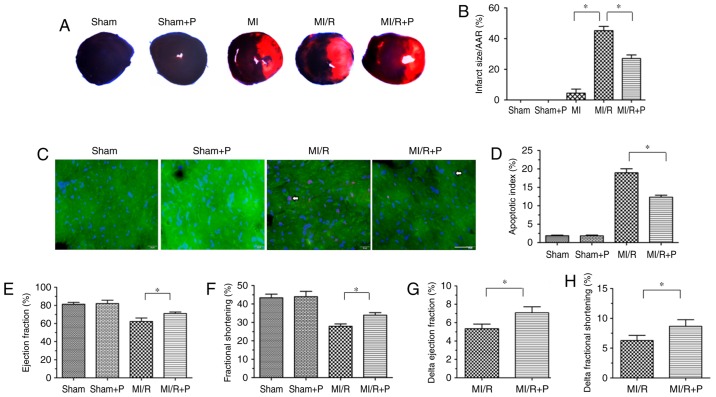
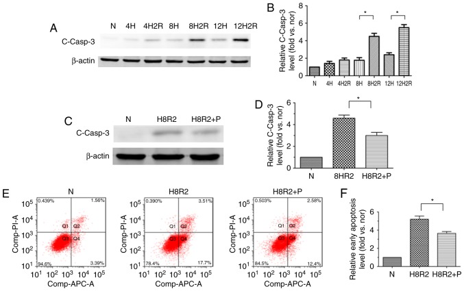
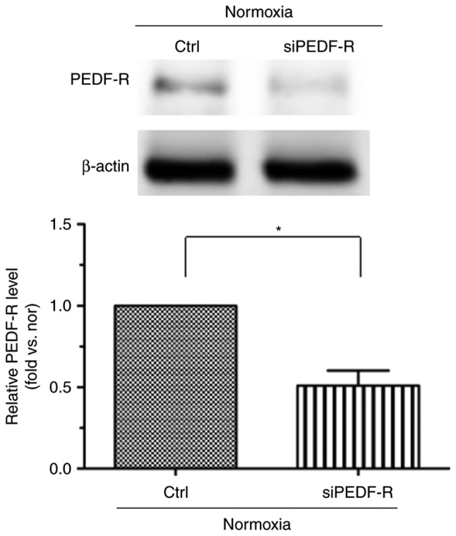
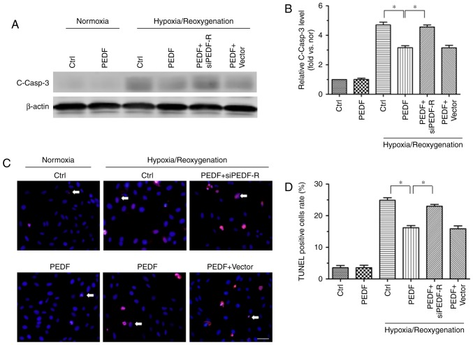
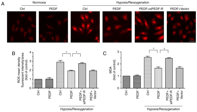
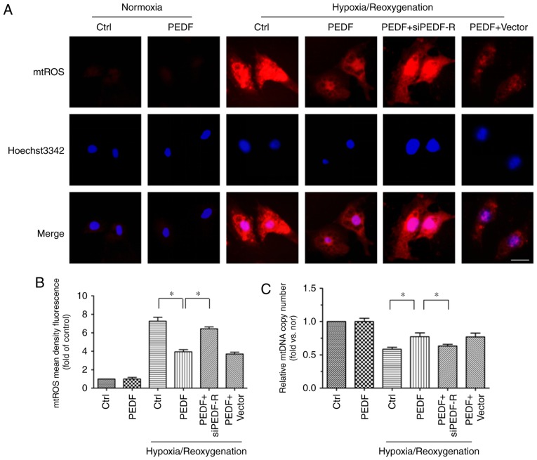
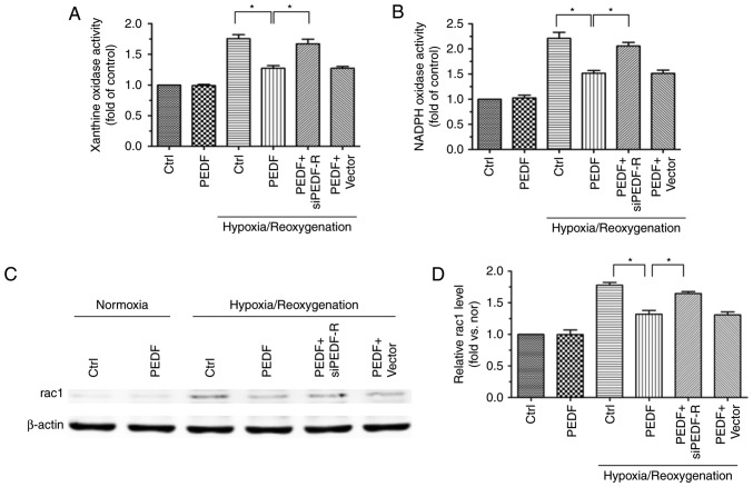
The prevention and management of myocardial ischemia/reperfusion (MI/R) injury is an essential part of coronary heart disease surgery and is becoming a major clinical problem in the treatment of ischemic heart disease. Previous studies by our group have demonstrated that pigment epithelium‑derived factor (PEDF) improves cardiac function in rats with acute myocardial infarction and reduces hypoxia‑induced cell injury. However, the protective function and mechanisms underlying the effect of PEDF in MI/R injury remain to be fully understood. In the present study, the positive effect of PEDF in MI/R injury was confirmed by construction of the adult Sprague‑Dawley rat MI/R model. PEDF reduced myocardial infarct size and downregulated cardiomyocyte apoptosis in the I/R myocardium in this model. In addition, PEDF improved cardiac function and increased cardiac functional reserve in rats subjected to MI/R Injury. To further study the protective effect of PEDF and the underlying mechanisms in MI/R injury, a H9c2 cardiomyocyte hypoxia/reoxygenation (H/R) model was constructed. PEDF was confirmed to decrease H/R‑induced apoptosis in H9c2 cells, and this anti‑apoptotic function was abolished by pigment epithelium‑derived factor‑receptor (PEDF R) small interfering (si)RNA. Furthermore, administration of PEDF decreased the levels of reactive oxygen species (ROS) and malondialdehyde (MDA) in H/R H9c2 cells. Compared with the H/R group, PEDF decreased mitochondrial ROS, increased the mitochondrial DNA copy number, reduced xanthine oxidase and NADPH oxidase activity, as well as RAC family small GTPase 1 protein expression. However, these effects of PEDF were markedly attenuated by PEDF‑R siRNA. To the best of our knowledge, the present study is the first to identify the protective effect of PEDF in MI/R injury, and confirm that the antioxidative effect PEDF occurred via inhibition of ROS generation via PEDF‑R under MI/R conditions.


Figures

Figure 1
Figure 1
PEDF reduces myocardial infarct size and improves cardiac function under MI/R conditions. (A) Representative figures of the Evans Blue/TTC-stained myocardial tissues in each indicated experimental condition, with (B) quantification of the infarct size (n=6). (C) Representative images of TUNEL staining of cardiomyocyte apoptosis (white arrow), with (D) quantification. Cardiomyocyte apoptosis was measured by TUNEL staining; cardiomyocytes were identified using α-sarcomeric actin antibodies. TUNEL staining for cardiomyocyte apoptosis (red), DAPI for nuclear staining (blue) and α-sarcomeric actin for cardiomyocytes (green) in the border zone of the infarcted left ventricle from all experimental groups (scale bar=20 μm; n=6). (E) Left ventricular ejection fraction prior to dobutamine injection determination by echocardiography (n=6). (F) Left ventricular fractional shortening prior to dobutamine injection determination by echocardiography (n=6). (G) Δ left ventricular ejection fraction following dobutamine injection by echocardiography (n=6). (H) Δ left ventricular fractional shortening following dobutamine injection by echocardiography (n=6). Data are expressed as the mean ± standard error of the mean. *P<0.05, with comparisons indicated by lines. PEDF, pigment epithelium-derived factor; MI/R, myocardial ischemia/reperfusion; TTC, 2,3,5-triphenyltetrazolium; TUNEL, terminal deoxynucleotidyl transferase dUTP nick end labeling.
Figure 2
Figure 2
PEDF protects H9c2 cells against H/R-induced apoptosis. H9c2 cells were exposed to H/R conditions for various durations (0/0, 4/0, 4/2, 8/0, 8/2, 12/0 and 12/2 h). In addition, H9c2 cells treated with or without PEDF (10 nM) were exposed to normoxic or hypoxic/reoxygenation condition for 8/2 h. (A) Western blotting detected the level of the cleaved casp3 protein, with (B) quantification (n=3). (C) Samples were collected and analyzed for the expression of the cleaved casp3 protein by western blotting analysis, with (D) quantification (n=4). (E) Flow cytometric detection of early apoptosis (APC+/PI), with (F) quantification (n=3). Data are expressed as the mean ± standard error of the mean. *P<0.05, with comparisons indicated by lines. PEDF, pigment epithelium-derived factor; H/R, hypoxia/reoxygenation; casp3, caspase-3; APC, allophycocyanin; PI, propidium iodide; N, negative control.
Figure 3
Figure 3
siPEDF-R reduces the levels of PEDF-R under normoxic conditions in H9c2 cells. RNA interference assays were used to silence PEDF-R. Western blot analysis of PEDF-R expression was then performed (n=4). Data are expressed as the mean ± standard error of the mean. *P<0.05, with comparisons indicated by lines. si, small interfering; PEDF-R, pigment epithelium-derived factor receptor.
Figure 4
Figure 4
PEDF decreases H/R-induced apoptosis via PEDF-R in cultured H9c2 cells. H9c2 cells were maintained in normoxic or H/R conditions for 8/2 h with or without PEDF (10 nM). RNA interference assays were used to silence PEDF-R. (A) Samples were collected for western blotting to analyze the expression of cleaved casp3 protein, with (B) quantification performed using ImageJ software (n=4). (C) Effect of PEDF on H9c2 cells apoptosis, with (D) quantification. TUNEL (red) staining was performed for each group. Nuclei were stained with DAPI (blue). Cells that were TUNEL and DAPI-positive were apoptotic (indicated by the arrows), while DAPI positive were control cells (scale bar=50 μm; n=4). Data are expressed as the mean ± standard error of the mean. *P<0.05, with comparisons indicated by lines. PEDF, pigment epithelium-derived factor; H/R, hypoxia/reoxygenation; PEDF-R, pigment epithelium-derived factor receptor; TUNEL, terminal deoxynucleotidyl transferase dUTP nick end labeling; si, small interfering.
Figure 5
Figure 5
PEDF suppresses the levels of ROS and MDA via PEDF-R during H/R. H9c2 cells were maintained in normoxic or H/R conditions for 8/2 h with or without PEDF (10 nM). RNA interference assays were used to silence PEDF-R. (A) ROS level was measured with dihydroethidium fluorescence probes and (B) quantified with Image-Pro Plus software (Scale bar=50 μm; n=4). (C) MDA levels were assessed (n=3). Data are expressed as the mean ± standard error of the mean. *P<0.05, with comparisons indicated by lines. PEDF, pigment epithelium-derived factor; ROS, reactive oxygen species; MDA, malondialdehyde; PEDF-R, pigment epithelium-derived factor receptor; H/R, hypoxia/reoxygenation; si, small interfering.
Figure 6
Figure 6
PEDF reduces H/R-induced mtROS generation via PEDF-R. H9c2 cells were maintained in normoxic or H/R conditions for 8/2 h with or without PEDF (10 nM). RNA interference assays were used to silence PEDF-R. (A) mtROS production was monitored by MitoSOX™ Red in H9c2 cells, with (B) quantification. ROS production was observed by red fluorescence of MitoSOX™ by fluorescence microscopy and analyzed by Image-Pro Plus software (scale bar=20 μm; n=5). (C) mtDNA copy number was quantified by comparing D-loop expression to 18s RNA content using quantitative polymerase chain reaction analysis (n=4). Data are expressed as the mean ± standard error of the mean. *P<0.05, with comparisons indicated by lines. PEDF, pigment epithelium-derived factor; H/R, hypoxia/reoxygenation; mtROS, mitochondrial reactive oxygen species; mtDNA, mitochondrial DNA; si, small interfering; PEDF-R, pigment epithelium-derived factor receptor.
Figure 7
Figure 7
PEDF decreases H/R-induced cytoplasmic ROS generation via PEDF-R. H9c2 cells were maintained in normoxic or H/R conditions for 8/2 h with or without PEDF (10 nM). RNA interference assays were used to silence PEDF-R. (A) XO activity was assessed in all experimental groups using the XO activity assay kit (n=4). (B) NOX activity was assessed in all experimental groups using the NOX activity assay kit (n=4). (C) Western blot analysis of rac1 protein expression, with (D) quantification (n=4). Data are expressed as the mean ± standard error of the mean. *P<0.05, with comparisons indicated by lines. PEDF, pigment epithelium-derived factor; H/R, hypoxia/reoxygenation; ROS, reactive oxygen species; PEDF-R, pigment epithelium-derived factor receptor; XO, xanthine oxidase; NOX, NADPH oxidase; rac1, RAC family small GTPase 1; si, small interfering.

Similar articles

Cited by

References

    1. Damiani G, Salvatori E, Silvestrini G, Ivanova I, Bojovic L, Iodice L, Ricciardi W. Influence of socioeconomic factors on hospital readmissions for heart failure and acute myocardial infarction in patients 65 years and older: Evidence from a systematic review. Clin Interv Aging. 2015;10:237–245. doi: 10.2147/CIA.S71165. - DOI - PMC - PubMed
    1. Crea F, Battipaglia I, Andreotti F. Sex differences in mechanisms, presentation and management of ischaemic heart disease. Atherosclerosis. 2015;241:157–168. doi: 10.1016/j.atherosclerosis.2015.04.802. - DOI - PubMed
    1. Hearse DJ. Myocardial protection during ischemia and reperfusion. Mol Cell Biochem. 1998;186:177–184. doi: 10.1023/A:1006808507605. - DOI - PubMed
    1. Hearse DJ, Bolli R. Reperfusion induced injury: Manifestations, mechanisms, and clinical relevance. Cardiovasc Res. 1992;26:101–108. doi: 10.1093/cvr/26.2.101. - DOI - PubMed
    1. Turer AT, Hill JA. Pathogenesis of myocardial ischemia-reperfusion injury and rationale for therapy. Am J Cardiol. 2010;106:360–368. doi: 10.1016/j.amjcard.2010.03.032. - DOI - PMC - PubMed