Comparative Study
. 2018 May;17(5):6551-6559.
doi: 10.3892/mmr.2018.8725. Epub 2018 Mar 9.

Comparative characterization of SHED and DPSCs during extended cultivation in vitro

Affiliations
Comparative Study

Comparative characterization of SHED and DPSCs during extended cultivation in vitro

Huihui Wang et al. Mol Med Rep. 2018 May.

Abstract

Dental pulp stem cells (DPSCs) and stem cells from human exfoliated deciduous teeth (SHED) are types of human dental tissue‑derived mesenchymal stem cells (MSCs). These cells possess a capacity for self‑renewal, multilineage differentiation potential and immunomodulatory functions. Previous studies have reported that DPSCs and SHED may be beneficial in regenerative treatments and immunotherapy. The substantial expansion of cells in vitro is a prerequisite to obtaining adequate cell numbers required for cell‑based therapy. However, the regeneration and clinical potential of MSCs diminishes with long‑term cell culture amplification. To assess the alterations in SHED and DPSCs characteristics that underlie cellular senescence and result from extended in vitro amplification, the biological properties of SHED and DPSCs at passages 4 (P4) and 20 (P20) were compared. The cells underwent senescence following serial expansion to P20, as determined by altered cell morphology, decreased proliferation and migration capacity, attenuated differentiation potential, elevated senescence‑associated β‑galactosidase (SA‑β‑gal)‑positive rates and increased apoptosis. The phenotypic changes were also accompanied by a marked increase in the expression of p53, p21 and p16Ink4a. The present study also identified that senescent DPSCs exhibited an increased number of positive cells in SA‑β‑gal staining and demonstrated varying expressions of p53, p21 and p16Ink4a in comparison with SHED, indicating the involvement of diverse pathways in cellular senescence during long‑term sequential in vitro culture and passage. Furthermore, at early and late passages, SHED exhibited a higher proliferation rate and osteogenic differentiation capability when compared with DPSCs. In addition, both cell types maintained their characteristic immunophenotype during long‑term cultivation, while the expression levels of CD73 were higher in SHED at P20. The present study concluded that notable alterations were exhibited in SHED and DPSCs during the process of extensive expansion in vitro and the results may provide guidance for the selection of safe and effective expanded SHED and DPSCs for regenerative medicine and therapy.

Keywords: dental pulp stem cells; stem cells from human exfoliated deciduous teeth; biological characteristics; cellular senescence; in vitro cultivation.


Figures

Figure 1.
Figure 1.
SHED and DPSCs morphology, proliferation and immunophenotype. Representative morphological features of SHED and DPSCs at (A) P4 and (B) P20. Scale bars, 100 µm. (C) Proliferation curves of SHED and DPSCs at P4 and P20. (D) Statistical analysis of the percentage of CD73-, CD90- or CD105-positive cells in SHED and DPSCs groups at P4 and P20. (E) Statistical analysis of the percentage of CD34-, CD11b-, CD19-, CD45- and HLA-DR-positive cells of the SHED and DPSCs groups at P4 and P20. Data are presented as the mean ± standard error of the mean (n=3). *P<0.05, **P<0.01 and ***P<0.001, as indicated. SHED, stem cells from human exfoliated deciduous teeth; DPSCs, dental pulp stem cells; P4/20, passage 4/20; HLA-DR, human leukocyte antigen D-related.
Figure 2.
Figure 2.
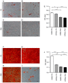
In vitro differentiation of SHED and DPSCs. Adipogenesis was assessed by Oil Red O staining of (A) SHED at P4, (B) DPSCs at P4, (C) SHED at P20 and (D) DPSCs at P20. Scale bars, 100 µm. (E) Oil Red O staining quantification of adipogenic differentiation of SHED and DPSCs at P4 and P20). Osteogenesis was assessed by Alizarin Red staining of (F) SHED at P4, (G) DPSCs at P4, (H) SHED at P20 and (I) DPSCs at P20. Scale bars, 100 µm. (J) Alizarin Red staining quantification of osteogenic differentiation of SHED and DPSCs at P4 and P20. Data are presented as the mean ± standard error of the mean (n=4). *P<0.05 and ***P<0.001, as indicated. SHED, stem cells from human exfoliated deciduous teeth; DPSCs, dental pulp stem cells; P4/20, passage 4/20; OD, optical density.
Figure 3.
Figure 3.
Quantification of osteogenic and adipogenic markers. Reverse transcription-quantitative polymerase chain reaction was performed to determine the mRNA expression levels of (A) ALP, (B) Runx2 and (C) PPARγ2 in SHED and DPSCs at P4 and P20 following in vitro differentiation. ALP and Runx2 were considered to be osteogenic markers, while PPARγ2 was considered to indicate adipogenic differentiation. Data are presented as the mean ± standard error of the mean (n=3). *P<0.05, **P<0.01 and ***P<0.001, as indicated. ALP, alkaline phosphatase; Runx2, runt-related transcription factor 2; PPARγ2, peroxisome proliferator-activated receptor γ2; SHED, stem cells from human exfoliated deciduous teeth; DPSCs, dental pulp stem cells; P4/20, passage 4/20.
Figure 4.
Figure 4.
Senescence-associated alterations in SHED and DPSCs during prolonged culture in vitro. Crystal violet staining revealed the migratory cells of SHED and DPSCs at (A) P4 and (B) P20. Scale bars, 200 µm. (C) Quantitative analysis of migratory cells in each group. (D) Representative flow cytometry plots indicating the apoptotic cell proportions of SHED and DPSCs groups (upper and lower right-hand side quadrants combined). (E) Statistical analysis of the apoptotic cell proportions of SHED and DPSCs groups. Data are presented as the mean ± standard error of the mean (n=3). *P<0.05 and **P<0.01, as indicated. SHED, stem cells from human exfoliated deciduous teeth; DPSCs, dental pulp stem cells; P4/20, passage 4/20; FITC, fluorescein isothiocyanate.
Figure 5.
Figure 5.
Phenotypic and molecular characterizations of SHED and DPSCs in senescence. Representative images of SA-β-gal-positive cells in SHED and DPSCs at (A) P4 and (B) P20. Scale bar, 50 µm. (C) The percentage of SA-β-gal-positive cells in SHED and DPSCs. (D) Western blot analysis of p53, p21 and p16Ink4a expression in SHED and DPSCs. Reverse transcription-quantitative polymerase chain reaction analysis of p53, p21 and p16Ink4a mRNA expression normalized to GAPDH in (E) SHED and (F) DPSCs at P4 and P20. Data are presented as the mean + standard error of the mean (n=3). *P<0.05, **P<0.01 and ***P<0.001, as indicated. SHED, stem cells from human exfoliated deciduous teeth; DPSCs, dental pulp stem cells; SA-β-gal, senescence-associated β-galactosidase; P4/20, passage 4/20.

References

    1. Salem HK, Thiemermann C. Mesenchymal stromal cells: Current understanding and clinical status. Stem Cells. 2010;28:585–596. - PMC - PubMed
    1. Reddy BY, Xu DS, Hantash BM. Mesenchymal stem cells as immunomodulator therapies for immune-mediated systemic dermatoses. Stem Cells Dev. 2012;21:352–362. doi: 10.1089/scd.2011.0404. - DOI - PubMed
    1. Liu J, Yu F, Sun Y, Jiang B, Zhang W, Yang J, Xu GT, Liang A, Liu S. Concise reviews: Characteristics and potential applications of human dental tissue-derived mesenchymal stem cells. Stem Cells. 2015;33:627–638. doi: 10.1002/stem.1909. - DOI - PubMed
    1. Gronthos S, Mankani M, Brahim J, Robey PG, Shi S. Postnatal human dental pulp stem cells (DPSCs) in vitro and in vivo; Proc Natl Acad Sci USA; 2000; pp. 13625–13630. - DOI - PMC - PubMed
    1. Gronthos S, Brahim J, Li W, Fisher LW, Cherman N, Boyde A, DenBesten P, Robey PG, Shi S. Stem cell properties of human dental pulp stem cells. J Dent Res. 2002;81:531–535. doi: 10.1177/154405910208100806. - DOI - PubMed

Publication types

Substances