. 2018 Apr;52(4):1149-1164.
doi: 10.3892/ijo.2018.4293. Epub 2018 Feb 28.

Role of the EZH2/miR-200 axis in STAT3-mediated OSCC invasion

Affiliations

Role of the EZH2/miR-200 axis in STAT3-mediated OSCC invasion

Yu Wang et al. Int J Oncol. 2018 Apr.

Erratum in

Retraction in

Abstract

Abnormal activation of signal transducer and activator of transcription 3 (STAT3) serves a pivotal role in oral squamous cell carcinoma (OSCC) tumor cell invasion into normal tissues or distant organs. However the downstream regulatory network of STAT3 signaling remains unclear. The present study aimed to investigate the potential mechanism underlying how STAT3 triggers enhancer of zeste homolog 2 (EZH2) expression and inhibits microRNA (miR)-200a/b/429 expression in SCC25 and SCC15 cells in vitro and in vivo. Western blotting and reverse transcription-quantitative polymerase chain reaction were performed to detect expression, and numerous functional tests were conducted to explore cancer metastasis. The results indicated that when STAT3 signaling activity was attenuated by Stattic or enhanced with a STAT3 plasmid, the EZH2/miR-200 axis was markedly altered, thus resulting in modulation of the invasion and migration of OSCC cell lines. In addition, loss of function of EZH2 compromised the oncogenic role of STAT3 in both cell lines. F-actin morphology and the expression of epithelial-mesenchymal transition markers were also altered following disruption of the STAT3/EZH2/miR-200 axis. An orthotopic tumor model derived from SCC15 cells was used to confirm that targeting STAT3 or EZH2 suppressed OSCC invasion in vivo. In conclusion, the EZH2/miR-200 axis was revealed to mediate antitumor effects by targeting STAT3 signaling; these findings may provide a novel therapeutic strategy for the treatment of OSCC.


Conflict of interest statement

Competing interests

The authors declare that they have no competing interests.

Figures
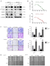
Figure 1
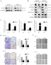
Figure 1
STAT3 contributes to invasion and migration of OSCC cells. (A) Western blot detection of STAT3, p-STAT3 (Tyr705), MMP2 and MMP9 expression. GAPDH was used as a loading control. (B) MTT analysis was used to determine the IC50 values for both OSCC cell lines. (C) Targeting STAT3 significantly inhibited SCC25 and SCC15 cell invasion and migration ability, as determined by Transwell assay (magnification, ×40). (D) Cell migration ability was measured by a wound-healing assay (magnification, ×40). ***P<0.001 vs. EV group; ###P<0.001 EV + Stattic group vs. STAT3 group. EV, empty vector; IC50, half maximal inhibitory concentration; MMP, matrix metalloproteinase; OSCC, oral squamous cell carcinoma; p-STAT3, phosphorylated-STAT3; STAT3, signal transducer and activator of transcription 3.
Figure 2
Figure 2
EZH2/miR-200/b/a/429 axis regulates the invasiveness of OSCC cells in vitro. (A) Following transfection with EZH2 siRNAs (si#1 and si#2) for 3 days, the expression levels of EZH2 were detected using western blotting. (B) OSCC cells underwent western blot analysis using antibodies specific to STAT3, p-STAT3 (Tyr705), EZH2, H3K27me3, H3 and GAPDH. EZH2 siRNA markedly inhibited EZH2 and H3K27me3 expression. In addition, p-STAT3 (Tyr705), but not STAT3, was suppressed by EZH2 depletion. (C) EZH2 siRNA induced miR-200b/a/429 expression. (D) Results of a Transwell assay demonstrated that EZH2 knockdown markedly attenuated migration (without Matrigel) and invasion (with Matrigel) of SCC25 and SCC15 cells (scale bar, 100 μm; magnification, ×40). (E) Scratch assay demonstrated that EZH2 silencing markedly delayed wound healing in SCC25 and SCC15 cells (magnification, ×40). *P<0.05 and ***P<0.001 vs. si-NC group. EZH2, enhancer of zeste homolog 3; H3, histone 3; H3K27me3, trimethylation of lysine 27 in H3; miR-200b/a/429, microRNA-200b, -200a and -429; NC, negative control; OSCC, oral squamous cell carcinoma; p-STAT3, phosphorylated-STAT3; siRNA/si, small interfering RNA; STAT3, signal transducer and activator of transcription 3.
Figure 3
Figure 3
EZH2 silencing affects the EMT process in OSCC cells. (A) Protein expression levels of EMT-associated markers were analyzed by western blotting and were normalized to GAPDH. (B) F-actin staining exhibited a stress-fiber pattern in NC siRNA-transfected cells, whereas a cortical pattern was observed in EZH2 siRNA-transfected cells, as assessed by immunofluorescence (scale bar, 20 μm). (C) E-cadherin, N-cadherin and Vimentin staining of OSCC cells transfected with si-EZH2 or si-NC. Cell nuclei were stained with DAPI (scale bar, 20 μm). EMT, epithelial-mesenchymal transition; EZH2, enhancer of zeste homolog 3; MMP, matrix metalloproteinase; NC, negative control; OSCC, oral squamous cell carcinoma; siRNA/si, small interfering RNA.
Figure 4
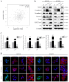
Figure 4
EZH2/miR-200/b/a/429 axis mediates the prometastatic role of STAT3 in an EMT-dependent manner. (A) The Cancer Genome Atlas data were used to determine the correlation between STAT3 and EZH2 in head and neck squamous cell carcinoma (P<0.05). (B) Western blot analysis of EZH2, H3K27me3 and EMT markers following Stattic treatment or STAT3 transduction. (C) Stattic upregulated miR-200b/a/429 expression, whereas ectopic STAT3 overexpression significantly attenuated mir-200b/a/429 expression. (D) Expression of the epithelial marker E-cadherin, and the mesenchymal markers N-cadherin and Vimentin, were detected by immunofluorescence staining (scale bar, 20 μm). ***P<0.001 vs. EV group; ###P<0.001 EV + Stattic group vs. STAT3 group. EMT, epithelial-mesenchymal transition; EV, empty vector; EZH2, enhancer of zeste homolog 3; H3, histone 3; H3K27me3, trimethylation of lysine 27 in H3; miR-200b/a/429, microRNA-200b, -200a and -429; STAT3, signal transducer and activator of transcription 3.
Figure 5
Figure 5
EZH2 silencing counteracts STAT3-induced invasion by targeting miR-200b/a/429. (A) STAT3 and EZH2 expression levels were evaluated by western blotting. (B) EZH2 depletion markedly reduced the inhibitory effects of STAT3 on miR-200b/a/429 expression. (C and D) EZH2 knockdown reduced the invasion and migration of oral squamous cell carcinoma cells overexpressing STAT3 (magnification, ×40). *P<0.05 and ***P<0.001 vs. si-NC + EV group; ###P<0.001 si-NC + STAT3 group vs. si-EZH2 + STAT3 group. EV, empty vector; EZH2, enhancer of zeste homolog 3; H3, histone 3; H3K27me3, tri-methylation of lysine 27 in H3; miR-200b/a/429, microRNA-200b, -200a and -429; p-STAT3, phosphorylated-STAT3; siRNA/si, small interfering RNA; STAT3, signal transducer and activator of transcription 3.
Figure 6
Figure 6
EZH2 silencing impairs STAT3-induced EMT-mediated metastasis. (A) Protein expression levels of EMT-associated markers were analyzed using western blotting and were normalized to GAPDH. (B) Subcellular location and expression of the epithelial marker E-cadherin, and the mesenchymal markers N-cadherin and Vimentin, in oral squamous cell carcinoma cells. F-actin distribution was rearranged to a cortical pattern following EZH2 knockdown (scale bar, 20 μm). EZH2, enhancer of zeste homolog 3; siRNA/si, small interfering RNA; STAT3, signal transducer and activator of transcription 3.
Figure 7
Figure 7
Targeting STAT3 and EZH2 inhibits tumorigenesis and invasiveness in an orthotopic OSCC model. (A) Representative bioluminescence images of mice implanted with orthotopic tumors, which were intraperitoneally treated with 3.75 mg/kg Stattic, 1 mg/kg DZNeP or DMSO every 3 days. (B) Based on body weight, no detectable toxicity was observed at the tested dose. (C) Representative photographs of the tumor resection procedure. Stattic and DZNeP treatment inhibited local invasion of orthotopic OSCC tumors. (D) Tumor diameter and volume were measured. (E) Quantitative polymerase chain reaction was used to detect miR-200b/a/429 expression in OSCC tumor sections. (F) Tumor samples from three distinct groups underwent immunohistochemistry for STAT3, p-STAT3 (Tyr705), EZH2, H3K27me3, MMP2, MMP6, E-cadherin, N-cadherin and Vimentin expression (scale bar, 100 μm; magnification, ×200). ***P<0.001 vs. DMSO group. DMSO, dimethyl sulfoxide; DZNeP, 3-deazaneplanocin A; EZH2, enhancer of zeste homolog 3; H3K27me3, trimethylation of lysine 27 in histone 3; H&E, hematoxylin and eosin; miR, microRNA; MMP, matrix metalloproteinase; p-STAT3, phosphorylated-STAT3; STAT3, signal transducer and activator of transcription 3.
Figure 8
Figure 8
miR-429 inhibits tumor progression in the orthotopic mouse model of OSCC. (A) Representative bioluminescence images of mice implanted with orthotopic tumors and treated intraperitoneally with miR-Ctrl (10 nM/mice) or miR-429 (10 nM/mice) every 3 days. (B) Quantification of body weight in control and miR-429-treated mice. (C) miR-429 inhibits OSCC local invasion in an orthotopic mouse model. (D) Tumor diameter and volume were measured. (E) Quantitative polymerase chain reaction was used to detect the expression levels of mir-429 in OSCC tumor sections. (F) Tumor samples from control and miR-429-treated mice underwent immunohistochemistry for MMP2, MMP9, E-cadherin, N-cadherin and Vimentin (scale bar, 100 μm magnification, ×200). ***P<0.001 vs. miR-Ctrl group. Ctrl, control; H&E, hematoxylin and eosin; miR, microRNA; MMP, matrix metalloproteinase; NC, negative control.

References

    1. Pulte D, Brenner H. Changes in survival in head and neck cancers in the late 20th and early 21st century: A period analysis. Oncologist. 2010;15:994–1001. doi: 10.1634/theoncologist.2009-0289. - DOI - PMC - PubMed
    1. Lam L, Logan RM, Luke C. Epidemiological analysis of tongue cancer in South Australia for the 24-year period, 1977–2001. Aust Dent J. 2006;51:16–22. doi: 10.1111/j.1834-7819.2006.tb00395.x. - DOI - PubMed
    1. Beasley NJ, Prevo R, Banerji S, Leek RD, Moore J, van Trappen P, Cox G, Harris AL, Jackson DG. Intratumoral lymphangiogenesis and lymph node metastasis in head and neck cancer. Cancer Res. 2002;62:1315–1320. - PubMed
    1. Patel V, Marsh CA, Dorsam RT, Mikelis CM, Masedunskas A, Amornphimoltham P, Nathan CA, Singh B, Weigert R, Molinolo AA, et al. Decreased lymphangiogenesis and lymph node metastasis by mTOR inhibition in head and neck cancer. Cancer Res. 2011;71:7103–7112. doi: 10.1158/0008-5472.CAN-10-3192. - DOI - PMC - PubMed
    1. Roepman P, Kemmeren P, Wessels LF, Slootweg PJ, Holstege FC. Multiple robust signatures for detecting lymph node metastasis in head and neck cancer. Cancer Res. 2006;66:2361–2366. doi: 10.1158/0008-5472.CAN-05-3960. - DOI - PubMed

Publication types

MeSH terms