Review
. 2018 Jun 5;57(22):3105-3114.
doi: 10.1021/acs.biochem.8b00079. Epub 2018 Mar 21.

Polyamine Deacetylase Structure and Catalysis: Prokaryotic Acetylpolyamine Amidohydrolase and Eukaryotic HDAC10

Affiliations
Review

Polyamine Deacetylase Structure and Catalysis: Prokaryotic Acetylpolyamine Amidohydrolase and Eukaryotic HDAC10

Stephen A Shinsky et al. Biochemistry. .

Abstract

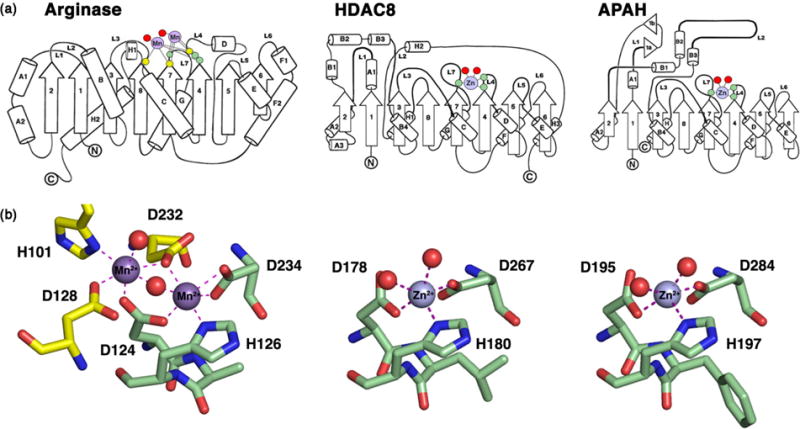
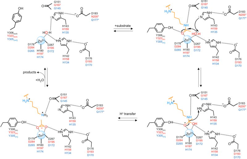
Polyamines such as putrescine, spermidine, and spermine are small aliphatic cations that serve myriad biological functions in all forms of life. While polyamine biosynthesis and cellular trafficking pathways are generally well-defined, only recently has the molecular basis of reversible polyamine acetylation been established. In particular, enzymes that catalyze polyamine deacetylation reactions have been identified and structurally characterized: histone deacetylase 10 (HDAC10) from Homo sapiens and Danio rerio (zebrafish) is a highly specific N8-acetylspermidine deacetylase, and its prokaryotic counterpart, acetylpolyamine amidohydrolase (APAH) from Mycoplana ramosa, is a broad-specificity polyamine deacetylase. Similar to the greater family of HDACs, which mainly serve as lysine deacetylases, both enzymes adopt the characteristic arginase-deacetylase fold and employ a Zn2+-activated water molecule for catalysis. In contrast with HDACs, however, the active sites of HDAC10 and APAH are sterically constricted to enforce specificity for long, slender polyamine substrates and exclude bulky peptides and proteins containing acetyl-l-lysine. Crystal structures of APAH and D. rerio HDAC10 reveal that quaternary structure, i.e., dimer assembly, provides the steric constriction that directs the polyamine substrate specificity of APAH, whereas tertiary structure, a unique 310 helix defined by the P(E,A)CE motif, provides the steric constriction that directs the polyamine substrate specificity of HDAC10. Given the recent identification of HDAC10 and spermidine as mediators of autophagy, HDAC10 is rapidly emerging as a biomarker and target for the design of isozyme-selective inhibitors that will suppress autophagic responses to cancer chemotherapy, thereby rendering cancer cells more susceptible to cytotoxic drugs.


Figures

Figure 1
Figure 1. Eukaryotic and prokaryotic polyamine metabolism
ADC, arginine decarboxylase; APAO, N1-acetylpolyamine oxidase; APAH, acetylpolyamine amidohydrolase; HDAC10, histone deacetylase 10; ODC, ornithine decarboxylase; P/CAF, P300/CBP associated factor; SMOX, spermine oxidase; SMS, spermine synthase; SRM, spermidine synthase; SSAT, spermidine/spermine acetyltransferase.
Figure 2
Figure 2. The arginase-deacetylase fold
(a) Arginase, HDAC8, and APAH share a conserved α/β fold consisting of a central 8-stranded parallel β-sheet flanked by a-helices. Residues that coordinate to catalytic Mn2+ ions in arginase are located on loops L3, L4, and L7 (yellow and green circles), and residues that coordinate to the catalytic Zn2+ ion in HDAC8 and APAH are conserved on loops L4 and L7 (green circles). Red circles signify metal-bound water molecules or hydroxamate inhibitor atoms. (b) The Mn2+B site of arginase is conserved in HDACs and APAH as D(A,V,L,F)HX~100D (boldface indicates metal ligands). The Mn2+A site of arginase is not conserved in the deacetylase family. Reprinted from ref. , copyright 2011, with permission from Elsevier.
Figure 3
Figure 3. Proposed mechanism of acetylpolyamine hydrolysis
Residue labels for HDAC8, APAH, and HDAC10 are color-coded black, red, and blue, respectively. Note that for APAH and HDAC10, the substrate is N8-acetylspermidine as shown; for HDAC8, the substrate would be an acetyl-L-lysine residue on a peptide or protein substrate (not shown). Also note that the residue that hydrogen bonds with the second of the tandem histidine residues differs in each of these enzymes. This feature may influence the chemical function of this histidine residue in general base-general acid catalysis.
Figure 4
Figure 4. Stereoviews of polyamine deacetylase complexes
(a) Structure of the H159A APAH-N8-acetylspermidine complex reveals that the substrate carbonyl group coordinates to Zn2+ and accepts a hydrogen bond from Y323in (the Y323out conformer is not shown for clarity). Reprinted from Ref. . Copyright 2015 American Chemical Society. (b) Structure of the APAH-AAT complex reveals the binding of the trifluoromethylketone as a gem-diolate that mimics the tetrahedral intermediate and flanking transition states for the hydrolysis of N8-acetylspermidine. Reprinted from Ref. . Copyright 2015 American Chemical Society. (c) Structure of the HDAC10-AAT complex similarly reveals the binding of the trifluoromethylketone as a gem-diolate transition state analogue. E274 plays a critical electrostatic role in enzyme-substrate recognition, and the P(E,A)CE motif (purple) sterically constricts the active site to favor the binding of the slender acetylpolyamine substrate. Reprinted from ref. .
Figure 5
Figure 5. Catalytic activity of human HDAC10 (hHDAC10) and zebrafish HDAC10 (zHDAC10)
(a,b) Steady-state kinetics measured using acetylpolyamines and acetyl-L-lysine (K(Ac)) peptides reveal a clear preference for N8-acetylspermidine and acetylputrescine hydrolysis, with little to no acetyl-L-lysine peptide hydrolysis. Abbreviations: AcPUT, acetylputrescine; N8-AcSPD, N8-acetylspermidine; AcCAD, acetylcadaverine; AcDAO, N-(aminooctyl)acetamide; N1,N8-diAcSPD, N1,N8 –diacetylspermidine; N1-AcSPD, N1-acetylspermidine; N1-AcSPM, N1-acetylspermine; AcPAD, N-(3-aminopropyl)acetamide; BTA, N-butylacetamide. (c) Ratio of catalytic efficiencies (kcat/KM) for polyamine deacetylase (PDAC) activity measured with N8-acetylspermidine and acetyl-L-lysine deacetylase (HDAC) activity measured with RGK(ac)-AMC (AMC = aminomethylcoumarin). APAH, hHDAC10, and zHDAC10 exhibit a clear catalytic preference for PDAC activity. The E274L mutation converts zHDAC10 from a PDAC into an HDAC, and the zHDAC10 ΔηA2 mutant is a bifunctional PDAC-HDAC. Abbreviations: zHDAC10Δ, proteolytically nicked zHDAC10 used for crystal structure determination; hHDAC6 CD12, human HDAC6 construct containing both catalytic domains; zHDAC6 CD1 or CD2, zebrafish HDAC6 catalytic domain 1 or catalytic domain 2. Reprinted from ref. .
Figure 6
Figure 6. Structural determinants of polyamine substrate specificity
(a) Assembly of the APAH dimer constricts the approach to the active site through an “L”-shaped tunnel indicated by red dotted line. Reprinted from ref. . Copyright 2011 American Chemical Society. (b) Stereoview showing the superposition of the polyamine deacetylase domain of HDAC10 with HDAC6 catalytic domains CD1 and CD2. The P(E,A)CE motif (purple) is conserved in HDAC10 orthologs and constricts the approach to the active site, as indicated by the binding of the transition state analogue AAT (stick figure). A close-up view of the P(E,A)CE motif is also visible in Figure 4c. Reprinted from Ref. .
Figure 7
Figure 7. Domain architecture of class IIb HDACs
(a) The HDAC6 structure reported by Miyake and colleagues (PDB 5G0J) consists of two catalytically active domains, CD1 (cyan) and CD2 (mauve), connected by an interdomain linker (green). Active sites are indicated by red arrows. Helices that mediate domain-domain association are indicated. (b) The HDAC10 structure reported by Hai and colleagues (PDB 5TD7) consists of a catalytically active polyamine deacetylase domain (PDAC, blue) and a catalytically inactive pseudo-deacetylase domain (ΨDAC, green). The active site in the PDAC domain is indicated by the binding of transition state analogue AAT (stick figure). The interdomain linker is proteolytically nicked and is not observed in the crystal structure.
Figure 8
Figure 8. HDAC10 as a mediator of autophagy
(a) As reported by Oehme and colleagues, BE(2)-C human neuroblastoma cells were stably transfected with short hairpin RNAs targeting HDAC10 (shR-1, -2, -3, -4) or a negative control (shR-NC). Cells were treated with 0.05 μg/mL doxorubicin (a cytotoxic cancer chemotherapy drug) or normal culture medium. After 10 days, colonies were stained and results were quantified (bar diagram; shR-NC, open bars; shR-HDAC10, filled bars). The Western Blot shows HDAC10 expression in stably transfected cells, and β-actin was used as a loading control. Knockdown of HDAC10 clearly enhances the cytotoxicity of doxorubicin relative to the negative control treated with shR-NC. Reprinted with permission from Ref. . (b) Summary of proposed roles of HDAC10 and N8-acetylspermidine in promoting autophagy. A selective inhibitor of HDAC10 (HDACi) will blunt the autophagic response to cytotoxic cancer drugs such as doxorubicin and thus render cancer cells more susceptible to chemotherapy.

References

    1. Tabor CW, Tabor H. Polyamines. Annu Rev Biochem. 1984;53:749–790. - PubMed
    1. Thomas T, Thomas TJ. Polyamines in cell growth and cell death: molecular mechanisms and therapeutic applications. Cell Mol Life Sci. 2001;58:244–258. - PMC - PubMed
    1. Casero RA, Jr, Pegg AE. Polyamine catabolism and disease. Biochem J. 2009;421:323–338. - PMC - PubMed
    1. Michael AJ. Polyamines in eukaryotes, bacteria, and archaea. J Biol Chem. 2016;291:14896–14903. - PMC - PubMed
    1. Michael AJ. Evolution of biosynthetic diversity. Biochem J. 2017;474:2277–2299. - PubMed

Publication types

MeSH terms

LinkOut - more resources