Review
. 2018 Mar 27;57(12):1821-1832.
doi: 10.1021/acs.biochem.7b00937. Epub 2018 Mar 13.

Template-Independent Enzymatic Oligonucleotide Synthesis (TiEOS): Its History, Prospects, and Challenges

Affiliations
Review

Template-Independent Enzymatic Oligonucleotide Synthesis (TiEOS): Its History, Prospects, and Challenges

Michael A Jensen et al. Biochemistry. .

Abstract

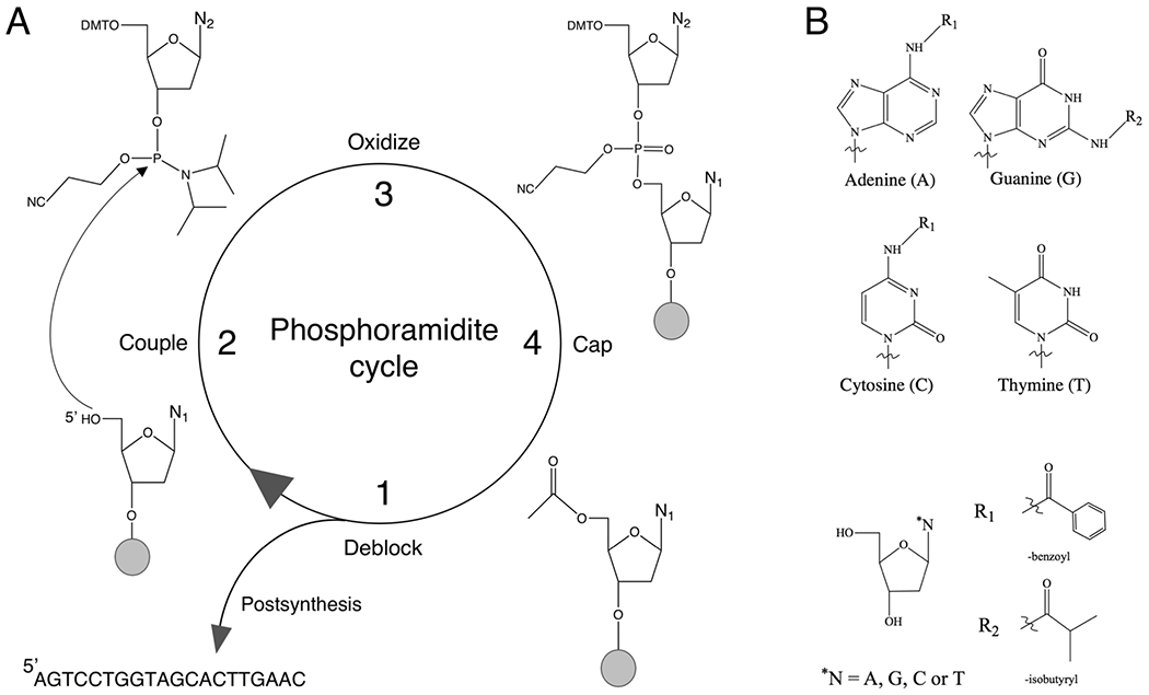
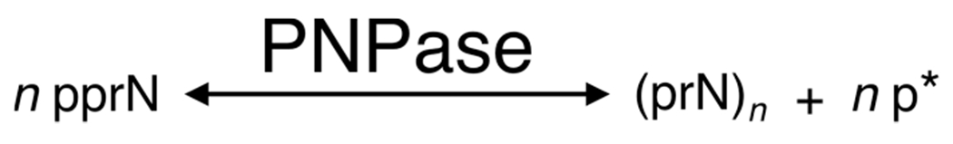
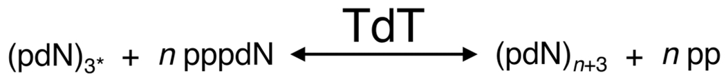
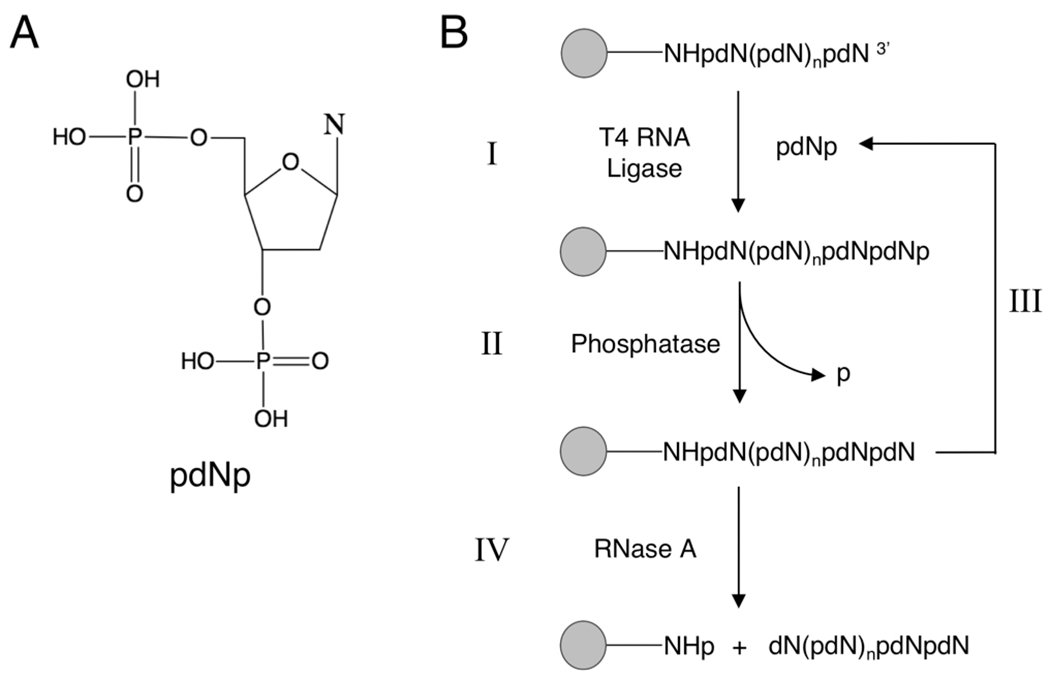
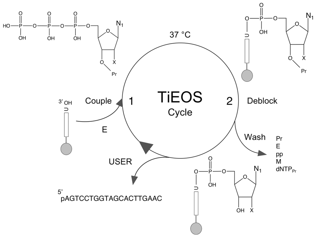
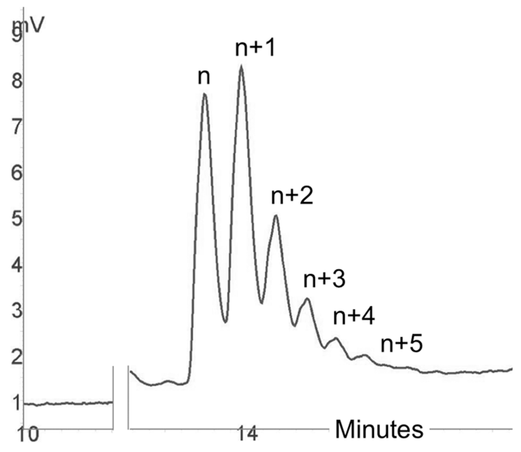
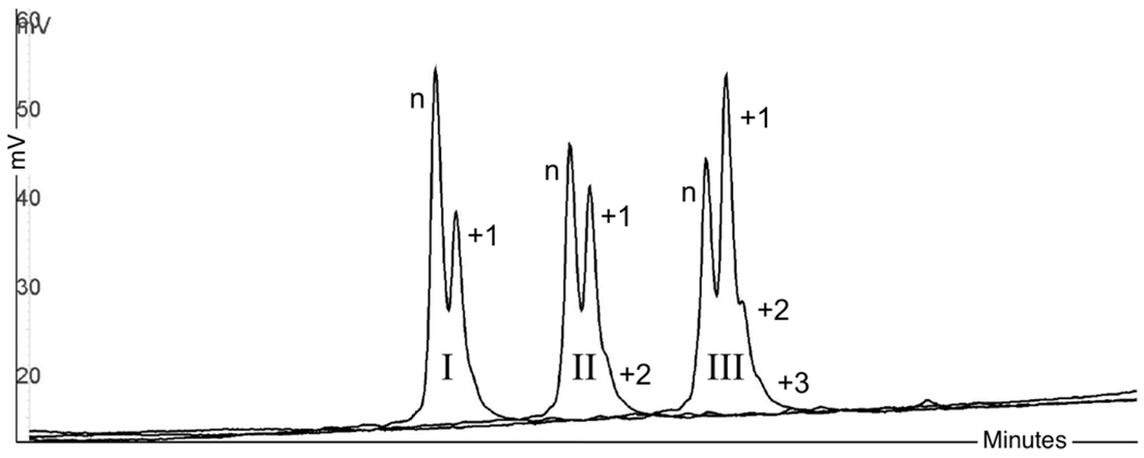
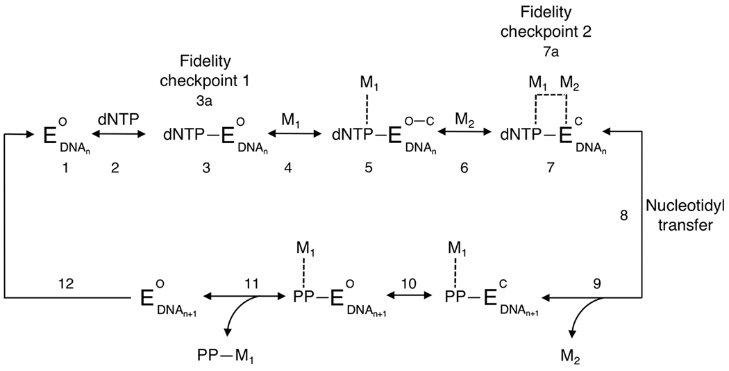
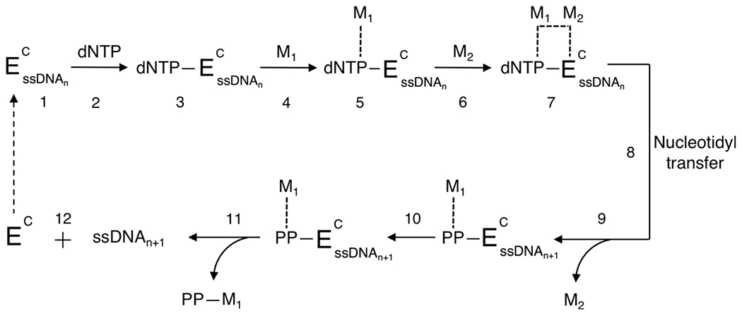
There is a growing demand for sustainable methods in research and development, where instead of hazardous chemicals, an aqueous medium is chosen to perform biological reactions. In this Perspective, we examine the history and current methodology of using enzymes to generate artificial single-stranded DNA. By using traditional solid-phase phosphoramidite chemistry as a metric, we also explore criteria for the method of template-independent enzymatic oligonucleotide synthesis (TiEOS). As its key component, we delve into the biology of one of the most enigmatic enzymes, terminal deoxynucleotidyl transferase (TdT). As TdT is found to exponentially increase antigen receptor diversity in the vertebrate immune system by adding nucleotides in a template-free manner, researchers have exploited this function as an alternative to the phosphoramidite synthesis method. Though TdT is currently the preferred enzyme for TiEOS, its random nucleotide incorporation presents a barrier in synthesis automation. Taking a closer look at the TiEOS cycle, particularly the coupling step, we find it is comprised of additions > n+1 and deletions. By tapping into the physical and biochemical properties of TdT, we strive to further elucidate its mercurial behavior and offer ways to better optimize TiEOS for production-grade oligonucleotide synthesis.


Conflict of interest statement

The authors declare no competing financial interest.

Figures

Figure 1.
Figure 1.
(A) Traditional 3′ → 5′ solid-phase phosphoramidite chemical synthesis cycle. The cycle begins with the first nucleoside tethered to a solid substrate by a cleavable succinate linker (alkaline-labile); the 5′ region, protected by dimethoxytrityl (DMT), is deblocked with acid (e.g., trichloroacetic acid/dichloromethane) to yield a hydroxyl group (step 1). In the coupling reaction (step 2), the phosphorus atom of an incoming nucleoside phosphoramidite is activated (e.g., with ethylthio-1H-tetrazole) for nucleophilic attack by the 5′ oxygen of the N1 nucleoside. Here, exocyclic amines for adenine, guanine, and cytosine are also reactive and must be blocked to prevent branching (panel B, R1 and R2). Once the nucleoside phosphoramidite is added, the phosphite bond (also protected with cyanoethyl) is oxidized with iodine/pyridine to generate a phosphate linkage to stabilize the sugar backbone (step 3). In step 4, oligonucleotide DNA strands (n) that fail to couple (n−1) are capped (acetylated) at the 5′ end to prevent any further reactions in successive cycles; n − 1 strands that remain uncapped will present with internal deletions. Wash steps are introduced using acetonitrile (petroleum byproduct) to clear the reaction well/column of waste. The cycle is repeated until the full-length product is generated, and then it is released from the solid support with ammonium hydroxide (methylamine); incubation at an elevated temperature (for ≤16 h) will remove all protecting groups. N1 and N2 denote nucleobases; n is the full-length oligonucleotide. (B) Nucleobase structures and exocyclic amine protecting groups. R1 and R2 denote benzoyl and isobutyryl, respectively.
Figure 2.
Figure 2.
Proposed pathway of PNPase RNA synthesis and degradation. From left to right, 5′ diphosphate ribonucleosides (pprN) drive the reaction toward PNPase catalysis of RNA (n monomers long), with the release of orthophosphate. At high concentrations, phosphate byproduct (p*) drives the reaction (right to left) toward phosphorolysis, degrading RNA into its 5′ diphosphate ribonucleoside monomers.
Figure 3.
Figure 3.
TdT polymerization of ssDNA. Here, a 3′-hydroxylated initiator strand (also termed an acceptor) is required [in the presence of a divalent cation (e.g., Mg2+)] for TdT to catalyze ssDNA using a deoxyribonucleoside triphosphate monomer (dNTP, also termed a donor). Here dNTP (pppdN) monomers are the substrate for addition of TdT to the 3′ end of an initiator strand (pdN)3* to generate ssDNA (pdN)n+3 with the release of pyrophosphate (pp). 3* indicates the length must be at least 3 nt. There are two primary isoforms of the TdT gene, TdTL (3′ exonuclease activity only, arrow right to left) and TdTS (3′ terminal transferase only, arrow left to right).
Figure 4.
Figure 4.
PNPase synthesis of polyribonucleotides of defined sequence. (A) Ribonucleotide representation, either adenosine (A) or uridine (U), diphosphate (pp), with 2′ (3′)-O-(α-methoxyethyl) blocking group (ME). (B) Solution-phase enzymatic RNA synthesis with 2′-protected ribonucleotides. (I) Adenosine trinucleotide (initiator) is coupled to 5′ diphosphate uridine protected at the 2′ position with α-methoxyethyl in the presence of PNPase at 37 °C for 7 h to generate 2′-protected tetranucleotide. (II) The α-methoxyethyl protecting group is removed with acid [pH 2 (3) for 15 min]. (III) The next nucleotide, 2′-protected 5′ ADP, is coupled to the tetranucleotide initiator (conditions are the same as those in step I). (IV) The structure of a blocked pentanucleotide is then confirmed by hydrolysis with pancreatic ribonuclease.
Figure 5.
Figure 5.
Solid-phase T4 RNA ligation of bisphosphate monomers to generate ssDNA. (A) Structure of 5′, 3′ bisphosphate-2′-deoxynucleoside (pdNp), where dN is any 2′ deoxynucleoside (A, G, C, or T). (B) Enzymatic synthesis performed in the solid phase, where 5′-phosphorylated initiator is covalently attached to Tentagel. (I) pdNp is ligated to the initiator at the 3′ hydroxyl in the presence of T4 RNA ligase. (II) The 3′ phosphate (p) is removed with alkaline phosphatase. (III) The process is repeated until the full-length product is established. (IV) The target strand is enzymatically released from the support with RNase A, yielding 5′, 3′ dephosphorylated product.
Figure 6.
Figure 6.
Proposed cycle for template-independent enzymatic oligonucleotide synthesis (TiEOS). In step 1, an incoming 3′-protected (Pr) dNTPPr (or NTPPr) is coupled to a 20 nt initiator (e.g., 37 °C, 30 s), which allows for enzyme (TdT) attachment and polymerization. Here the initiator is chemically presynthesized and covalently tethered {e.g., carbodiimide chemistry to a solid substrate [e.g., superparamagnetic beads (SPMB), silicon, or glass]}. A 20 nt long initiator was chosen to prevent steric hindrance at the surface of the beads during polymerization. In step 2, newly added dNTPPr is deblocked at the 3′ to hydroxyl (dNTP), followed by a wash step to remove protecting groups, enzyme, pyrophosphate, metal ions, and unincorporated dNTPPr. The cycle is repeated until the FLP is completed. For the purposes of enzymatic target strand release, uracil is placed at the 3′ end of the initiator for a point of cleavage; uracil DNA glycosylase and endonuclease VIII (USER) cleave the target strand [now 5′-phosphorylated (p)] from the support. Legend: gray spheres, solid support; vertical bars, initiator strands (>20 nt); E, enzyme (e.g., terminal transferase); U, uridine; N, nucleobase (see Figure 1B); p, phosphate; O, oxygen; pp, pyrophosphate; M, metal (divalent cation); X, either H (dNTP), OH (NTP for RNA synthesis), or a protecting group (Figure 8).
Figure 7.
Figure 7.
Homopolymeric tract formation due to 3′ unblocked dNTP contamination during TiEOS. Here, n represents the initiator strand; n + 1, n+2, etc., represent single-thymidine nucleotide additions, where anything greater than n+1 is considered a failure. This sample was generated using 60 units of TdT (M0315, NEB), 2 μL of buffer, 2 μL of CoCl2, 1 μL of MgCl2 (50 mM), 500 μM dTTP (3′ unblocked), and 50 pmol of T20 initiator (water added to a final volume of 20 μL), for 10 min at 37 °C. The reaction was terminated using 5% 0.4 M EDTA. The chromatogram for this figure was generated using reverse-phase high-performance liquid chromatography (RP-HPLC); conditions included a DNASep column (C-18) using reverse-phase buffers (ADS Biotec). Samples were processed at 80 °C with ultraviolet detection at 260 nm.
Figure 8.
Figure 8.
3′ reversible terminator protecting groups. (A) General structure for a 3′ O-triphosphate nucleotide, where N is any one of the nucleobases shown in Figure 1B. (B) Methyl, (C) 2-nitrobenzyl, (D) 3′-O-(2-cyanoethyl), (E) allyl, (F) amine, (G) azidomethyl, and (H) tert-butoxy ethoxy (TBE).
Figure 9.
Figure 9.
Incorporation of dTTPTBE by TdT over time. n → n+1 conversion after (I) 5, (II) 10, and (III) 15 min. Reactions were stopped immediately after each time point with 5% 0.4 M EDTA. Reaction conditions included 3 μL (60 units) of TdT (M0315, NEB), 2 μL of buffer, 2 μL of CoCl2, 1 μL of MgCl2 (50 mM), 1 μL of initiator (20 μM T20), 1 μL of dTTPTBE (TriLink Biotechnologies, 50 mM), and 10 μL of water (all stock reagents from NEB). See Figure 7 for HPLC conditions.
Figure 10.
Figure 10.
Kinetic pathway of Pol β processive (template-dependent) enzymatic synthesis. Components of the pathway: EO, enzyme (Pol β) in the open binary conformation; EC, closed ternary conformation; dNTP, any deoxynucleotide triphosphate; M1 and M2, metal ions [e.g., magnesium (Mg2+)]; DNAn, dsDNA strand ≥10 (n) nt (template and primer); DNAn+1, primer extended by 1 nt; PP, pyrophosphate. Dashed lines indicate interaction of the metal ion with the nucleotide triphosphate, enzyme binding site, and pyrophosphate. Pol β is in an open binary complex bound to the template and primer strand (step 1). The first incoming nucleotide (dNTP) binds to the active site, converting the polymerase complex into the ternary conformation (step 2). The triphosphate, of which the charge is neutralized by binding pocket side chain residues, is in the extended orientation and stabilized by hydrogen bonding via surrounding water molecules (step 3). dNTP is now paired with the template nucleotide in a buckled conformation. The first metal ion (M1) binds to the active site (steps 4 and 5). M1 coordinates the geometry of α-, β-, and γ-phosphate oxygens, including carboxylates, Asp190, and Asp192, partially closing the ternary complex (step 5). The catalytic ion, M2, then binds (steps 6 and 7) and coordinates the α-phosphate of the incoming nucleotide and 3′ oxygen of the primer strand (step 7) at a distance of 3.4 Å. The ternary complex is now in the fully closed conformation. With the correct nucleotide incorporated, the closed enzyme complex is now poised for nucleotidyl transfer (step 8). This slow, rate-limiting chemical step involves 3′ OH proton abstraction from the primer followed by 3′ O-nucleophilic attack on the dNTP α-phosphate (DNAn → DNAn+1). The catalytic ion (M2) is next expelled (step 9), and Pol β undergoes a rapid transition to the open conformation (step 10). Finally, the pyrophosphate–M1 complex dissociates from the enzyme active site (step 11), and with the Pol β enzyme still bound to the template–primer DNAn+1, it continues the processive addition of the second incoming dNTP through the translocation step (step 12).
Figure 11.
Figure 11.
Proposed kinetic pathway of TdT distributive (template-independent) enzymatic synthesis. See Figure 10 for nomenclature. Here “n” is any strand ≥3 nt in length. Also, M1 and M2 can be either divalent cation, Mg2+ or Co2+, whereas Zn2+ may act as an allosteric cofactor. This pathway represents transferase catalytic activity for the TdTS variant of the TdT gene. The dashed arrow following step 12 indicates TdT either dissociates from the initiator and binds to another strand or remains bound to the original initiator during polymerization.

References

    1. Beadle GW, and Tatum EL (1941) Genetic Control of Biochemical Reactions in Neurospora. Proc. Natl. Acad. Sci. U. S. A 27, 499–506. - PMC - PubMed
    1. Hershey AD, and Chase M (1952) Independent functions of viral protein and nucleic acid in growth of bacteriophage. J. Gen. Physiol 36, 39–56. - PMC - PubMed
    1. Watson JD, and Crick FH (1953) Molecular structure of nucleic acids; a structure for deoxyribose nucleic acid. Nature 171, 737–738. - PubMed
    1. Lehman IR, Bessman MJ, Simms ES, and Kornberg A (1958) Enzymatic synthesis of deoxyribonucleic acid. I. Preparation of substrates and partial purification of an enzyme from Escherichia coli. J. Biol. Chem 233, 163–170. - PubMed
    1. Khorana HG (1968) Synthetic nucleic acids and the genetic code. JAMA, J. Am. Med. Assoc 206, 1978–1982. - PubMed

MeSH terms

Substances