. 2018 Jul;32(7):4016-4030.
doi: 10.1096/fj.201701424R. Epub 2018 Mar 13.

OC-STAMP promotes osteoclast fusion for pathogenic bone resorption in periodontitis via up-regulation of permissive fusogen CD9

Affiliations

OC-STAMP promotes osteoclast fusion for pathogenic bone resorption in periodontitis via up-regulation of permissive fusogen CD9

Takenobu Ishii et al. FASEB J. 2018 Jul.

Abstract

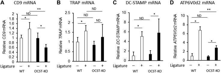
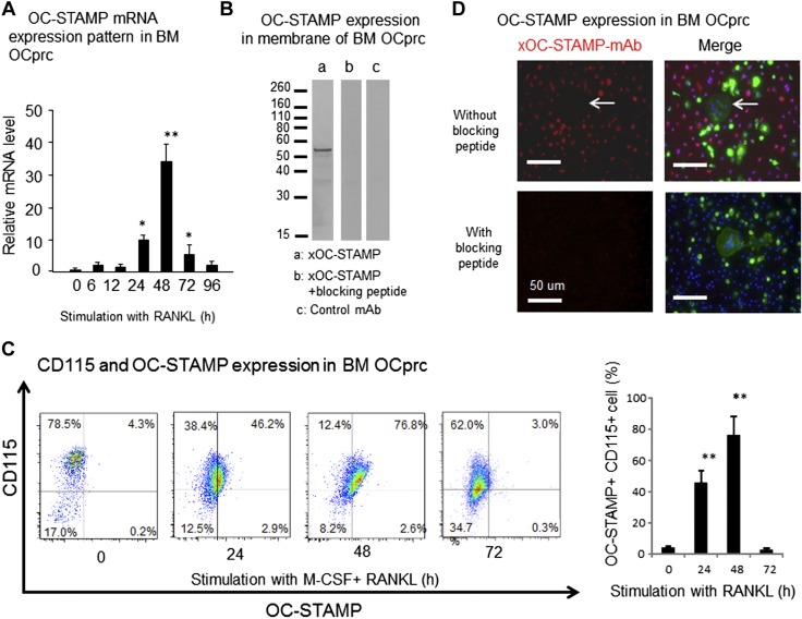
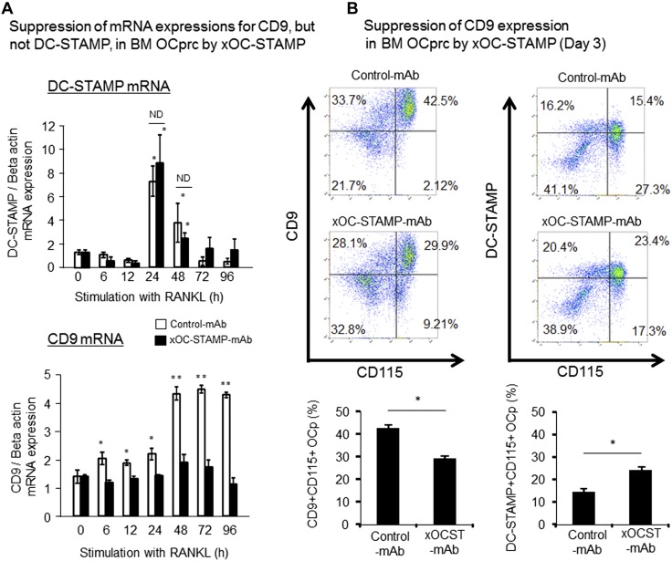
Cell fusion-mediated formation of multinuclear osteoclasts (OCs) plays a key role in bone resorption. It is reported that 2 unique OC-specific fusogens [ i.e., OC-stimulatory transmembrane protein (OC-STAMP) and dendritic cell-specific transmembrane protein (DC-STAMP)], and permissive fusogen CD9, are involved in OC fusion. In contrast to DC-STAMP-knockout (KO) mice, which show the osteopetrotic phenotype, OC-STAMP-KO mice show no difference in systemic bone mineral density. Nonetheless, according to the ligature-induced periodontitis model, significantly lower level of bone resorption was found in OC-STAMP-KO mice compared to WT mice. Anti-OC-STAMP-neutralizing mAb down-modulated in vitro: 1) the emergence of large multinuclear tartrate-resistant acid phosphatase-positive cells, 2) pit formation, and 3) mRNA and protein expression of CD9, but not DC-STAMP, in receptor activator of NF-κB ligand (RANKL)-stimulated OC precursor cells (OCps). While anti-DC-STAMP-mAb also down-regulated RANKL-induced osteoclastogenesis in vitro, it had no effect on CD9 expression. In our mouse model, systemic administration of anti-OC-STAMP-mAb suppressed the expression of CD9 mRNA, but not DC-STAMP mRNA, in periodontal tissue, along with diminished alveolar bone loss and reduced emergence of CD9+ OCps and tartrate-resistant acid phosphatase-positive multinuclear OCs. The present study demonstrated that OC-STAMP partners CD9 to promote periodontal bone destruction by up-regulation of fusion during osteoclastogenesis, suggesting that anti-OC-STAMP-mAb may lead to the development of a novel therapeutic regimen for periodontitis.-Ishii, T., Ruiz-Torruella, M., Ikeda, A., Shindo, S., Movila, A., Mawardi, H., Albassam, A., Kayal, R. A., Al-Dharrab, A. A., Egashira, K., Wisitrasameewong, W., Yamamoto, K., Mira, A. I., Sueishi, K., Han, X., Taubman, M. A., Miyamoto, T., Kawai, T. OC-STAMP promotes osteoclast fusion for pathogenic bone resorption in periodontitis via up-regulation of permissive fusogen CD9.

Keywords: DC-STAMP; RANKL; mouse model; osteoclastogenesis; periodontal bone loss.


Conflict of interest statement

This work was supported, in part, by U.S. National Institutes of Health (NIH) National Institute of Dental and Craniofacial Research Grants DE-018499, DE-019917, and T32 DE 007327-12, NIH National Institute on Aging Grant AG-053615, and a research grant from King Abdulaziz University. The authors declare no conflicts of interest.

Figures

Figure 1
Figure 1
OC-STAMP-KO mice showed diminished alveolar bone loss in ligature-induced mouse model of periodontitis. A) Alveolar bone resorption induced in mice by attachment of ligature. Total alveolar bone loss was calculated by summing CEJ-AC distances of distal root of first molar; mesial, midbuccal, and distal, root of second molar; and mesial root of third molar. Seven days after ligature attachment, OC-STAMP KO demonstrated significantly diminished level of periodontal bone loss compared to WT mice [WT: n = 6, OC-STAMP-KO: n = 5, means ± sd. *P < 0.05, no difference (ND)]. B) Frozen sections of decalcified maxilla isolated from humanely killed mice (d 7) were subjected to TRAP staining. Scale bars, 50 µm. C) Histogram showing numbers of TRAP+ OCs counted in section (means ± sd. *P < 0.05, ND).
Figure 2
Figure 2
Expression profile of gene-associated OC cell fusion in OC-STAMP-KO and WT mice. CD9 (A), TRAP (B), DC-STAMP (C), and ATP6V0d2 (D) mRNA were measured by qPCR in periodontal tissue sampled from site that received ligature and control site without ligature from WT and OC-STAMP-KO mice on d 3 [WT: n = 4, OC-STAMP-KO: n = 4. *P < 0.05, **P < 0.01, no difference (ND)]. All data shown in histograms are means ± sd.
Figure 3
Figure 3
Expressions of OC-STAMP mRNA and protein in RANKL-stimulated BM-derived OCps in vitro. In order to induce CD115+ OCps, mononuculear cells isolated from mouse BM were preincubated with M-CSF (20 ng/ml) for 3 d and then used for following experiments. A) According to qPCR, OC-STAMP mRNA expression was induced in OCps derived from BM by stimulation with RANKL (50 ng/ml). Peak expression of OC-STAMP mRNA was detected at 48 h, followed by gradual reduction. B) OCps derived from BM were stimulated in vitro with RANKL for 3 d. Cell membrane fraction isolated from cultured OCps was subjected to SDS-PAGE. After transblotting to nitrocellulose membrane, biotinylated anti–OC-STAMP-mAb (1 μg/ml) was reacted to membrane. Positive band reactive to anti–OC-STAMP-mAb was detected at MW 56 kDa (a). In same Western blot experiment, addition of blocking peptide, which was used as immune antigen for generation of anti–OC-STAMP-mAb, inhibited development of positive band reactive to anti–OC-STAMP-mAb (b). Isotype-matched control mAb did not show any positive reaction by Western blot (c). C) Temporal change of OC-STAMP expressed on RANKL-stimulated OCps was evaluated by flow cytometry. OCps were stimulated with RANKL for 0, 24, 48, and 72 h. After reaction with Alexa Fluor 546–labeled anti–OC-STAMP-mAb and Alexa Fluor 647–labeled anti-CD115 (M-CSF)-mAb, RANKL-stimulated OCps were subjected to flow cytometry. Histogram shows incidence (%) of OC-STAMP+ OCps among CD115+ OCps. D) Expression of OC-STAMP protein on RANKL-stimulated OCps was monitored by Alexa Fluor 647–labeled anti–OC-STAMP-mAb, along with Alexa Fluor 488–conjugated phalloidin and DAPI, which stain actin fiber and nuclei, respectively, by laser scanning confocal microscope. Red, OC-STAMP; green, F-actin; blue, nuclei. Scale bars, 50 μm. *P < 0.05, **P < 0.01, compared to 0 h (statistically significantly different, by Tukey’s test).
Figure 4
Figure 4
Effects of anti–OC-STAMP-mAb on RANKL-induced OC-genesis in vitro. OCps derived from BM cells were stimulated with RANKL in presence or absence of anti–OC-STAMP-mAb or control mAb for 7 d, and TRAP staining was performed. A) Images of TRAP staining for RANKL-stimulated OCps. Scale bars, 50 μm. B) Number of TRAP+ multinuclear OCs was calculated. Based on number of nuclei in each OC, OCs were classified into large OCs (>10 nuclei/cell) and small OCs (10 ≥ nuclei/cell > 3). C) OCps were incubated in presence or absence of anti–OC-STAMP-mAb or control mAb for 7 d in Corning Osteo Assay Surface plate for 7 d. Control group received no conditioned media. Pit area in each well was quantified by ImageJ software. Data are means ± sd (n = 3). *P < 0.05, **P < 0.01 (statistically significantly different, by Tukey’s test).
Figure 5
Figure 5
Effects of OC-STAMP on expression of CD9 and DC-STAMP during in vitro RANKL-stimulated OC-genesis. After preincubation with M-CSF (20 ng/ml) for 3 d, mononuclear cells isolated form BM were stimulated with RANKL (50 ng/ml) and M-CSF (20 ng/ml) in presence or absence of anti–OC-STAMP-mAb or control mAb for 3 d, and temporal changes of mRNA and cell surface expression of DC-STAMP and CD9 were examined. A) In response to stimulation with RANKL, OCps increased expression of DC-STAMP mRNA, which reached peak at 24 h, followed by gradual reduction with time. However, no significant difference was observed in level of DC-STAMP mRNA between groups treated with anti–OC-STAMP-mAb and control mAb at 24 and 48 h. Statistically significantly different at *P < 0.05 by Tukey’s test, compared to 0 h; ND, no significant difference. B) Cell surface expressions of CD9 and DC-STAMP on OCps stimulated with RANKL in presence or absence of anti–OC-STAMP-mAb or control mAb for 3 d were monitored using flow cytometer. Data in histograms show means ± sd of CD9+CD115+ double-positive cells among mononuclear cells. Statistically significantly different at *P < 0.05 or ND by Tukey’s test (n = 3/group).
Figure 6
Figure 6
CD9 expression on RANKL-stimulated OCps isolated from OC-STAMP-KO mice. OCps derived from BM cells of WT and OC-STAMP-KO mice were stimulated with RANKL in presence or absence of anti–OC-STAMP-mAb or control mAb. TRAP staining for differentiated OCs on d 7 (A) and flow cytometry for detection of CD9 expressed on OCps on d 3 (B) were carried out. Data in histograms show means ± sd of TRAP+ multinuclear OCs (A) and CD9+CD115+ double-positive cells among mononuclear cells (B). Statistically significantly different at *P < 0.05 or no difference (ND) by Tukey’s test (n = 3/group).
Figure 7
Figure 7
Anti–DC-STAMP-mAb, but not anti–OC-STAMP-mAb, showed additive effects on anti–OC-STAMP-mAb–mediated suppression of formation of large TRAP+ OCs. A) OCps derived from BM cells of WT mice were stimulated with RANKL in presence or absence of anti–DC-STAMP-mAb or control mAb, and expression of CD9 mRNA was monitored by qPCR. Significantly elevated CD9 mRNA expression was detected 72 and 96 h after RANKL stimulation at 0 h. *P < 0.05, **P < 0.01 (Tukey’s test). No difference (ND) in CD9 mRNA was detected between groups treated with anti–DC-STAMP-mAb and control mAb. B) OCps stimulated with RANKL in presence or absence of anti–CD9-mAb, anti–DC-STAMP-mAb, and anti–OC-STAMP-mAb for 7 d were stained for TRAP expression. C) Number of small OCs (10 ≥ nuclei/cell > 3) and large OCs (>10 nuclei/cell) in assay (B) were counted and are presented in histograms. Tukey’s test, compared to stimulation with RANKL alone. *P < 0.05, **P < 0.01, ***P < 0.001; #P < 0.05, ##P < 0.01 (statistical difference between 2 columns by Tukey’s test).
Figure 8
Figure 8
xOCSTAMP-mAb suppressed periodontal bone loss induced in ligatured experimental mice. Mice (6–8 wk old, males, C57BL/6J, n = 5/group) received silk ligature on upper left second molar, whereas upper right second molar without ligature was used as control. Immediately after attachment of ligature, mice received systemic injection of anti–OC-STAMP-mAb (5 mg per mouse, i.p.) or control mAb (5 mg per mouse, i.p.). A) Seven days after ligature placement, gingival tissue was collected from humanely killed mice for detection of OC-STAMP mRNA using qPCR. B) Expression levels of RANKL mRNA and OPG mRNA detected in gingival tissue (d 3 and 7) were expressed as RANKL/OPG mRNA ratio. C) Images of defleshed alveolar bones from humanely killed mice (d 7) are shown from buccal aspect. Scale bar, 1 mm. Quantitative data of bone loss (d 7) are shown in histogram. D) Frozen sections of decalcified maxilla isolated from humanely killed mice (d 7) were subjected to TRAP staining. Scale bars, 100 µm. Low-magnification image is shown in Supplemental Fig. 3. Histogram shows numbers of TRAP+ OCs counted in section (means ± sd, n = 5). **P < 0.01. Column and bar data indicate means ± sd. *P < 0.05, **P < 0.01 (statistically significant differences, by Tukey’s test).
Figure 9
Figure 9
Effects of anti–OC-STAMP-mAb on expression of CD9 in periodontal tissue of induced periodontitis. A) At d 3 and 7 after attachment of ligature, periodontal tissue was isolated from humanely killed mice, and mRNA levels for DC-STAMP and CD9 were monitored by qPCR (Fig. 8A). B) Decalcified jaw sections were subjected to fluorescent microscopy to detect CD9+ cells among CD115+ OCps on alveolar bone surface of ligature-induced periodontitis. Column and bar data indicate means ± sd of CD9+/CD115+ double-positive cells in microscopic area (100 × 100 μm). *P < 0.05 (statistically significant difference, by Tukey’s test).

Similar articles

Cited by

References

    1. Xing L., Xiu Y., Boyce B. F. (2012) Osteoclast fusion and regulation by RANKL-dependent and independent factors. World J. Orthop. 3, 212–222 https://doi.org/10.5312/wjo.v3.i12.212 - DOI - PMC - PubMed
    1. Yagi M., Miyamoto T., Sawatani Y., Iwamoto K., Hosogane N., Fujita N., Morita K., Ninomiya K., Suzuki T., Miyamoto K., Oike Y., Takeya M., Toyama Y., Suda T. (2005) DC-STAMP is essential for cell–cell fusion in osteoclasts and foreign body giant cells. J. Exp. Med. 202, 345–351 https://doi.org/10.1084/jem.20050645 - DOI - PMC - PubMed
    1. Witwicka H., Hwang S. Y., Reyes-Gutierrez P., Jia H., Odgren P. E., Donahue L. R., Birnbaum M. J., Odgren P. R. (2015) Studies of OC-STAMP in osteoclast fusion: a new knockout mouse model, rescue of cell fusion, and transmembrane topology. PLoS One 10, e0128275 https://doi.org/10.1371/journal.pone.0128275 - DOI - PMC - PubMed
    1. Yang M., Birnbaum M. J., MacKay C. A., Mason-Savas A., Thompson B., Odgren P. R. (2008) Osteoclast stimulatory transmembrane protein (OC-STAMP), a novel protein induced by RANKL that promotes osteoclast differentiation. J. Cell. Physiol. 215, 497–505 https://doi.org/10.1002/jcp.21331 - DOI - PMC - PubMed
    1. Miyamoto H., Suzuki T., Miyauchi Y., Iwasaki R., Kobayashi T., Sato Y., Miyamoto K., Hoshi H., Hashimoto K., Yoshida S., Hao W., Mori T., Kanagawa H., Katsuyama E., Fujie A., Morioka H., Matsumoto M., Chiba K., Takeya M., Toyama Y., Miyamoto T. (2012) Osteoclast stimulatory transmembrane protein and dendritic cell–specific transmembrane protein cooperatively modulate cell–cell fusion to form osteoclasts and foreign body giant cells. J. Bone Miner. Res. 27, 1289–1297 https://doi.org/10.1002/jbmr.1575 - DOI - PubMed

Publication types