. 2018 Mar 12;44(5):582-596.e4.
doi: 10.1016/j.devcel.2018.02.010.

PAF-Myc-Controlled Cell Stemness Is Required for Intestinal Regeneration and Tumorigenesis

Affiliations

PAF-Myc-Controlled Cell Stemness Is Required for Intestinal Regeneration and Tumorigenesis

Moon Jong Kim et al. Dev Cell. .

Abstract

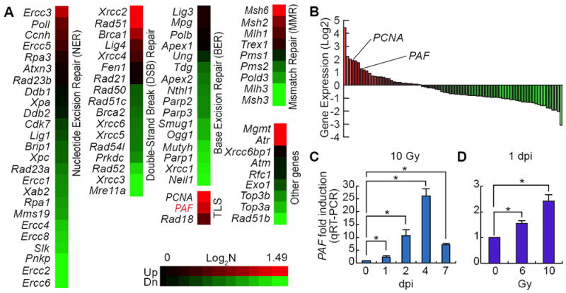
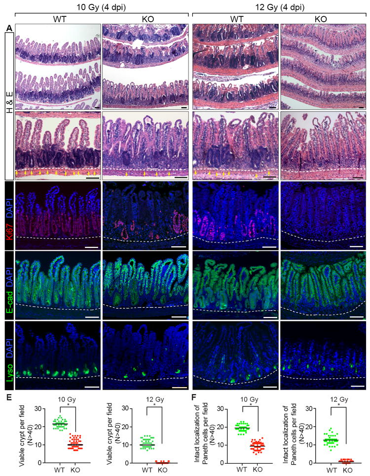
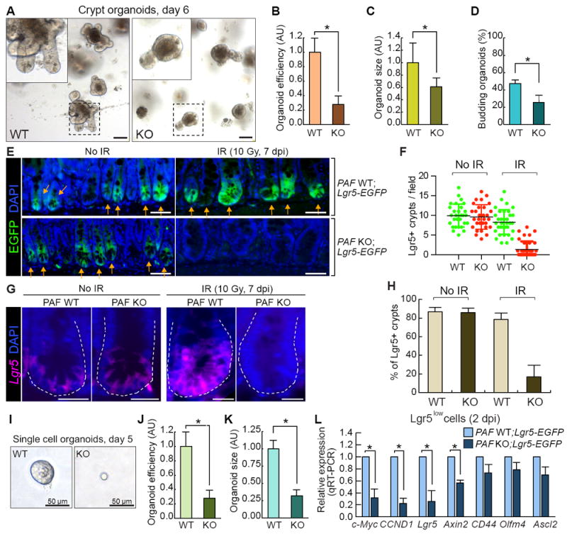
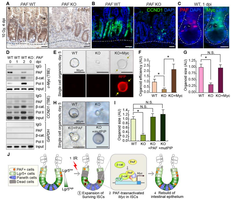
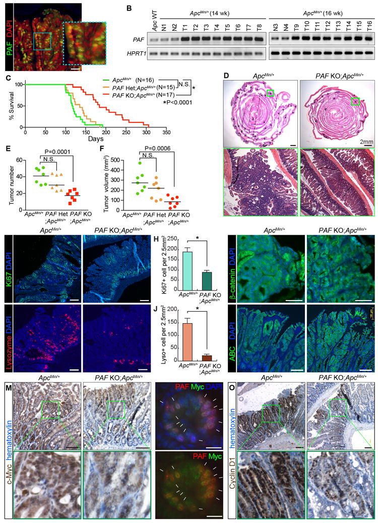
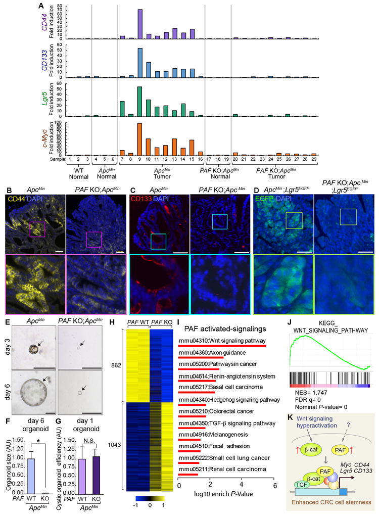
The underlying mechanisms of how self-renewing cells are controlled in regenerating tissues and cancer remain ambiguous. PCNA-associated factor (PAF) modulates DNA repair via PCNA. Also, PAF hyperactivates Wnt/β-catenin signaling independently of PCNA interaction. We found that PAF is expressed in intestinal stem and progenitor cells (ISCs and IPCs) and markedly upregulated during intestinal regeneration and tumorigenesis. Whereas PAF is dispensable for intestinal homeostasis, upon radiation injury, genetic ablation of PAF impairs intestinal regeneration along with the severe loss of ISCs and Myc expression. Mechanistically, PAF conditionally occupies and transactivates the c-Myc promoter, which induces the expansion of ISCs/IPCs during intestinal regeneration. In mouse models, PAF knockout inhibits Apc inactivation-driven intestinal tumorigenesis with reduced tumor cell stemness and suppressed Wnt/β-catenin signaling activity, supported by transcriptome profiling. Collectively, our results unveil that the PAF-Myc signaling axis is indispensable for intestinal regeneration and tumorigenesis by positively regulating self-renewing cells.

Keywords: Myc; PAF; Wnt signaling; cancer stem cells; colorectal cancer; intestinal regeneration; intestinal stem cells; stem/progenitor cells; β-catenin.


Conflict of interest statement

DECLARATION OF INTERESTS

The authors declare no competing interests.

Figures

Figure 1
Figure 1. Upregulation of PAF Expression upon Radiation Injury
(A, B) Gene expression profiling of DNA repair genes upon radiation injury in mouse small intestine. After treatment of 10 Gy irradiation (1 day post-injury [1 dpi]), the whole small intestine samples were analyzed by qRT-PCR (N=3). PCNA is the fourth and PAF are the seventh upregulated genes among the 79 genes related to DNA repair. (C) Time-dependent upregulation of PAF expression upon IR injury. At 0, 1, 2, 4, and 7 dpi, the small intestine samples were collected and analyzed by qRT-PCR (N=3). (D) Dose-dependent upregulation of PAF expression upon injury. 6 and 10 Gy irradiation were used (1 dpi). Student’s t-test; error bars = S.E.M; asterisks: P< 0.05.
Figure 2
Figure 2. PAF Expression in Normal and Regenerating Intestinal Crypts
(A) PAF expression in the small intestine. PAF WT and KO mice were analyzed for immunofluorescent (IF) staining of PAF (arrows). Asterisks mark non-specific staining signals. (B) Quantification of PAF+ cells in the small intestinal crypts. (C) Co-immunostaining of mouse small intestine (PAF WT and KO) for PAF and Ki67. White arrows: PAF+:Ki67+ cells; yellow arrows: PAF+:Ki67+ CBC cells. Asterisks mark non-specific staining signals. (D) Co-immunostaining of mouse small intestine (PAF WT and KO) for PAF and PCNA. White arrows: PAF+:PCNA+ cells; yellow arrows: PAF+:PCNA+ CBC cells; Asterisks mark non-specific staining signals. (E) Quantification of PAF+ cells in K67+ or PCNA+ cell population. Of note, PAF+:Ki67- cells were rarely found (approximately 1/50 crypts). (F) No effects of PAF KO on PCNA expression pattern. IF staining of the mouse small intestine for PCNA. (G) No expression of PAF in the Paneth cells. Co-immunostaining of the small intestine for PAF and Lysozyme. White arrows: PAF+ cells; yellow arrows: PAF+ CBC cell. (H) Co-expression of PAF and Lgr5 in the small intestine. Co-immunostaining of the small intestine of Lgr5-EGFP-CreERT2 mouse strain. White arrows: PAF+:Lgr5+ cells; yellow arrows: PAF+:Lgr5+ CBC cells. Asterisks indicate non-specific staining signals. (I) PAF expression in Lgr5+ cells. FACS-isolated Lgr5+ cells were analyzed for qRT-PCR. Asterisk=P<0.05. (J, K) PAF expression in TERT+ and Bmi1+ cells. Co-immunostaining of TERT-Tdtomato-CreERT2 or Bmi1-EGFP knock-in mouse intestine samples for PAF. Arrowheads: PAF+:TERT+ (J) and PAF+:Bmi1+ (K) cells; arrows: PAF+ cells. Asterisks mark non-specific staining signals. (L, M) The increase of PAF+ cells upon radiation injury. Immunostaining of mouse intestine (0, 1, 2, 4, and 7 dpi; 10 Gy) for PAF (L). White arrows: PAF+ cells; yellow arrows: PAF+ CBC cells. Quantification of PAF+ cells in the regenerating crypts (M). The representative images were shown from at least three independent experiments. Scale bars=20μm. See also Figures S1 and S2.
Figure 3
Figure 3. Impaired Intestinal Regeneration by PAF KO
(A-D) Impaired intestinal regeneration after irradiation (4 dpi; 10 and 12 Gy). Hematoxylin and eosin staining of the mouse small intestine samples of PAF+/+ (WT) or PAF-/- (KO) mice (A); IHC of the small intestine samples for Ki67 (B); IHC of the small intestine samples for E-cadherin, a marker for epithelial cell (C); IHC of the small intestine samples for Lysozyme, a marker for Paneth cell (D); Yellow arrows: crypts. (E) Quantification of the viable crypts. Crypts that showed five successive Ki67+ cells were counted as a viable crypt. (D) Quantification of the intact localization of Lysozyme. Crypts possessing at least three Lyso+ cells localized at the crypt bottom were counted as intact localization. The representative images were shown from at least three animals for each condition. Scale bars=100μm. See also Figures S3 and S4.
Figure 4
Figure 4. PAF Is Required for ISCs/IPCs Expansion in Regenerating Crypts
(A-D) Reduced organoid development by PAF KO. The crypt organoid growth assays from the small intestine samples of PAF WT and KO mice (A); quantification of organoid efficiency (N≥2000 from the three independent experiments) (B); size (5 days after seeding; N≥50) (C); budding efficiency (N≥50) (D). Asterisks=P< 0.01. Scale bars=100μm. (E, F) Impaired ISCs/IPCs expansion by PAF KO. Visualization of Lgr5+ EGFP cells by GFP immunostaining of the small intestines from PAF WT;Lgr5-EGFP and PAF KO;Lgr5-EGFP mice at 0 and 7 dpi (E). Scale bars=50μm; Quantification Lgr5+ (EGFP+) existing crypts per field (F). At least 30 fields of view were counted; Arrows (EGFP+ crypts); asterisks (EGFP-regenerating crypts). (G, H) Analysis of Lgr5 expression by FISH in PAF WT and PAF KO regeneration crypts. The representative images were shown. (G). Scale bars=20μm; % of Lgr5+ (EGFP+) crypts in the field (H). At least 10 fields of view were counted. (I-K) Reduced single cell organoid development by PAF KO. Representative images of single cell (Lgr5+) organoids (day 5) derived from PAF WT;Lgr5-EGFP and PAF KO;Lgr5-EGFP mice (I). Scale bars=50μm; quantification of organoid development efficiency (J) (N≥5000 cells were analyzed for from three independent experiments); size (K) (N≥30 single cell organoids were analyzed). (L) Gene expression analysis of FACS-isolated Lgr5low (GFP+) cells in the IR-treated intestine from PAF WT;Lgr5-EGFP and PAF KO;Lgr5-EGFP mice (2 dpi, 10 Gy). qRT-PCR analysis. Asterisks=P< 0.05. See also Figure S5.
Figure 5
Figure 5. Requirement of PAF-Myc Axis for ISCs/IPCs Expansion
(A, B) Downregulation of c-Myc and Cyclin D1 in PAF KO crypts (4 dpi, 10 Gy). IHC for c-Myc or Cyclin D1. Hematoxylin or DAPI for nuclear counterstaining (blue). (C) Co-expression of PAF and c-Myc in surviving cells in the regenerating crypts. Arrows: PAF+:Myc+ cells; asterisks: disappeared CBC cells. (D) Conditional recruitment of PAF and β-catenin to TCF-binding elements (TBEs) in c-Myc and CCND1 (Cyclin D1) proximal promoter. Chromatin immunoprecipitation (ChIP) assays of the mouse small intestine (PAF WT and KO; 1 and 2 dpi; 10 Gy). IgG ChIP and PAF KO small intestine samples served as negative control. RNA Pol II ChIP (positive control for gene transactivation). GAPDH promoter served as negative control. (E-G) Rescue of PAF KO-induced organoid growth failure by c-Myc expression. The Lgr5+ cells isolated from (PAF WT and KO) were transduced with Retroviruses encoding c-Myc and RFP (red fluorescent protein) and cultured for organoid development. Arrows indicate the budding. The representative images (E); quantification of organoid efficiency (F) and size (G). Asterisks: P<0.05 (N≥30). N.S. (not significant; P≥0.05). (H, I) Rescue of PAF KO-induced organoid growth failure by ectopic expression of wild-type PAF and PIP mutant PAF (mutPIP-PAF). The Lgr5+ cells isolated from PAF KO;Lgr5-EGFP were transduced with retroviruses encoding wild-type PAF and mutPIP-PAF and cultured for organoid development. The representative images (H); Quantification of organoid size (I) Asterisks: P<0.05 (N≥20). (J) Illustration of the working model. Upon irradiation injury, the highly proliferative cells (Lgr5high ISCs and some of Lgr5low TA cells) undergo apoptosis. PAF and β-catenin transactivate c-Myc in the surviving ISCs/IPCs (Lgr5low), which leads to the expansion of ISCs/IPCs and the subsequent rebuilding of the intestinal epithelium. Scale bars=50μm (A, B, E, H) and 20μm (C); the representative images were shown from at least three independent experiments. See also Figure S5
Figure 6
Figure 6. Attenuation of Intestinal Tumorigenesis by PAF KO
(A, B) Expression of PAF in ApcMin adenomas. Immunostaining of ApcMin/+ intestinal adenoma (16 wk old) (A), scale bar=50μm; semi-QT-RT-PCR (B). Apc WT: Wild-type (Apc+/+) intestine sample; N1-4: normal adjacent intestine samples; T1-16: intestinal adenomas. (C) The extended life span of ApcMin/+ mice by PAF KO. Kaplan-Meier survival curve of ApcMin/+ (N=16), PAF Het;ApcMin/+ (N=15), and PAF KO;ApcMin/+ (N=17). (D) H&E staining of the small intestines from ApcMin/+ and PAF KO;ApcMin/+ (age of 16 weeks). Asterisks indicate intestinal adenomas. Scale bars=2mm. (E, F) Decreased tumor burden of ApcMin/+ mice by PAF KO. The number of tumors (≥1.5mm) (E) and tumor volumes (mm3) (F) were quantified; 16 wk old; N=7 for each group. (G, H) Reduced cell proliferation of ApcMin tumors by PAF KO. Ki67 staining of small intestine adenomas from ApcMin/+ or PAF KO;ApcMin/+ (16 wk old) (G); quantification (H). Asterisk: P<0.001. (I, J) Decreased differentiation of the Paneth cells of ApcMin tumors by PAF KO. Lysozyme staining of small intestine adenomas from ApcMin/+ or PAF KO;ApcMin/+ (16 wk old) (I); quantification (J). Asterisk: P<0.001. (K, L) No change in β-catenin level and activity of ApcMin tumors by PAF KO. Immunostaining of small intestine adenomas from ApcMin/+ or PAF KO;ApcMin/+ (16wk old) for total β-catenin (K) and active (unphosphorylated) β-catenin (ABC) (L). Scale bars=100μm. (M) Downregulation of c-Myc of ApcMin tumors by PAF KO. c-Myc IHC; 16 wk old. Scale bars=100μm. (N) Co-expression of c-Myc and PAF in ApcMin tumors. Arrows: PAF+:Myc+ cells. Scale bars=20μm. (O) Downregulation of Cyclin D1 of ApcMin tumors by PAF KO. Cyclin D1 IHC; 16 wk old. Scale bars=100μm. The representative images (N≥3) were shown. See also Figure S6.
Figure 7
Figure 7. Decreased CRC Cell Stemness by PAF KO
(A) Gene expression analysis of CD44, CD133, Lgr5, and c-Myc in the normal intestine or adenomas from ApcMin/+ and PAF KO;ApcMin/+ (16 wk old). At least three individual samples from WT and normal region of ApcMin/+ and PAF KO;ApcMin/+ were used as the control. (B, C) Downregulation of CD44 and CD133 expression in ApcMin tumors by PAF KO. IF staining for CD44 and CD133; 16 wk old. Scale bars=100μm. (D) Downregulation of Lgr5 in ApcMin tumors by PAF KO. IF for GFP (adenomas of ApcMin/+;Lgr5-EGFP and PAF KO;ApcMin/+;Lgr5-EGFP [16 wk old]). Scale bars=100 μm. (E) Reduced cystic organoid development by PAF KO. Representative images of organoids (day 5) derived from ApcMin and PAF KO;ApcMin adenomas (F, G) Quantification of organoid size (F) (N≥30 cystic organoids were analyzed); efficiency (G) (N≥5000 cells were analyzed for from three independent experiments). Scale bars=100μm. Asterisk: P<0.001. (H) Heatmap gene expression profile generated by significant differential expression (P<0.05) of ApcMin/+ and PAF KO;ApcMin adenomas by RNA sequencing (N=2 per group). (I) Significantly PAF upregulated signaling pathways identified by GSEA analysis (KEGG) using RNA-seq result. P<0.05. (J) Gene set enrichment analysis (GSEA) for Wnt signaling pathway. NES: normalized enrichment score; FDR: False detection rate; P-value: Nominal P-value. (K) Illustration of the working model. During tumorigenesis, Wnt/β-catenin signaling is hyperactivated and PAF is upregulated. PAF and β-catenin transactivate c-Myc and CRC stemness-related genes (CD44, CD133, and Lgr5), which leads to the increase of CRC stemness. See also Figure S7, Tables S1 and S2.

References

    1. Araki R, Hoki Y, Uda M, Nakamura M, Jincho Y, Tamura C, Sunayama M, Ando S, Sugiura M, Yoshida MA, et al. Crucial role of c-Myc in the generation of induced pluripotent stem cells. Stem Cells. 2011;29:1362–1370. - PubMed
    1. Asfaha S, Hayakawa Y, Muley A, Stokes S, Graham TA, Ericksen RE, Westphalen CB, von Burstin J, Mastracci TL, Worthley DL, et al. Krt19(+)/Lgr5(-) Cells Are Radioresistant Cancer-Initiating Stem Cells in the Colon and Intestine. Cell Stem Cell. 2015;16:627–638. - PMC - PubMed
    1. Ashton GH, Morton JP, Myant K, Phesse TJ, Ridgway RA, Marsh V, Wilkins JA, Athineos D, Muncan V, Kemp R, et al. Focal adhesion kinase is required for intestinal regeneration and tumorigenesis downstream of Wnt/c-Myc signaling. Dev Cell. 2010;19:259–269. - PMC - PubMed
    1. Barker N, Van Es JH, Kuipers J, Kujala P, Van Den Born M, Cozijnsen M, Haegebarth A, Korving J, Begthel H, Peters PJ, et al. Identification of stem cells in small intestine and colon by marker gene Lgr5. Nature. 2007;449:1003–1007. - PubMed
    1. Bettess MD, Dubois N, Murphy MJ, Dubey C, Roger C, Robine S, Trumpp A. c-Myc is required for the formation of intestinal crypts but dispensable for homeostasis of the adult intestinal epithelium. Mol Cell Biol. 2005;25:7868–7878. - PMC - PubMed

Publication types

MeSH terms