Review
. 2018 Mar 12;33(3):347-354.
doi: 10.1016/j.ccell.2018.02.001.

Unique Metabolic Adaptations Dictate Distal Organ-Specific Metastatic Colonization

Affiliations
Review

Unique Metabolic Adaptations Dictate Distal Organ-Specific Metastatic Colonization

Tanya Schild et al. Cancer Cell. .

Abstract

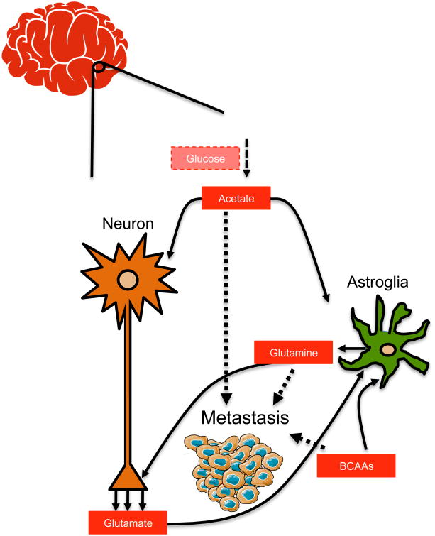
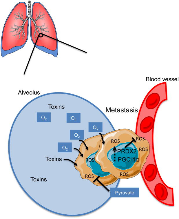
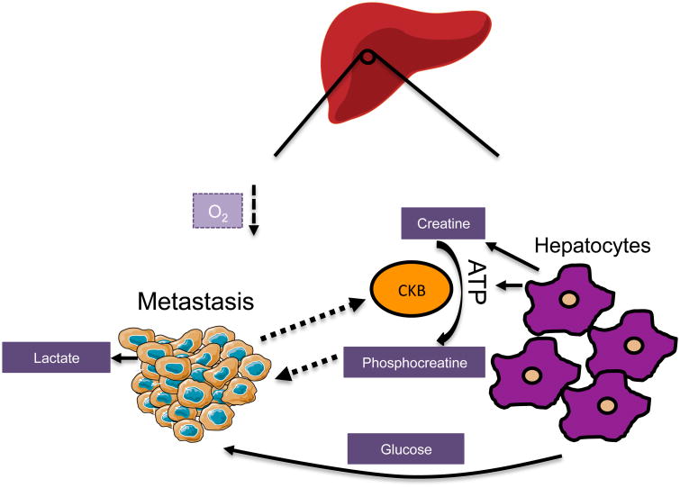
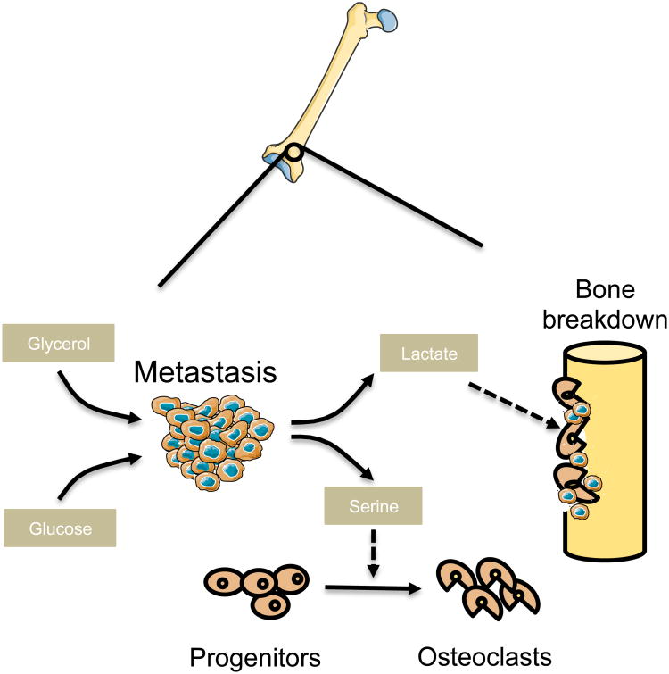
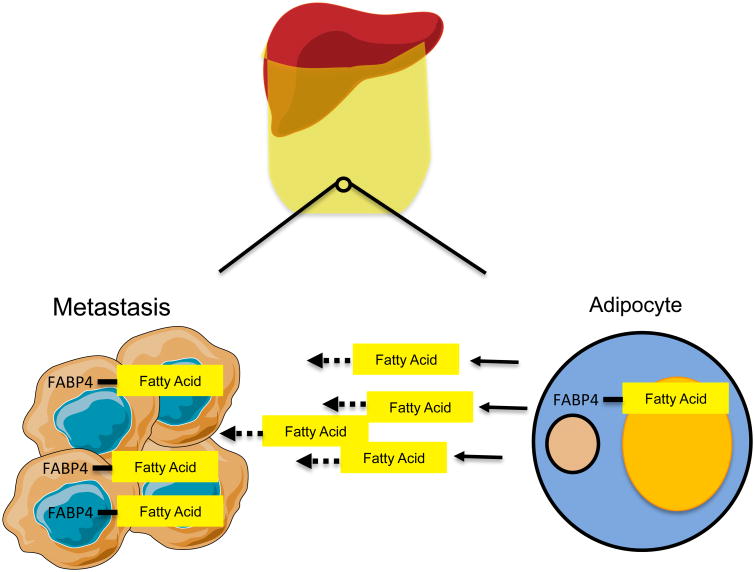
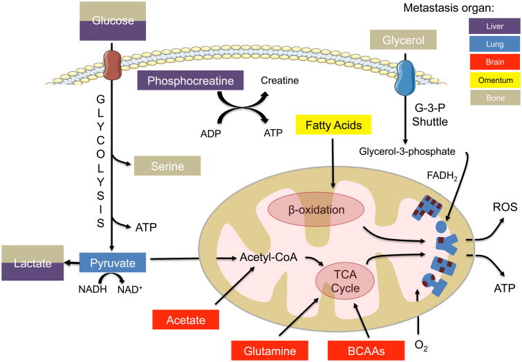
Metastases arising from tumors have the proclivity to colonize specific organs, suggesting that they must rewire their biology to meet the demands of the organ colonized, thus altering their primary properties. Each metastatic site presents distinct metabolic challenges to a colonizing cancer cell, ranging from fuel and oxygen availability to oxidative stress. Here, we discuss the organ-specific metabolic adaptations that cancer cells must undergo, which provide the ability to overcome the unique barriers to colonization in foreign tissues and establish the metastatic tissue tropism phenotype.

Keywords: cancer; metabolism; metastasis; tissue tropism.


Figures

Figure 1
Figure 1. Brain metastases are able to use acetate, branched chain amino acids and glutamine as alternative sources of fuel |
As metastatic cells colonize the brain, they adapt their metabolism to be able to use locally available nutrients—acetate, branched chain amino acids and glutamine—when glucose becomes limiting.
Figure 2
Figure 2. Metastases to the lung must survive in a pro-oxidant environment |
In order to cope with increased ROS, lung metastases upregulate PGC-1α and PRDX2. Additionally, metastases increase pyruvate consumption to fuel the TCA through anaplerosis and to alleviate strain on the mitochondrial electron chain.
Figure 3
Figure 3. Metastases to the liver face a unique biosynthetic milieu and must compete for resources |
Liver metastases become highly glycolytic through high consumption of local glucose. To replenish their ATP, metastases start expressing and secreting CKB which generates phosphocreatine from local ATP and creatine. Phosphocreatine is then imported into the metastatic cells and catabolized for energy.
Figure 4
Figure 4. Bone metastases remodel the stroma to free up nutrients and space for growth |
Osteotropic metastases secrete serine and lactate to facilitate osteoclastogenesis which results in bone resorption thereby releasing spatial and nutritional resources. Metastatic cells can also take on properties of local cells through the expression of bone-specific secreted, cell-surface and matrix markers, which can rewire their glucose and glycerol metabolism.
Figure 5
Figure 5. Metastases to omentum reprogram omental adipocytes to release fatty acids which supply metastatic cells with energy |
Omental metastases stimulate lipolysis in adipocytes which releases fatty acids into the environment. Metastatic cells also upregulate FABP4 which is allows them to import and catabolize newly available fatty acids.
Figure 6
Figure 6. Metastases in different organs utilize distinct metabolic pathways to generate energy |
Metastases that arise in different organs adapt to the specific conditions of their particular environment in order to generate ATP. In the brain (red), metastases are able to utilize glutamine, BCAAs and acetate as carbon sources to fuel the TCA cycle. In the lung (blue), pyruvate fuels the TCA cycle and additionally can replenish NAD+ pools for anaplerosis. The liver (purple) supports a glycolytic profile, and metastases to this location also use phosphocreatine to generate ATP. Omental (yellow) metastases bind fatty acids from adipocytes and oxidize them in order to fuel anaplerosis. Bone (gray) metastases rewire their metabolism through expression of osteopontin to differentially utilize glucose and glycerol to meet energetic demands and produce serine.

Similar articles

Cited by

References

    1. Albrecht J, Sidoryk-Wegrzynowicz M, Zielinska M, Aschner M. Roles of glutamine in neurotransmission. Neuron Glia Biol. 2010;6:263–276. - PubMed
    1. Alvarado A, Arce I. Metabolic Functions of the Lung, Disorders and Associated Pathologies. J Clin Med Res. 2016;8:689–700. - PMC - PubMed
    1. Andrzejewski S, Klimcakova E, Johnson RM, Tabaries S, Annis MG, McGuirk S, Northey JJ, Chenard V, Sriram U, Papadopoli DJ, et al. PGC-1alpha Promotes Breast Cancer Metastasis and Confers Bioenergetic Flexibility against Metabolic Drugs. Cell Metab. 2017;26:778–787 e775. - PubMed
    1. Brastianos PK, Carter SL, Santagata S, Cahill DP, Taylor-Weiner A, Jones RT, Van Allen EM, Lawrence MS, Horowitz PM, Cibulskis K, et al. Genomic Characterization of Brain Metastases Reveals Branched Evolution and Potential Therapeutic Targets. Cancer Discov. 2015;5:1164–1177. - PMC - PubMed
    1. Carlinfante G, Vassiliou D, Svensson O, Wendel M, Heinegard D, Andersson G. Differential expression of osteopontin and bone sialoprotein in bone metastasis of breast and prostate carcinoma. Clin Exp Metastasis. 2003;20:437–444. - PubMed

Publication types

LinkOut - more resources