. 2018 Jun 1;128(6):2281-2296.
doi: 10.1172/JCI93910. Epub 2018 Apr 30.

Circadian clock component REV-ERBα controls homeostatic regulation of pulmonary inflammation

Affiliations

Circadian clock component REV-ERBα controls homeostatic regulation of pulmonary inflammation

Marie Pariollaud et al. J Clin Invest. .

Abstract

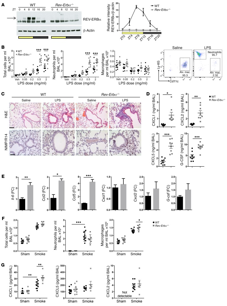
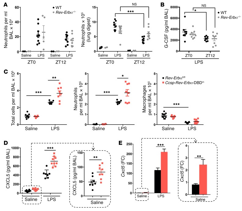
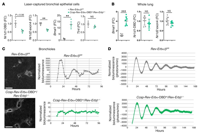
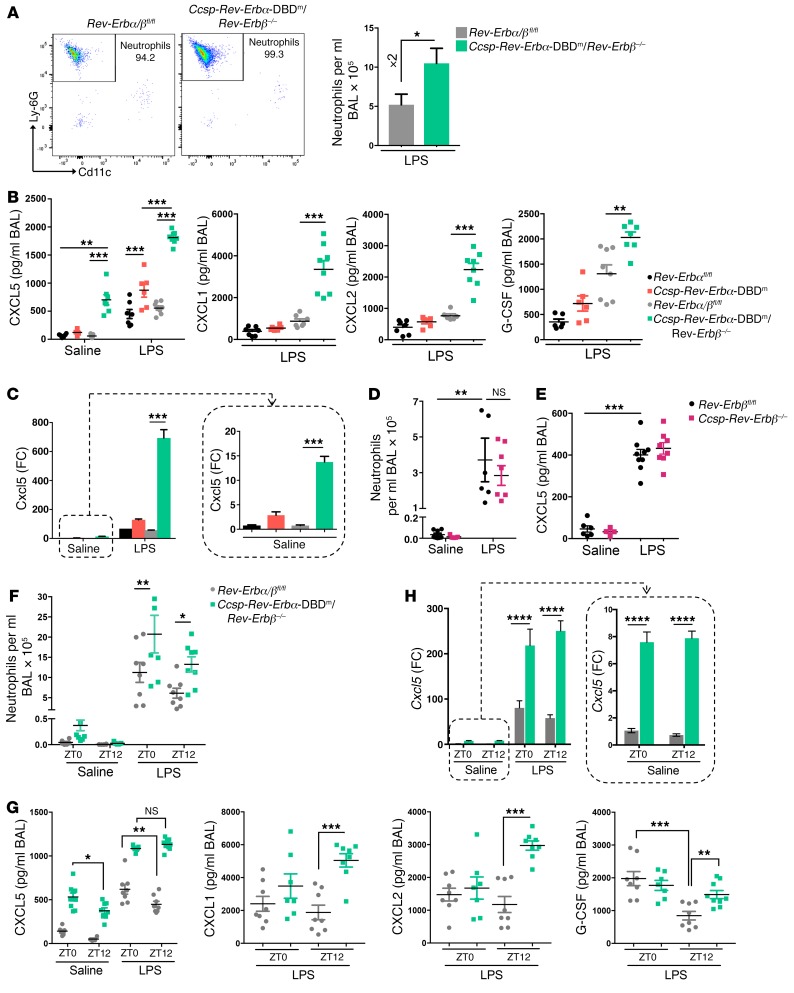
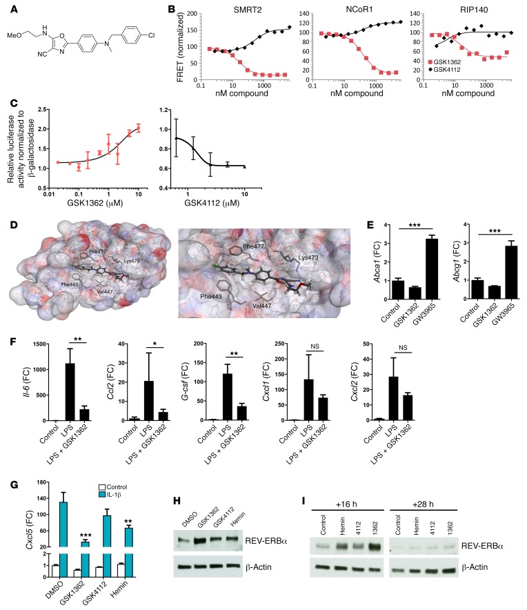
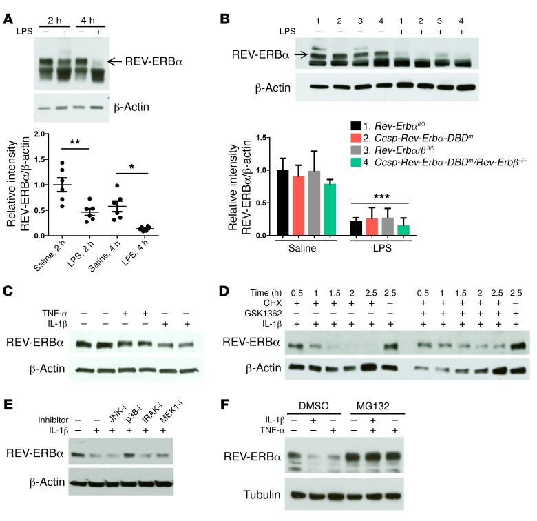
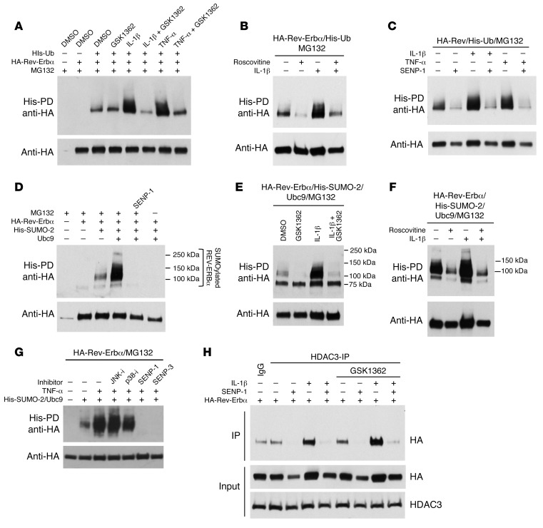
Recent studies reveal that airway epithelial cells are critical pulmonary circadian pacemaker cells, mediating rhythmic inflammatory responses. Using mouse models, we now identify the rhythmic circadian repressor REV-ERBα as essential to the mechanism coupling the pulmonary clock to innate immunity, involving both myeloid and bronchial epithelial cells in temporal gating and determining amplitude of response to inhaled endotoxin. Dual mutation of REV-ERBα and its paralog REV-ERBβ in bronchial epithelia further augmented inflammatory responses and chemokine activation, but also initiated a basal inflammatory state, revealing a critical homeostatic role for REV-ERB proteins in the suppression of the endogenous proinflammatory mechanism in unchallenged cells. However, REV-ERBα plays the dominant role, as deletion of REV-ERBβ alone had no impact on inflammatory responses. In turn, inflammatory challenges cause striking changes in stability and degradation of REV-ERBα protein, driven by SUMOylation and ubiquitination. We developed a novel selective oxazole-based inverse agonist of REV-ERB, which protects REV-ERBα protein from degradation, and used this to reveal how proinflammatory cytokines trigger rapid degradation of REV-ERBα in the elaboration of an inflammatory response. Thus, dynamic changes in stability of REV-ERBα protein couple the core clock to innate immunity.

Keywords: Inflammation; Innate immunity; Mouse models; Neutrophils; Pulmonology.


Conflict of interest statement

Conflict of interest: The authors have declared that no conflict of interest exists.

Figures

Figure 1
Figure 1. REV-ERBα plays a critical role in regulation of lung inflammation.
(A) Whole-lung REV-ERBα protein across the day (ZT, time from lights on). REV-ERBα densitometry (mean ± SEM) was normalized to β-actin and to WT at ZT0; n = 5 for WT and n = 3 for Rev-Erbα–/– per time point. (B) Mice were exposed to aerosolized LPS at ZT4 and culled 5 hours later; cellular infiltrates were quantified in BAL using flow cytometry. Data presented as mean ± SEM; n = 6–8, ***P < 0.001 (2-way ANOVA, post hoc Bonferroni). Veh, vehicle. (C) H&E staining and immunohistochemistry for the neutrophil maker (NIMP/R14) of lung sections from mice after LPS challenge at 2 mg/ml. Representative of n = 4; scale bars: 50 μm. (D) Cytokine/chemokine levels in BAL fluid from mice exposed to aerosolized LPS (2 mg/ml). Representative of n = 8, Student’s t test with Welch’s correction. (E) Quantitative PCR (qPCR) analysis of cytokine transcripts in alveolar macrophages isolated from mice and stimulated ex vivo with LPS at 100 ng/ml for 2 hours. Data normalized to WT and presented as mean ± SEM; n = 3, *P < 0.05, **P < 0.01, ***P < 0.001 (Student’s t test). FC, fold change. (F) Ten-day cigarette smoke exposures were performed between ZT8 and ZT10, and animals were culled 20 hours after the last exposure. Cellular infiltrates were quantified in BAL using a hemocytometer for total cell number and cytospin for neutrophil and macrophage counts. Data presented as mean ± SEM; n = 6–10, *P < 0.05, ***P < 0.001 (2-way ANOVA, post hoc Bonferroni). (G) Chemokine levels in BAL fluid after 10-day cigarette smoke exposures. Data presented as mean ± SEM; n = 6–10, **P < 0.01 (2-way ANOVA, post hoc Bonferroni).
Figure 2
Figure 2. REV-ERBα in myeloid and airway epithelium regulates pulmonary inflammation.
(A and B) Bone marrow cells from Rev-Erbα–/– or littermate controls were transplanted into WT recipient mice, which were then exposed to aerosolized LPS at 2 mg/ml or saline at indicated times for 20 minutes. (A) Neutrophil numbers in BAL samples or lung digests collected 5 hours after challenge, determined by flow cytometry analyses. (B) Chemokine protein levels in BAL samples. Data presented as mean ± SEM; n = 2 (saline) or 5–8 (LPS), *P < 0.05, ***P < 0.001 (2-way ANOVA, post hoc Bonferroni). (CE) Ccsp-Rev-Erbα-DBDm and littermate control mice were exposed to aerosolized LPS at 2 mg/ml or saline at ZT4 for 20 minutes. (C) Total cell counts in BAL samples collected 5 hours after challenge. Neutrophil and macrophage numbers in the same samples were determined by flow cytometry analyses. (D) Chemokine protein levels in BAL samples, measured using multiplex assay. (E) qPCR analysis of Cxcl5 mRNA in lung tissues. Data normalized to saline Rev-Erbαfl/fl control group. Data presented as mean ± SEM; n = 5–9, *P < 0.05, **P < 0.01, ***P < 0.001 (2-way ANOVA, post hoc Bonferroni).
Figure 3
Figure 3. Both REV-ERB paralogs are required for circadian rhythms in the airway epithelium.
(A) qPCR analysis of mRNA in bronchial epithelial cells, laser-captured from lung tissues collected at ZT9. Data normalized to Rev-Erbα/βfl/fl control group and presented as mean ± SD; n = 3, **P < 0.01, Student’s t test. (B) qPCR analysis of mRNA in whole lung collected at ZT9. Data normalized to Rev-Erbα/βfl/fl control group and presented as mean ± SEM; n = 7, ***P < 0.001, Student’s t test. (C) Snapshots of PER2 oscillations in bronchioles within precision-cut lung slices. Scale bars: 500 μm. Bioluminescence intensity from bronchioles was quantified, normalized to a 24-hour moving average. Traces are representative of 2 biological replicates. (D) Bioluminescence recordings of whole-lung PER2 oscillations in precision-cut lung slices. Photon counts per minute were normalized to a 24-hour moving average, and traces are representative of 3 biological replicates.
Figure 4
Figure 4. Loss of both REV-ERBα DBD and REV-ERBβ in the airway epithelium further exaggerates inflammation.
(A) Flow analysis of neutrophils in BAL samples after aerosolized LPS (2 mg/ml) at ZT4 for 20 minutes. Data presented as mean ± SEM; n = 5–8, *P < 0.05 (Student’s t test). (B) Cytokine/chemokine protein levels in BAL samples after aerosolized LPS or saline at ZT4 for 20 minutes. Data presented as mean ± SEM; n = 5–9, **P < 0.01, ***P < 0.001 (2-way ANOVA, post hoc Bonferroni). (C) qPCR analysis of Cxcl5 levels in lung tissues from the same mice as above. Data normalized to saline Rev-Erbαfl/fl control group and presented as mean ± SEM; n = 5–9, ***P < 0.001 (2-way ANOVA, post hoc Bonferroni). (D) Neutrophil numbers in BAL samples after aerosolized LPS or saline at ZT4 for 20 minutes. Data presented as mean ± SEM; n = 5–9, **P < 0.01 (2-way ANOVA, post hoc Bonferroni). (E) CXCL5 protein levels in the same BAL samples as above. Data presented as mean ± SEM; n = 5–9, ***P < 0.001 (2-way ANOVA, post hoc Bonferroni). (F) Neutrophil numbers in BAL samples collected 5 hours after aerosolized LPS challenge at ZT0 or ZT12. Data presented as mean ± SEM; n = 7–9, *P < 0.05, **P < 0.01 (2-way ANOVA, post hoc Bonferroni). (G) Cytokine/chemokine protein levels in the same BAL samples as above. Data presented as mean ± SEM; n = 7–9, *P < 0.05, **P < 0.01, ***P < 0.001 (2-way ANOVA, post hoc Bonferroni). (H) qPCR analysis of Cxcl5 levels in lung tissues from the same mice as above. Data normalized to saline Rev-Erbα/βfl/fl control group and presented as mean ± SEM; n = 7–9, ****P < 0.0001 (2-way ANOVA, post hoc Bonferroni).
Figure 5
Figure 5. REV-ERBα ligand GSK1362 represses inflammatory genes in macrophages and epithelial cells and stabilizes REV-ERBα protein.
(A) Chemical structure of GSK1362. (B) Effect of GSK1362 and GSK4112 on peptide fragment recruitment to REV-ERBα. (C) Cotransfection of HEK293 cells with HA–Rev-Erbα and Bmal1-Luc reporter. Cells were treated with GSK1362 or GSK4112 at different concentrations for 24 hours before luciferase assay. Values plotted relatively to 0.1% DMSO; error bars indicate mean ± SD. Data representative of n = 3. (D) Models showing GSK1362 docked in REV-ERBα ligand-binding domain. (E) qPCR analysis of LXR target genes in peritoneal exudate cells treated ex vivo with GSK1362 at 10 μM or GW3965, a standard LXR agonist, at 2 μM for 4 hours. Data presented as mean ± SD; n = 3, ***P < 0.001 (1-way ANOVA, post hoc Bonferroni). (F) qPCR analysis of cytokine mRNA in alveolar macrophages collected at ZT8, seeded into plates and directly treated with GSK1362 at 10 μM in the presence or absence of LPS at 100 ng/ml for 4 hours. Data presented as mean ± SD; n = 3, *P < 0.05, **P < 0.01 (1-way ANOVA, post hoc Bonferroni). (G) qPCR analysis of Cxcl5 in LA-4 cells synchronized by serum shock and treated 16 hours later with ligands at 10 μM, followed 2 hours later by IL-1β at 1 ng/ml for 2 additional hours. Data normalized to unstimulated control cells and presented as mean ± SD; representative of n = 3, **P < 0.01, ***P < 0.001 (2-way ANOVA, post hoc Bonferroni). (H) REV-ERBα protein in LA-4 cells synchronized by serum shock and treated 16 hours later with ligands at 10 μM for 4 hours. Representative of n = 3. (I) REV-ERBα protein in NHBE cells synchronized by serum shock and treated 16 or 28 hours later with ligands at 10 μM for 4 hours. Representative of n = 3.
Figure 6
Figure 6. Inflammatory stimuli promote REV-ERBα degradation.
(A) Whole-lung REV-ERBα protein in WT mice 2 or 4 hours after aerosolized LPS (2 mg/ml) or saline solution for 20 minutes at CT8 (see Methods). REV-ERBα densitometry (mean ± SEM) was normalized to β-actin and to saline at 2 hours; n = 6, *P < 0.05, **P < 0.01 (1-way ANOVA, post hoc Bonferroni). (B) Whole-lung REV-ERBα protein in mice after aerosolized LPS (2 mg/ml) or saline solution for 20 minutes at ZT4 for 5 hours. REV-ERBα densitometry (mean ± SD) was normalized to β-actin and to group 1; n = 3, ***P < 0.001 (2-way ANOVA, post hoc Bonferroni). (C) REV-ERBα protein in NHBE cells synchronized by serum shock and treated 18 hours later with TNF-α or IL-1β at 10 ng/ml for 1 hour. (D) REV-ERBα protein in NHBE cells synchronized by serum shock and treated 18 hours later with GSK1362 or DMSO at 10 μM followed 15 minutes later by cycloheximide (CHX) at 10 μM and IL-1β at 1 ng/ml. Cells were lysed at different times as indicated. (E) REV-ERBα protein in SW1353 cells synchronized by serum shock and treated 23 hours later with kinase inhibitors for 30 minutes followed by IL-1β at 5 ng/ml for 1 hour. (F) REV-ERBα protein in SW1353 cells synchronized by serum shock and treated 23 hours later with PBS, TNF-α, or IL-1β at 5 ng/ml for 1 hour in the absence and presence of MG132 at 5 μM. All blots are representative of at least n = 3.
Figure 7
Figure 7. Requirement of posttranslational modifications for REV-ERBα degradation.
(AC) Ubiquitinated REV-ERBα protein in HEK293T cells transfected with HA–Rev-Erbα, His-Ub, and SENP-1 plasmids and treated with GSK1362 at 10 μM, roscovitine at 25 μM, and TNF-α or IL-1β at 5 ng/ml for 4 hours in the presence of MG132 at 5 μM. (DG) SUMO-2 ligation to REV-ERBα protein in HEK293T cells transfected with HA–Rev-Erbα, His-SUMO2, Ubc9, and SENP-1 plasmids and treated with GSK1362 at 10 μM and kinase inhibitors and IL-1β or TNF-α at 5 ng/ml for 4 hours in the presence of MG132 at 5 μM. (H) Coimmunoprecipitation of HDAC3 and REV-ERBα protein in HEK293T cells transfected with HA–Rev-Erbα and SENP-1 plasmids and treated with GSK1362 at 10 μM and IL-1β at 5 ng/ml for 4 hours. All blots are representative of at least n = 3.

References

    1. Hussell T, Bell TJ. Alveolar macrophages: plasticity in a tissue-specific context. Nat Rev Immunol. 2014;14(2):81–93. doi: 10.1038/nri3600. - DOI - PubMed
    1. Gibbs J, et al. An epithelial circadian clock controls pulmonary inflammation and glucocorticoid action. Nat Med. 2014;20(8):919–926. doi: 10.1038/nm.3599. - DOI - PMC - PubMed
    1. Mohawk JA, Green CB, Takahashi JS. Central and peripheral circadian clocks in mammals. Annu Rev Neurosci. 2012;35:445–462. doi: 10.1146/annurev-neuro-060909-153128. - DOI - PMC - PubMed
    1. Bellet MM, et al. Circadian clock regulates the host response to Salmonella. Proc Natl Acad Sci U S A. 2013;110(24):9897–9902. doi: 10.1073/pnas.1120636110. - DOI - PMC - PubMed
    1. Gibbs JE, et al. The nuclear receptor REV-ERBα mediates circadian regulation of innate immunity through selective regulation of inflammatory cytokines. Proc Natl Acad Sci U S A. 2012;109(2):582–587. doi: 10.1073/pnas.1106750109. - DOI - PMC - PubMed

Publication types

Substances