Review
. 2018 Mar 13;8(1):19.
doi: 10.3390/diagnostics8010019.

Eyeing up the Future of the Pupillary Light Reflex in Neurodiagnostics

Affiliations
Review

Eyeing up the Future of the Pupillary Light Reflex in Neurodiagnostics

Charlotte A Hall et al. Diagnostics (Basel). .

Abstract

The pupillary light reflex (PLR) describes the constriction and subsequent dilation of the pupil in response to light as a result of the antagonistic actions of the iris sphincter and dilator muscles. Since these muscles are innervated by the parasympathetic and sympathetic nervous systems, respectively, different parameters of the PLR can be used as indicators for either sympathetic or parasympathetic modulation. Thus, the PLR provides an important metric of autonomic nervous system function that has been exploited for a wide range of clinical applications. Measurement of the PLR using dynamic pupillometry is now an established quantitative, non-invasive tool in assessment of traumatic head injuries. This review examines the more recent application of dynamic pupillometry as a diagnostic tool for a wide range of clinical conditions, varying from neurodegenerative disease to exposure to toxic chemicals, as well as its potential in the non-invasive diagnosis of infectious disease.

Keywords: acetylcholine; autism; chemicals; cholinergic system; infection; neurodegeneration; pupillometry; recreational drugs; toxins; trauma.


Conflict of interest statement

The authors declare no conflict of interest.

Figures

Figure 1
Figure 1
Schematic of the pupillogram (blue line) and associated PLR parameters. The light stimulus at time zero results in a rapid reduction in pupil diameter. Latency (tL) is calculated as the elapsed time between light onset and the start of constriction. The pupil then rapidly constricts (maximal constriction velocity; MCV) from the baseline (D0) pupil diameter to the minimum (Dmin) pupil diameter; the constriction time (tC) and maximum constriction amplitude (MCA) are calculated as the time interval and size difference between these two values, respectively. At offset of light stimulus or during sustained light stimulation the pupil undergoes a period of rapid redilation or pupillary “escape” to a partially constricted state. Subsequently the pupil slowly returns to the baseline diameter.
Figure 2
Figure 2
Simplified schematic view of retinal layers involved in the pupillary light reflex. Vertical signalling pathways in the retina are composed of the photoreceptors (rod and cone cells), bipolar cells and retinal ganglion cells (RGC), including intrinsically photosensitive retinal ganglion cells (ipRGCs). There are also two lateral pathways comprised of horizontal cells in the outer plexiform layer (OPL) and the amacrine cells in the inner plexiform layer (IPL). These cells modulate the activity of other retinal cells in the vertical pathway. The somata of the neurons are in three cellular layers. The rod and cone cells are located in the outer nuclear layer (ONL), which is adjacent to the retinal pigment epithelium (RPE). The horizontal cell, bipolar cell and amacrine cell somas are located in the inner nuclear layer (INL), whilst the ganglion cell somata are located in the ganglion cell layer (GCL). The axon terminals of the bipolar cells stratify at different depths of the inner plexiform layer, which is subdivided into the OFF outer sublamina (where OFF bipolar cells terminate) and the ON inner sublamina (where ON bipolar cells terminate). There are also ON and OFF bands of melanopsin dendrites from the ipRGCs, but both lie outside of the ON and OFF cholinergic bands within the IPL. The bipolar cells are photoreceptor specific and the bipolar dendrites synapse exclusively with either rod or cone cells.
Figure 3
Figure 3
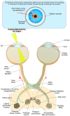
The parasympathetic nervous system is the main system responsible for pupil constriction in response to light. The integrated afferent input is transmitted along the axons of the retinal ganglion cells (RGC), which contribute to the optic nerve. At the optic chiasm, nerves from the nasal retina cross to the contralateral side, whilst nerves from the temporal retina continue ipsilaterally. The pupillary RGC axons exit the optic tract and synapse at the pretectal olivary nucleus. Pretectal neurons are projected either ipsilaterally or contralaterally, across the posterior commissure, to the Edinger-Westphal nucleus. From there, the pre-ganglionic parasympathetic fibres travel with the oculomotor, or III cranial nerve, and synapse at the ciliary ganglion. The post-ganglionic parasympathetic neurons (short ciliary nerves) travel to and innervate the contraction of the iris sphincter muscle via the release of acetylcholine at the neuromuscular junction, resulting in pupil constriction.
Figure 4
Figure 4
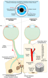
Both parasympathetic and sympathetic nervous systems are required for pupil dilation as part of the PLR. The parasympathetic innervation of the pupil sphincter is inhibited by central supranuclear inhibition of Edinger–Westphal nuclei via α2-adrenergic receptor activation, resulting in relaxation of the pupil sphincter muscle. The sympathetic influence on the iris dilator muscle consists of a paired three-neuron arc on both the right and left sides of the central and peripheral nervous system without decussations. The first-order (central) neuron originates in the hypothalamus and descends to synapse with the pre-ganglionic in the ciliospinal centre of Budge at C8-T1 of the spinal cord. The pre-ganglionic neuron ascends from the ciliospinal centre of Budge to synapse with the post-ganglionic neuron at the superior cervical ganglion, which is located at the periarterial plexus near the carotid artery bifurcation. Finally, long ciliary (post-ganglionic) nerves travel to and innervate the contraction of the iris dilator muscles, via a release of noradrenaline (NA) at the neuromuscular junction, resulting in pupil dilation. The synaptic transmission at the other junctions is mediated by acetylcholine.

References

    1. Hirata Y., Yamaji K., Sakai H., Usui S. Function of the pupil in vision and information capacity of retinal image. Syst. Comput. Jpn. 2003;34:48–57. doi: 10.1002/scj.10344. - DOI
    1. McDougal D.H., Gamlin P.D. Autonomic control of the eye. Compr. Physiol. 2015;5:439–473. doi: 10.1002/cphy.c140014. - DOI - PMC - PubMed
    1. Girkin C. Evaluation of the pupillary light response as an objective measure of visual function. Ophthalmol. Clin. N. Am. 2003;16:143–153. doi: 10.1016/S0896-1549(03)00002-6. - DOI - PubMed
    1. Loewenfeld I.E. The Pupil: Anatomy, Physiology, and Clinical Applications. 2nd ed. Butterworth-Heinemann; Boston, MA, USA: 1999.
    1. Winn B., Whitaker D., Elliott D.B., Phillips N.J. Factors affecting light-adapted pupil size in normal human subjects. Investig. Ophthalmol. Vis. Sci. 1994;35:1132–1137. - PubMed