. 2018 Mar 13;18(3):850.
doi: 10.3390/s18030850.

EquiMoves: A Wireless Networked Inertial Measurement System for Objective Examination of Horse Gait

Affiliations

EquiMoves: A Wireless Networked Inertial Measurement System for Objective Examination of Horse Gait

Stephan Bosch et al. Sensors (Basel). .

Abstract

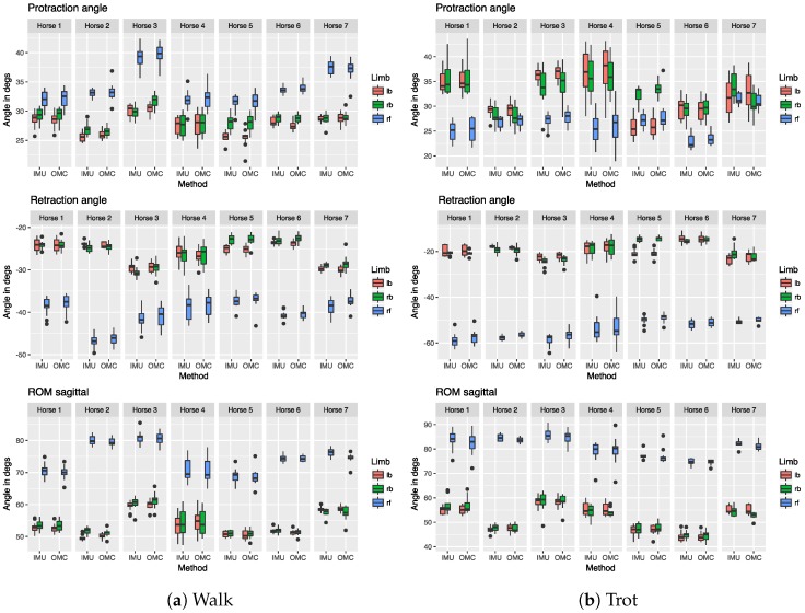
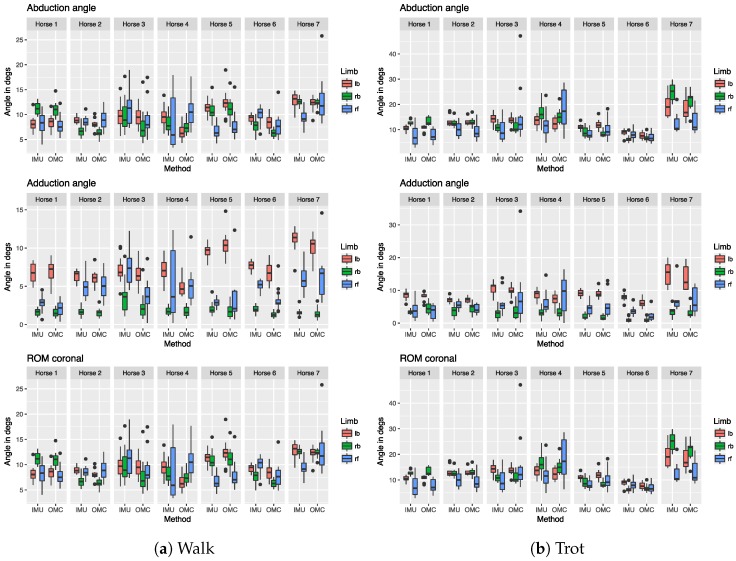
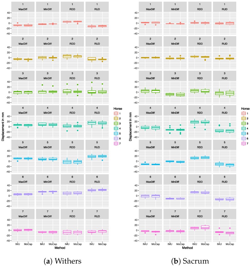
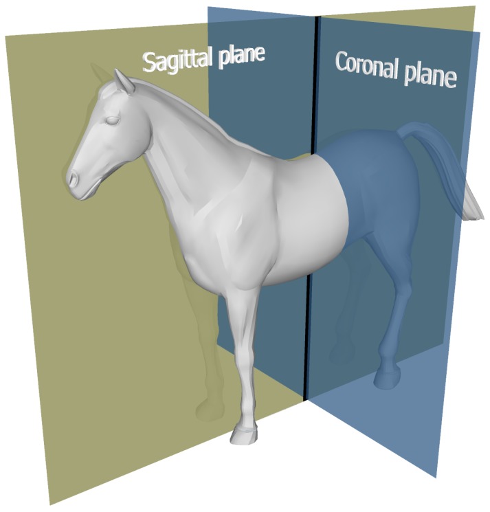
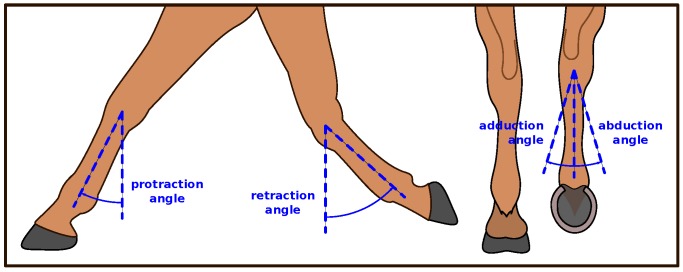
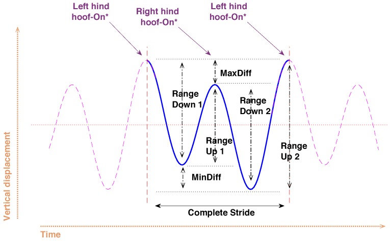
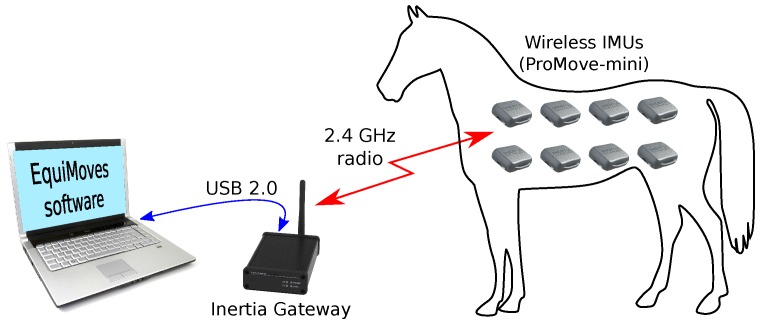
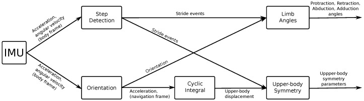
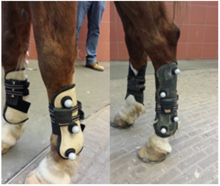
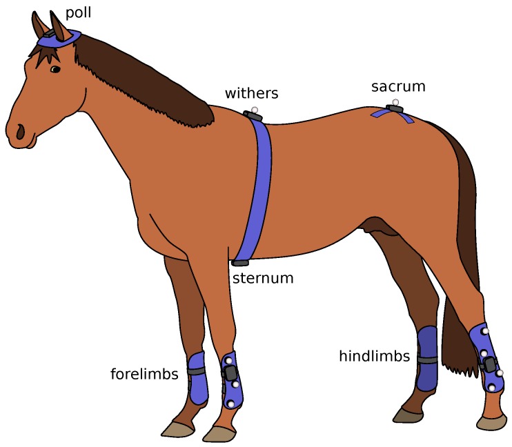
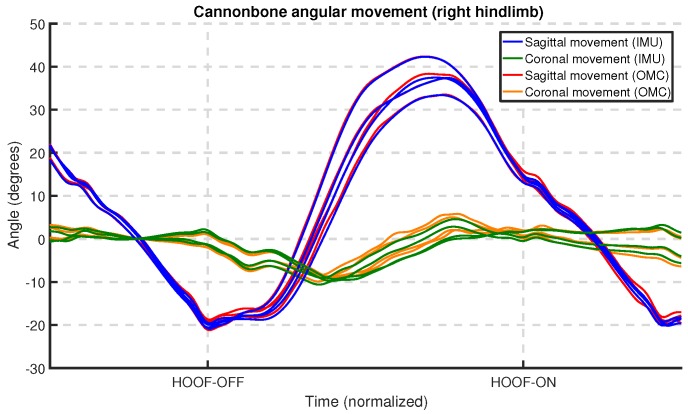
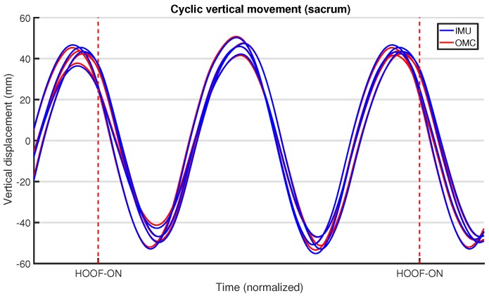
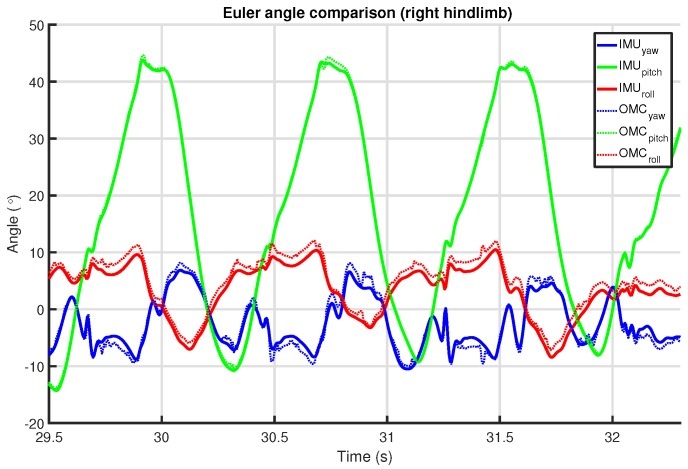
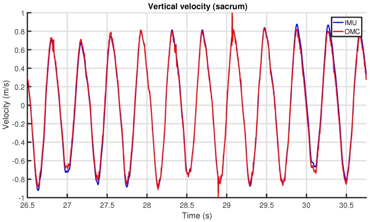
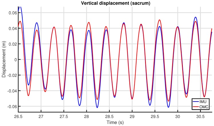
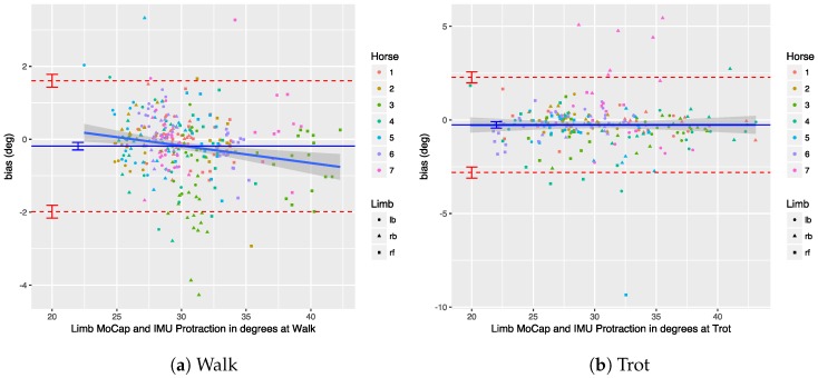
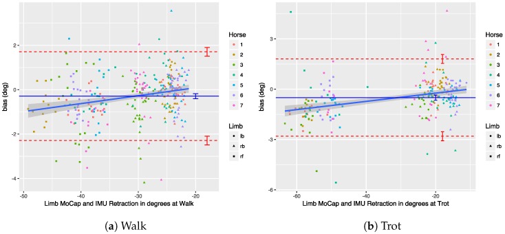
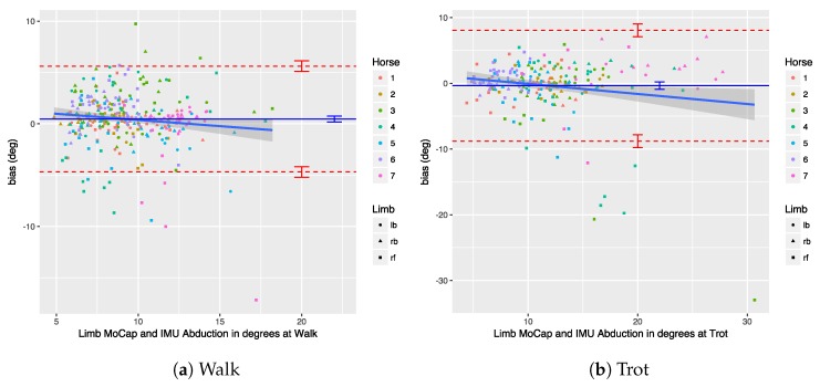
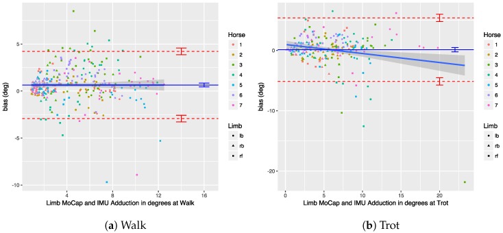
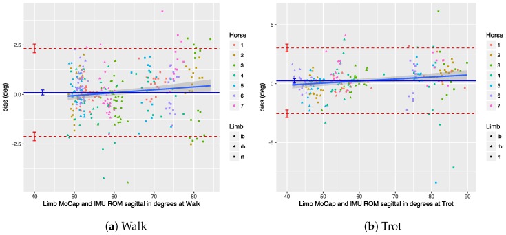
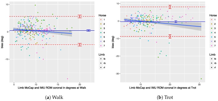
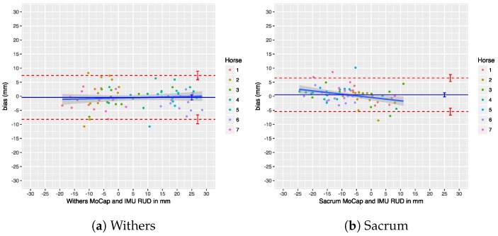
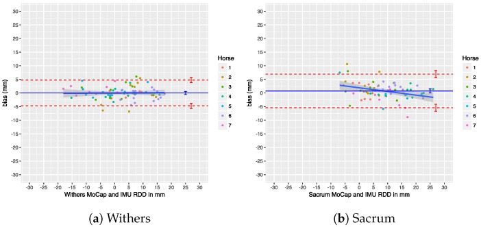
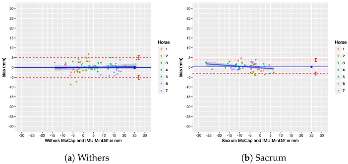
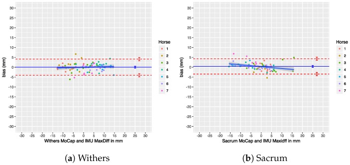
In this paper, we describe and validate the EquiMoves system, which aims to support equine veterinarians in assessing lameness and gait performance in horses. The system works by capturing horse motion from up to eight synchronized wireless inertial measurement units. It can be used in various equine gait modes, and analyzes both upper-body and limb movements. The validation against an optical motion capture system is based on a Bland-Altman analysis that illustrates the agreement between the two systems. The sagittal kinematic results (protraction, retraction, and sagittal range of motion) show limits of agreement of ± 2.3 degrees and an absolute bias of 0.3 degrees in the worst case. The coronal kinematic results (adduction, abduction, and coronal range of motion) show limits of agreement of - 8.8 and 8.1 degrees, and an absolute bias of 0.4 degrees in the worst case. The worse coronal kinematic results are most likely caused by the optical system setup (depth perception difficulty and suboptimal marker placement). The upper-body symmetry results show no significant bias in the agreement between the two systems; in most cases, the agreement is within ±5 mm. On a trial-level basis, the limits of agreement for withers and sacrum are within ±2 mm, meaning that the system can properly quantify motion asymmetry. Overall, the bias for all symmetry-related results is less than 1 mm, which is important for reproducibility and further comparison to other systems.

Keywords: IMU; agreement analysis; gait analysis; horse; lameness; optical motion capture.


Conflict of interest statement

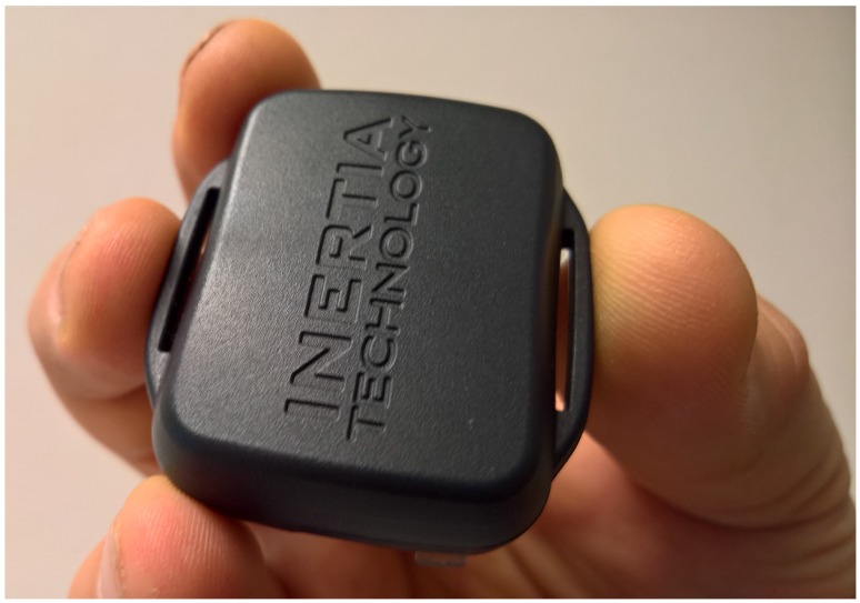
Mihai Marin-Perianu, Raluca Marin-Perianu and Paul Havinga founded Inertia-Technology B.V. (Enschede, The Netherlands), which sells the inertial sensor system (ProMove-mini) that is used as the basis of the EquiMoves system, which is evaluated in this study. Stephan Bosch and Berend-Jan van der Zwaag are employees of Inertia-Technology B.V.

Figures

Figure A1
Figure A1
Box plots for the sagittal limb angles measured by IMU and OMC.
Figure A2
Figure A2
Box plots for the coronal limb angles measured by IMU and OMC.
Figure A3
Figure A3
Box plot for the upper-body vertical displacement parameters of IMU and OMC.
Figure 1
Figure 1
Relevant anatomical positions on a horse.
Figure 2
Figure 2
Planes for horse motion.
Figure 3
Figure 3
Forelimb protraction and retraction angles, and hindlimb adduction and abduction angles.
Figure 4
Figure 4
Upper-body vertical displacement annotated with symmetry parameters. The timing of hoof-on instances is approximate.
Figure 5
Figure 5
System overview. IMU: inertial measurement unit.
Figure 6
Figure 6
ProMove-mini.
Figure 7
Figure 7
The EquiMoves motion processing framework.
Figure 8
Figure 8
The experiment venue at Utrecht University including the approximate path of the horses.
Figure 9
Figure 9
Placement of the IMU sensors and optical motion capture markers on fore- and hindlimbs.
Figure 10
Figure 10
Overview of the equipment attached to each horse.
Figure 11
Figure 11
Example of the cross-correlation between angular velocity magnitude between IMU and optical motion capture (OMC).
Figure 12
Figure 12
Example of the synchronization of angular velocity magnitude between IMU and OMC.
Figure 13
Figure 13
Example of the alignment of angular velocity between IMU and OMC.
Figure 14
Figure 14
Example of the cannon bone angular motion for IMU and OMC systems.
Figure 15
Figure 15
Example of the vertical displacement at the sacrum for IMU and OMC systems. The vertical dashed lines indicate the left hindlimb hoof-on moments used for stride segmentation.
Figure 16
Figure 16
Example of the limb orientation match between IMU and OMC systems for the right hindlimb.
Figure 17
Figure 17
Example of the vertical velocity match between IMU and OMC systems for the sacrum.
Figure 18
Figure 18
Example of the vertical displacement match between IMU and OMC systems for the sacrum.
Figure 19
Figure 19
Bland–Altman analysis for protraction angles at walk and trot.
Figure 20
Figure 20
Bland–Altman analysis for retraction angles at walk and trot.
Figure 21
Figure 21
Bland–Altman analysis for abduction angles at walk and trot.
Figure 22
Figure 22
Bland–Altman analysis for adduction angles at walk and trot.
Figure 23
Figure 23
Bland–Altman analysis for sagittal range of motion (ROM) at walk and trot.
Figure 24
Figure 24
Bland–Altman analysis for coronal ROM at walk and trot.
Figure 25
Figure 25
Bland–Altman analysis for Range_diffup parameter.
Figure 26
Figure 26
Bland–Altman analysis for Range_diffdown parameter.
Figure 27
Figure 27
Bland–Altman analysis for Min_diff parameter.
Figure 28
Figure 28
Bland–Altman analysis for Max_diff parameter.

References

    1. Van Weeren P.R., Pfau T., Rhodin M., Roepstorff L., Serra Bragança F., Weishaupt M.A. Do we have to redefine lameness in the era of quantitative gait analysis? Equine Vet. J. 2017;49:567–569. doi: 10.1111/evj.12715. - DOI - PubMed
    1. United States. National Animal Health Monitoring System (U.S.) National Economic Cost of Equine Lameness, Colic, and Equine Protozoal Myeloencephalitis (EPM) in the United States. U.S. Department of Agriculture, APHIS; Fort Collins, CO, USA: 2001.
    1. Loomans J.B.A., Stolk P.W.T., van Weeren P.R., Vaarkamp H., Barneveld A. A survey of the workload and clinical skills in current equine practices in The Netherlands. Equine Vet. Educ. 2007;19:162–168. doi: 10.2746/095777307X186875. - DOI
    1. Parkes R.S.V., Weller R., Groth A.M., May S., Pfau T. Evidence of the development of ‘domain-restricted’ expertise in the recognition of asymmetric motion characteristics of hindlimb lameness in the horse. Equine Vet. J. 2009;41:112–117. doi: 10.2746/042516408X343000. - DOI - PubMed
    1. Arkell M., Archer R.M., Guitian F.J., May S.A. Evidence of bias affecting the interpretation of the results of local anaesthetic nerve blocks when assessing lameness in horses. Vet. Rec. 2006;159:346–348. doi: 10.1136/vr.159.11.346. - DOI - PubMed