Review
. 2018 Mar 13;10(3):70.
doi: 10.3390/cancers10030070.

Any Place for Immunohistochemistry within the Predictive Biomarkers of Treatment in Lung Cancer Patients?

Affiliations
Review

Any Place for Immunohistochemistry within the Predictive Biomarkers of Treatment in Lung Cancer Patients?

Véronique Hofman et al. Cancers (Basel). .

Abstract

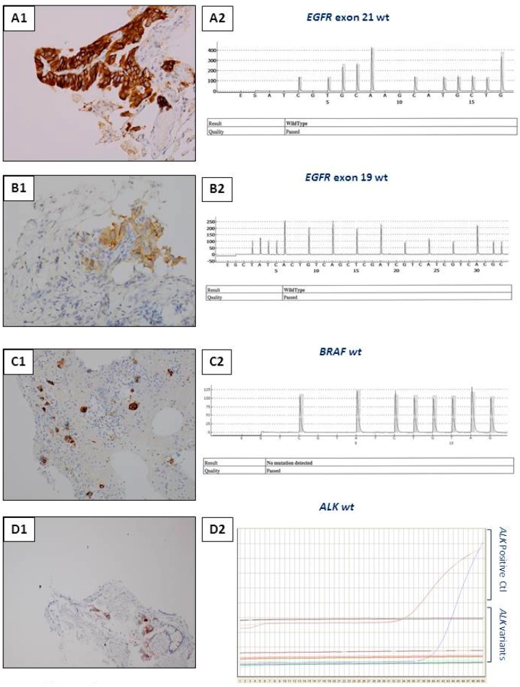
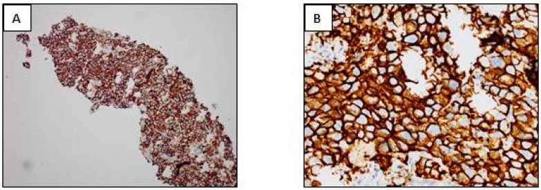
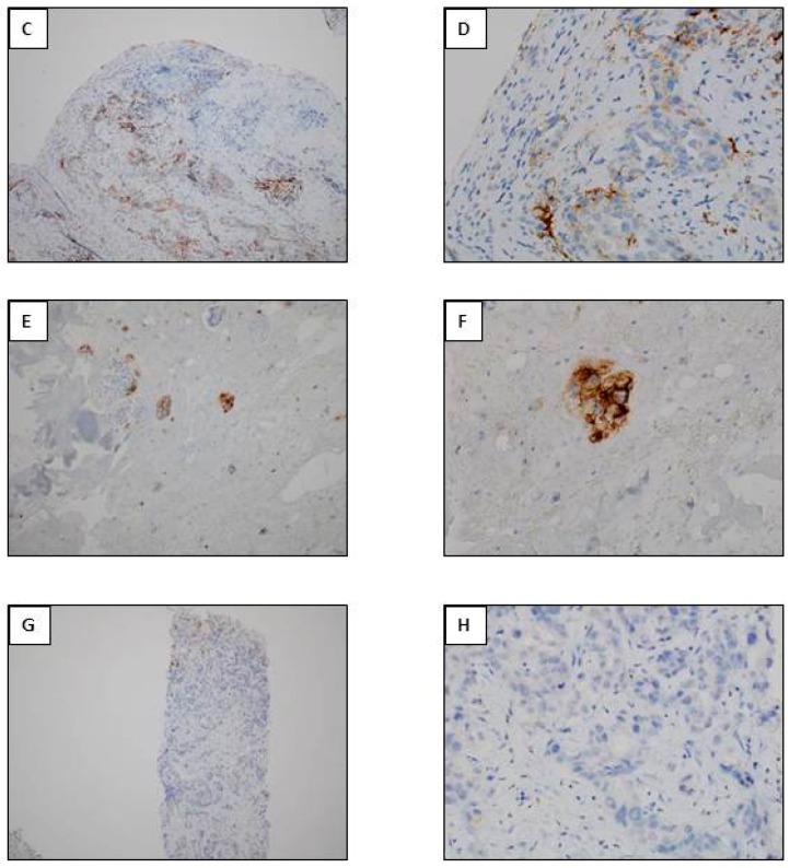
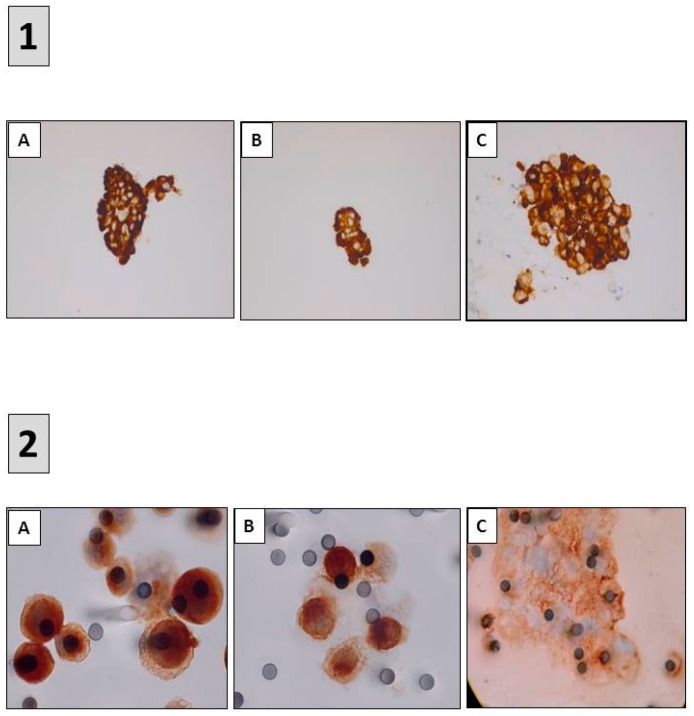
The identification of certain genomic alterations (EGFR, ALK, ROS1, BRAF) or immunological markers (PD-L1) in tissues or cells has led to targeted treatment for patients presenting with late stage or metastatic lung cancer. These biomarkers can be detected by immunohistochemistry (IHC) and/or by molecular biology (MB) techniques. These approaches are often complementary but depending on, the quantity and quality of the biological material, the urgency to get the results, the access to technological platforms, the financial resources and the expertise of the team, the choice of the approach can be questioned. The possibility of detecting simultaneously several molecular targets, and of analyzing the degree of tumor mutation burden and of the micro-satellite instability, as well as the recent requirement to quantify the expression of PD-L1 in tumor cells, has led to case by case development of algorithms and international recommendations, which depend on the quality and quantity of biological samples. This review will highlight the different predictive biomarkers detected by IHC for treatment of lung cancer as well as the present advantages and limitations of this approach. A number of perspectives will be considered.

Keywords: immune-oncology; immunocytochemistry; immunohistochemistry; lung cancer; predictive biomarkers.


Conflict of interest statement

The authors declare no conflict of interest.

Figures

Figure 1
Figure 1
Potential scenarios for detection of theranostic biomarkers for lung adenocarcinomas using thoracic biopsies. Depending on the quantity of tumor cells detected on hematoxylin and eosin stained sections, immunohistochemistry (IHC) and/or molecular biology approaches can be used (with or without panels). (A) Large biopsy with a high percentage of tumor cells (more than 50%). Paraffin block with one biopsy (a) and corresponding 4 tissue sections stained with hematoxylin eosin (b). Different magnifications of the same tissue section showing high number of adenocarcinoma cells (ce). In this latter case, the number of tumor cells should allow PD-L1 IHC to be performed first and then followed by NGS. Alternatively a workflow including successive PD-L1, ALK, ROS1, and BRAF IHC, and then NGS can be also adopted. (B) Small biopsies with a few tumor cells. Paraffin block with small biopsies (a) and corresponding 4 tissue sections stained with hematoxylin eosin (b). Different magnifications of the same tissue section showing in only one area (circle) a low number of tumor cells (no more than 15 tumor cells) (ce). IHC targeting PD-L1, then IHC for EGFR, ALK, ROS1, and BRAF should be probably done. (C) Large biopsies with a low percentage of tumor cells (less than 5%). Paraffin block with at least 5 large biopsies (a) and corresponding 4 tissue sections stained with hematoxylin eosin (b). Different magnifications of the same tissue section showing in only one area (circle) some tumor cells (ce). IHC for PD-L1 then successively for ALK, ROS1 and BRAF can be done and followed by targeted molecular biology for detection of EGFR mutations on a single tissue section or alternatively IHC for EGFR, according to the sensitivity of the MB test [1Ac, 1Bc, 1Cc, original magnification ×25; 1Ad, 1Bd, 1Cd, original magnification ×100; 1Ae, 1Be, 1Ce, original magnification ×400].
Figure 1
Figure 1
Potential scenarios for detection of theranostic biomarkers for lung adenocarcinomas using thoracic biopsies. Depending on the quantity of tumor cells detected on hematoxylin and eosin stained sections, immunohistochemistry (IHC) and/or molecular biology approaches can be used (with or without panels). (A) Large biopsy with a high percentage of tumor cells (more than 50%). Paraffin block with one biopsy (a) and corresponding 4 tissue sections stained with hematoxylin eosin (b). Different magnifications of the same tissue section showing high number of adenocarcinoma cells (ce). In this latter case, the number of tumor cells should allow PD-L1 IHC to be performed first and then followed by NGS. Alternatively a workflow including successive PD-L1, ALK, ROS1, and BRAF IHC, and then NGS can be also adopted. (B) Small biopsies with a few tumor cells. Paraffin block with small biopsies (a) and corresponding 4 tissue sections stained with hematoxylin eosin (b). Different magnifications of the same tissue section showing in only one area (circle) a low number of tumor cells (no more than 15 tumor cells) (ce). IHC targeting PD-L1, then IHC for EGFR, ALK, ROS1, and BRAF should be probably done. (C) Large biopsies with a low percentage of tumor cells (less than 5%). Paraffin block with at least 5 large biopsies (a) and corresponding 4 tissue sections stained with hematoxylin eosin (b). Different magnifications of the same tissue section showing in only one area (circle) some tumor cells (ce). IHC for PD-L1 then successively for ALK, ROS1 and BRAF can be done and followed by targeted molecular biology for detection of EGFR mutations on a single tissue section or alternatively IHC for EGFR, according to the sensitivity of the MB test [1Ac, 1Bc, 1Cc, original magnification ×25; 1Ad, 1Bd, 1Cd, original magnification ×100; 1Ae, 1Be, 1Ce, original magnification ×400].
Figure 2
Figure 2
Examples of different types of staining of immunohistochemistry (IHC) obtained with bronchial biopsies with antibodies used as theranostic tools for lung adenocarcinoma. (A,B) ALK IHC (D5F3, Ventana); (C,D) ROS1 IHC (D4D6, Cell Signaling); (E,F) EGFR IHC (L858 EGFR mutation; SP125, Ventana); (G,H) EGFR IHC (del 19 EGFR mutation; SP111, Ventana); (I,J) BRAF V600E IHC (VE1, Ventana); (K,L) pan Trk IHC (A7H6R, Cell Signaling). (A,C,E,G,I,K) Immunoperoxidase, magnification ×100; (B,D,F,H,J,L) Immunoperoxidase, magnification ×400.
Figure 2
Figure 2
Examples of different types of staining of immunohistochemistry (IHC) obtained with bronchial biopsies with antibodies used as theranostic tools for lung adenocarcinoma. (A,B) ALK IHC (D5F3, Ventana); (C,D) ROS1 IHC (D4D6, Cell Signaling); (E,F) EGFR IHC (L858 EGFR mutation; SP125, Ventana); (G,H) EGFR IHC (del 19 EGFR mutation; SP111, Ventana); (I,J) BRAF V600E IHC (VE1, Ventana); (K,L) pan Trk IHC (A7H6R, Cell Signaling). (A,C,E,G,I,K) Immunoperoxidase, magnification ×100; (B,D,F,H,J,L) Immunoperoxidase, magnification ×400.
Figure 3
Figure 3
Examples of interest for use of IHC as a unique tool for theranostic biomarker detection in biopsies with a few tumor cells. (A) L858R EGFR mutation detection. (A1) Positive EGFR IHC using the SP125 clone. (A2) A negative molecular biology result (using a pyrosequencing/therascreen EGFR panel from Qiagen, Hilden, Germany) for detection of the L858R EGFR mutation with the same biopsy. (B) Detection of the Del 19 EGFR mutation. (B1) Positive EGFR IHC using the SP111 clone. (B2) A negative molecular biology result (using a pyrosequencing/therascreen EGFR panel from Qiagen, Hilden, Germany) for the Del 19 mutation in EGFR detected in the same biopsy. (C) Detection of the BRAF V600E mutation. (C1) Positive BRAFV600E IHC using the VE1 clone. (C2) A negative molecular biology result (using a pyrosequencing/therascreen BRAF panel from Qiagen) for detection of a BRAF mutation. (D) ALK status detection. (D1) ALK status assessment. (D1) A positive ALK IHC using the D5F3 clone. (D2) A negative molecular result for detection of a ALK rearrangement [using RT-PCR targeting the variants 1 (E13; A20), 2 (E20; A20), 3A and 3B (E06; A20), 5 (E02; A20) and 7 (E17; A20) of ALK].
Figure 4
Figure 4
PD-L1 IHC (PD-L1 IHC 22C3 pharmDx, Agilent Technologies, Inc., Santa Clara, CA, USA). (A,B) Positivity of more than 50% of tumor cells. (C,D). Positivity of more than 1% and less than 50% of tumor cells. (E,F) Positivity of a few cells of a lymphangitis carcinoma. (G,H) Negative IHC. (A,C,E,G) immunoperoxisase, magnification ×100; (B,D,F,H) immunoperoxidase, magnification ×400).
Figure 4
Figure 4
PD-L1 IHC (PD-L1 IHC 22C3 pharmDx, Agilent Technologies, Inc., Santa Clara, CA, USA). (A,B) Positivity of more than 50% of tumor cells. (C,D). Positivity of more than 1% and less than 50% of tumor cells. (E,F) Positivity of a few cells of a lymphangitis carcinoma. (G,H) Negative IHC. (A,C,E,G) immunoperoxisase, magnification ×100; (B,D,F,H) immunoperoxidase, magnification ×400).
Figure 5
Figure 5
Detection of predictive biomarkers using immunocytochemistry (ICC). (1) ALK ICC (D5F3) (A) ROS1 ICC (D4D6) (B) and PD-L1 ICC (22C3) (C) performed on samples obtained after endobronchial ultrasound trans-bronchial needle aspirations (AC) immunoperoxidase, magnification ×400. (2) ALK ICC (D5F3) (A) MET ICC (SP44) (B) and PD-L1 ICC (22C3) (C) performed on circulating tumor cells isolated on filters after blood filtration (AC) immunoperoxidase, magnification ×1000.

References

    1. Hoseok I., Cho J.Y. Lung Cancer Biomarkers. Adv. Clin. Chem. 2015;72:107–170. - PubMed
    1. Lewis C., McQuaid S., Hamilton P.W., Salto-Tellez M., McArt D., James J.A. Building a ‘Repository of Science’: The importance of integrating biobanks within molecular pathology programmes. Eur. J. Cancer. 2016;67:191–199. doi: 10.1016/j.ejca.2016.08.009. - DOI - PubMed
    1. Oberndorfer F., Müllauer L. Molecular pathology of lung cancer: Current status and perspectives. Curr. Opin. Oncol. 2017;30:69–76. doi: 10.1097/CCO.0000000000000429. - DOI - PubMed
    1. Sholl L. Molecular diagnostics of lung cancer in the clinic. Transl. Lung Cancer Res. 2017;6:560–569. doi: 10.21037/tlcr.2017.08.03. - DOI - PMC - PubMed
    1. Aisner D.L., Sholl L.M., Berry L., Rossi M., Chen H., Fujimoto J., Moreira A.L., Ramalingam S., Villaruz L.C., Otterson G.A., et al. The Impact of Smoking and TP53 mutations in lung adenocarcinoma patients with targetable mutations—The Lung Cancer Mutation Consortium (LCMC2) Clin. Cancer Res. 2018;24:1038–1047. doi: 10.1158/1078-0432.CCR-17-2289. - DOI - PMC - PubMed