Review
. 2018 Mar 13;23(3):652.
doi: 10.3390/molecules23030652.

Seasonal Reproduction in Vertebrates: Melatonin Synthesis, Binding, and Functionality Using Tinbergen's Four Questions

Affiliations
Review

Seasonal Reproduction in Vertebrates: Melatonin Synthesis, Binding, and Functionality Using Tinbergen's Four Questions

Dax viviD et al. Molecules. .

Abstract

One of the many functions of melatonin in vertebrates is seasonal reproductive timing. Longer nights in winter correspond to an extended duration of melatonin secretion. The purpose of this review is to discuss melatonin synthesis, receptor subtypes, and function in the context of seasonality across vertebrates. We conclude with Tinbergen's Four Questions to create a comparative framework for future melatonin research in the context of seasonal reproduction.

Keywords: melatonin; seasonal reproduction; vertebrates.


Conflict of interest statement

The Authors declare that there is no conflict of interest.

Figures

Figure 1
Figure 1
Phototransduction pathway in mammals. Light is detected by the retina. Photic information is transduced via the retinohypothalamic tract to nuclei in the hypothalamus. The suprachiasmatic nucleus (SCN) projects to the paraventricular nucleus (PVN), which synapses in the superior cervical ganglion (SCG). The signal is transmitted from the SCG to the Pineal.
Figure 2
Figure 2
Transcription (a) and activation (b) of melatonin-synthesizing enzymes. Transcription factors of tph include Sp1 and CBF/NF-Y complex [17]. REV-ERBα inhibits tph transcription [18], implying some connection with circadian regulation, but it is not known through what mechanism. TPH is phosphorylated/activated by calmodulin (CaM), phosphokinase A (PKA) and phosphokinase C (PKC) [17]. Transcription factors of aadc can include AP-2 or octamer transcription factors [19], neither of which are known for circadian regulation. AADC depends on pyridoxal phosphate for functionality [20]. Transcription factors of aanat and hiomt include AP-1 and phosphorylated CREB (P-CREB). The aanat gene is likely regulated in a circadian fashion via the CLOCK/BMAL heterodimer [17]. AANAT and HIOMT have binding sites for casein kinase type II (CK-II) and PKC, and activation of HIOMT is enabled by tyrosine kinase (TK) [17].
Figure 3
Figure 3
Percent Identity Matrix of Melatonin Membrane Receptor Subtypes and Quinone Reductase 2. Analysis run by Clustal Omega 2.1 using NCBI GenBank. Red solid lines outline non-mammalian vertebrates (nmv), yellow dotted lines outline rodents, and blue dashed lines outline primates. Shades of grey indicate ranges of percent identity, black 100% and progressively lighter shades of grey down to 60%. Values <60% identity are white. Non-mammalian vertebrate (nmv) Mel1B sequences (lower right corner) do not have significant similarity with mammalian (m) Mel1B sequences (mid-upper left), suggesting that these are phylogenetically distinct melatonin membrane receptors. There is higher similarity within Mel1A receptor subtypes across vertebrates (mid-lower right). The quinone family in the upper-left [named NAD(P)H dehydrogenase quinone 2, ribosyldihydronicotinamide dehydrogenase, or NQO2] shows no significant similarity with other membrane melatonin receptor subtypes with percent identity across vertebrates and melatonin receptor subtypes <60%, supporting the position that QR2/NQO2 is not a putative MT3 membrane subtype receptor.
Figure 4
Figure 4
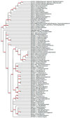
Cladogram of melatonin membrane receptor subtypes and quinone reductase 2. Modified from neighbor-joining tree (without distance corrections), generated by Phylogeny.fr (Dereeper et al., 2008 & 2010). Red numbers represent branch support values. Branch support values smaller than 50% are collapsed. The quinone family is named NAD(P)H dehydrogenase quinone 2, ribosyldihydronicotinamide dehydrogenase, or NQO2, based on how it is named in the NCBI database. Opioid Receptor for Homo sapiens (NCBI Accession No. L29301.1) served as the outgroup. NCBI Accession Numbers and full names of sequences are in Figure 4. The mRNA sequences of non-mammalian vertebrates (nmv) Mel1A diverged more recently than mammalian (m) Mel1B from mMel1A. The pharmacological evidence that Mel1A/MT1 in birds has one order of magnitude lower affinity for 2-[125I]iodomelatonin than MT1 in some mammals (rabbit, sheep and horse) and one order of magnitude higher than MT2 other mammals (Syrian and Siberian hamsters) supports the phylogenetic evidence presented here that avian Mel1A mRNA is not evolutionarily homologous with mammalian Mel1A/MT1 mRNA.
Figure 5
Figure 5
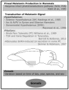
Summary of major findings on melatonin in the mammalian HPG axis.
Figure 6
Figure 6
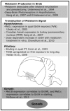
Summary of major findings on melatonin in the avian HPG axis.

Similar articles

Cited by

References

    1. Tan D.-X., Manchester L.C., Liu X., Rosales-Corral S.A., Acuna-Castroviejo D., Reiter R.J. Mitochondria and chloroplasts as the original sites of melatonin synthesis: A hypothesis related to melatonin’s primary function and evolution in eukaryotes. J. Pineal Res. 2013;54:127–138. doi: 10.1111/jpi.12026. - DOI - PubMed
    1. Reiter R.J. Pineal melatonin: Cell biology of its synthesis and of its physiological interactions. Endocr. Rev. 1991;12:151–180. doi: 10.1210/edrv-12-2-151. - DOI - PubMed
    1. Hoffman R.A., Reiter R.J. Pineal gland: influence on gonads of male hamsters. Science. 1965;148:1609–1611. doi: 10.1126/science.148.3677.1609. - DOI - PubMed
    1. Cassone V.M. Effects of melatonin on vertebrate circadian systems. Trends Neurosci. 1990;13:457–464. doi: 10.1016/0166-2236(90)90099-V. - DOI - PubMed
    1. Klein D.C., Moore R.Y. Pineal N-acetyltransferase and hydroxyindole-O-methyl-transferase: Control by the retinohypothalamic tract and the suprachiasmatic nucleus. Brain Res. 1979;174:245–262. doi: 10.1016/0006-8993(79)90848-5. - DOI - PubMed

LinkOut - more resources