. 2018 May 1;141(5):1422-1433.
doi: 10.1093/brain/awy054.

Artificial limb representation in amputees

Affiliations

Artificial limb representation in amputees

Fiona M Z van den Heiligenberg et al. Brain. .

Abstract

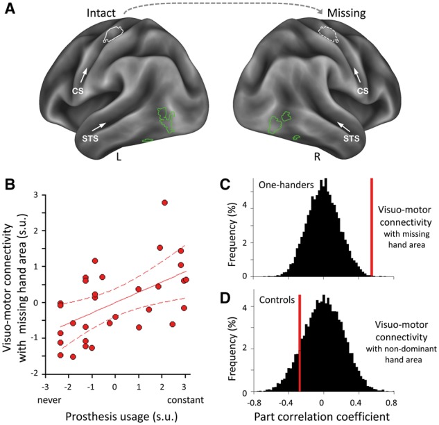
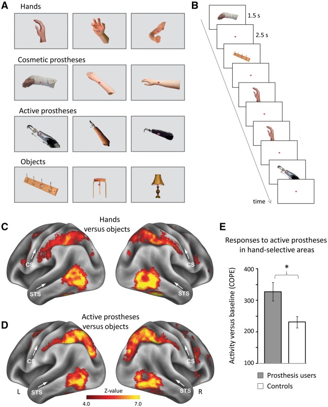
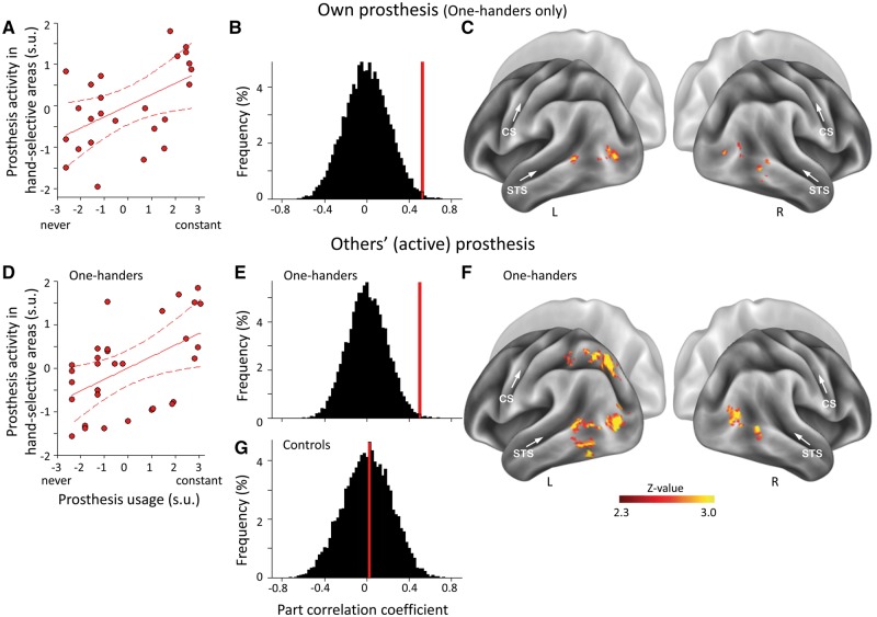
The human brain contains multiple hand-selective areas, in both the sensorimotor and visual systems. Could our brain repurpose neural resources, originally developed for supporting hand function, to represent and control artificial limbs? We studied individuals with congenital or acquired hand-loss (hereafter one-handers) using functional MRI. We show that the more one-handers use an artificial limb (prosthesis) in their everyday life, the stronger visual hand-selective areas in the lateral occipitotemporal cortex respond to prosthesis images. This was found even when one-handers were presented with images of active prostheses that share the functionality of the hand but not necessarily its visual features (e.g. a 'hook' prosthesis). Further, we show that daily prosthesis usage determines large-scale inter-network communication across hand-selective areas. This was demonstrated by increased resting state functional connectivity between visual and sensorimotor hand-selective areas, proportional to the intensiveness of everyday prosthesis usage. Further analysis revealed a 3-fold coupling between prosthesis activity, visuomotor connectivity and usage, suggesting a possible role for the motor system in shaping use-dependent representation in visual hand-selective areas, and/or vice versa. Moreover, able-bodied control participants who routinely observe prosthesis usage (albeit less intensively than the prosthesis users) showed significantly weaker associations between degree of prosthesis observation and visual cortex activity or connectivity. Together, our findings suggest that altered daily motor behaviour facilitates prosthesis-related visual processing and shapes communication across hand-selective areas. This neurophysiological substrate for prosthesis embodiment may inspire rehabilitation approaches to improve usage of existing substitutionary devices and aid implementation of future assistive and augmentative technologies.


Figures

Figure 1
Figure 1
Stronger connectivity between hand-selective areas in the visual and sensorimotor systems relates to greater prosthesis usage. (A) Individualized regions of interest in an example participant. Hand-selective voxels were identified in lateral occipitotemporal cortex bilaterally by contrasting responses to hand versus object images (green) or in SI/M1 unilaterally by contrasting intact-hand (or dominant hand in controls) versus feet movements (white). The putative sensorimotor missing-hand area was estimated (hatched white) by mirror projecting the intact hand region of interest across hemispheres. CS = central sulcus; R/L = right/left hemispheres; STS = superior temporal sulcus. (B) Visuo-motor functional connectivity (between bilateral lateral occipitotemporal cortex and missing-hand SI/M1): correlations with prosthesis usage in one-handers. Visuomotor connectivity with the intact hand sensorimotor region of interest was regressed out of the missing hand visuo-motor measure. Scatter diagram is fitted with regression line and associated 95% confidence intervals; s.u. = standardized units. (C and D) Correlations for visuomotor connectivity with prosthesis usage (C) and observance (D) in one-handers and controls, respectively. Permutation tests of the null distributions (black) show that the correlation between visuo-motor connectivity and prosthesis usage (red) is significantly greater in one-handers than chance, but not in controls.
Figure 2
Figure 2
Experimental design and brain activity. (A) Example stimuli of hands, cosmetic and active prostheses, and objects. Participants’ own prostheses were included (‘own’ condition) as well as prostheses from other participants (‘other’s’ condition). (B) Stimuli were presented in an event-related design, involving a one-back recognition task. (C and D) Whole-brain activity maps for (C) hands and (D) active prostheses versus objects across all participants. Prostheses images activated lateral occipito-temporal cortex, partially overlapping with hand-selective activity. CS = central sulcus; R/L = right/left hemispheres; STS = superior temporal sulcus. (E) Prosthesis users (n = 26) show stronger activity than controls in response to active prostheses in the visual hand area. COPE = contrasts of parameter estimates. Error bars indicate SEM. *P = 0.008.
Figure 3
Figure 3
Activity for prostheses in visual hand-selective areas relates to prosthesis usage. Correlations between prosthesis usage and prosthesis activity in lateral occipito-temporal cortex for one’s own prosthesis (A–C) and exemplars of unfamiliar (others’) active prostheses (D–F). Correlations within individuals’ visual hand-selective regions of interest (A and D) and whole-brain analysis (C and F) are presented. Correlation was calculated while controlling for objects activity to achieve prosthesis-specific variations in functional MRI signal. Correlations of prosthesis-related activity with prosthesis usage are significantly greater in one-handers than chance (B and E), but not with passive observation in controls (G). CS = central sulcus; R/L = right/left hemispheres; STS = superior temporal sulcus.

References

    1. Antfolk C, D’Alonzo M, Rosen B, Lundborg G, Sebelius F, Cipriani C. Sensory feedback in upper limb prosthetics. Expert Rev Med Devices 2013; 10: 45–54. - PubMed
    1. Astafiev SV, Stanley CM, Shulman GL, Corbetta M. Extrastriate body area in human occipital cortex responds to the performance of motor actions. Nat Neurosci 2004; 7: 542–8. - PubMed
    1. Behzadi Y, Restom K, Liau J, Liu TT. A component based noise correction method (CompCor) for BOLD and perfusion based fMRI. Neuroimage 2007; 37: 90–101. - PMC - PubMed
    1. Biswal B, Yetkin FZ, Haughton VM, Hyde JS. Functional connectivity in the motor cortex of resting human brain using echo-planar mri. Magn Reson Med 1995; 34: 537–41. - PubMed
    1. Bracci S, Cavina-Pratesi C, Ietswaart M, Caramazza A, Peelen MV. Closely overlapping responses to tools and hands in left lateral occipitotemporal cortex. J Neurophysiol 2012; 107: 1443–56. - PubMed

Publication types

LinkOut - more resources